

Selective induction of apoptosis in antigen-presenting cells in mice by Parapoxvirus ovis
- PMID: 11312341
- PMCID: PMC114224
- DOI: 10.1128/JVI.75.10.4699-4704.2001
Selective induction of apoptosis in antigen-presenting cells in mice by Parapoxvirus ovis
Abstract
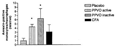
Viruses have evolved numerous mechanisms to avoid host immune reactions. Here we report a mechanism by which Parapoxvirus ovis (PPVO) interferes with antigen presentation. PPVO (orf virus) causes orf, an acute skin disease of sheep and goats worldwide. Importantly, PPVO can repeatedly infect its host in spite of a vigorous inflammatory and host immune response to the infection. We demonstrate in a mouse system that PPVO induces apoptosis in a significant number of antigen-presenting cells after intraperitoneal injection using the CD95 pathway, thus preventing a primary T-cell response. We also show that PPVO induces a compensatory activation of the immune system. Our results may help to explain the phenomenon that natural PPVO infections in sheep occur repeatedly even after short intervals. They also suggest that the combination of immunosuppressive and immunostimulatory mechanisms is an effective survival strategy that might be used in other viruses as well.
Figures


Similar articles
-
Inactivated parapoxvirus ovis (Orf virus) has antiviral activity against hepatitis B virus and herpes simplex virus.J Gen Virol. 2003 Jul;84(Pt 7):1843-1852. doi: 10.1099/vir.0.19138-0. J Gen Virol. 2003. PMID: 12810878
-
Immunomodulatory effects of inactivated parapoxvirus ovis (ORF virus) on human peripheral immune cells: induction of cytokine secretion in monocytes and Th1-like cells.J Virol. 2004 Sep;78(17):9400-11. doi: 10.1128/JVI.78.17.9400-9411.2004. J Virol. 2004. PMID: 15308734 Free PMC article.
-
CD95 (Fas) may control the expansion of activated T cells after elimination of bacteria in murine listeriosis.Infect Immun. 1997 May;65(5):1883-91. doi: 10.1128/iai.65.5.1883-1891.1997. Infect Immun. 1997. PMID: 9125576 Free PMC article.
-
Ovine diseases. Orf.Vet Res. 1998 May-Aug;29(3-4):311-26. Vet Res. 1998. PMID: 9689744 Review.
-
Immunity and counter-immunity during infection with the parapoxvirus orf virus.Virus Res. 2002 Sep;88(1-2):3-16. doi: 10.1016/s0168-1702(02)00117-x. Virus Res. 2002. PMID: 12297324 Review.
Cited by
-
Conventional bone marrow-derived dendritic cells contribute to toll-like receptor-independent production of alpha/beta interferon in response to inactivated parapoxvirus ovis.J Virol. 2009 Sep;83(18):9411-22. doi: 10.1128/JVI.02362-08. Epub 2009 Jul 1. J Virol. 2009. PMID: 19570869 Free PMC article.
-
Treatment with the Immunomodulator AIC649 in Combination with Entecavir Produces Antiviral Efficacy in the Woodchuck Model of Chronic Hepatitis B.Viruses. 2021 Apr 9;13(4):648. doi: 10.3390/v13040648. Viruses. 2021. PMID: 33918831 Free PMC article.
-
Modulation of macrophage functions by sheeppox virus provides clues to understand interaction of the virus with host immune system.Virol J. 2005 Mar 22;2:22. doi: 10.1186/1743-422X-2-22. Virol J. 2005. PMID: 15784144 Free PMC article.
-
Characterisation of parapoxviruses isolated from Norwegian semi-domesticated reindeer (Rangifer tarandus tarandus).Virol J. 2005 Sep 5;2:79. doi: 10.1186/1743-422X-2-79. Virol J. 2005. PMID: 16143041 Free PMC article.
-
Emergence of Salmon Gill Poxvirus.Viruses. 2022 Dec 1;14(12):2701. doi: 10.3390/v14122701. Viruses. 2022. PMID: 36560705 Free PMC article. Review.
References
-
- Bertin J, Armstrong R C, Ottilie S, Martin D A, Wang Y, Banks S, Wang G H, Senkevich T G, Alnemri E S, Moss B, Lenardo M J, Tomaselli K J, Cohen J I. Death effector domain-containing herpesvirus and poxvirus proteins inhibit both Fas- and TNFR1-induced apoptosis. Proc Natl Acad Sci USA. 1997;94:1172–1176. - PMC - PubMed
-
- Buddle B B, Dellers R W, Schurig G G. Contagious ecthyma virus-vaccination failures. Am J Vet Res. 1984;45:263–266. - PubMed
-
- Buettner M, Czerny C-P, Lehner K-H, Wertz K. Interferon induction in peripheral blood mononuclear leukocytes of man and farm animals by poxvirus vector candidates and some poxvirus constructs. Vet Immunol Immunopathol. 1995;46:237–260. - PubMed
-
- Foerster R, Wolf G, Mayr A. Highly attenuated poxviruses induce functional priming of neutrophils in vitro. Arch Virol. 1994;136:219–226. - PubMed
-
- Haig D M, Deane D L, Percival A, Myatt N, Thomson J, Inglis L, Rothel J, Seow H F, Wood P, Miller H R P, Reid H W. The cytokine response of afferent lymph following orf virus reinfection of sheep. Vet Dermatol. 1996;7:11–20. - PubMed
MeSH terms
Substances
LinkOut - more resources
Full Text Sources
Other Literature Sources
Research Materials
