

N-glycans of F protein differentially affect fusion activity of human respiratory syncytial virus
- PMID: 11312346
- PMCID: PMC114229
- DOI: 10.1128/JVI.75.10.4744-4751.2001
N-glycans of F protein differentially affect fusion activity of human respiratory syncytial virus
Abstract
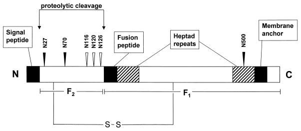
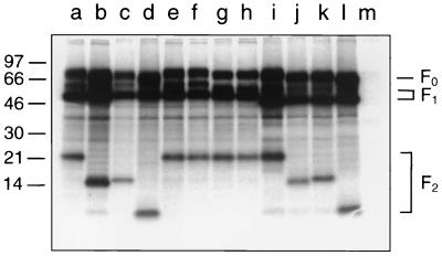
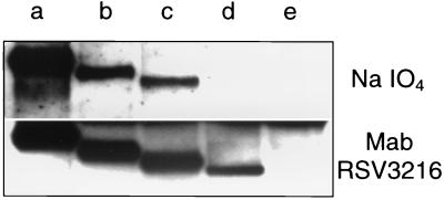
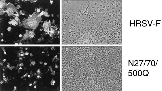
The human respiratory syncytial virus (Long strain) fusion protein contains six potential N-glycosylation sites: N27, N70, N116, N120, N126, and N500. Site-directed mutagenesis of these positions revealed that the mature fusion protein contains three N-linked oligosaccharides, attached to N27, N70, and N500. By introducing these mutations into the F gene in different combinations, four more mutants were generated. All mutants, including a triple mutant devoid of any N-linked oligosaccharide, were efficiently transported to the plasma membrane, as determined by flow cytometry and cell surface biotinylation. None of the glycosylation mutations interfered with proteolytic activation of the fusion protein. Despite similar levels of cell surface expression, the glycosylation mutants affected fusion activity in different ways. While the N27Q mutation did not have an effect on syncytium formation, loss of the N70-glycan caused a fusion activity increase of 40%. Elimination of both N-glycans (N27/70Q mutant) reduced the fusion activity by about 50%. A more pronounced reduction of the fusion activity of about 90% was observed with the mutants N500Q, N27/500Q, and N70/500Q. Almost no fusion activity was detected with the triple mutant N27/70/500Q. These data indicate that N-glycosylation of the F2 subunit at N27 and N70 is of minor importance for the fusion activity of the F protein. The single N-glycan of the F1 subunit attached to N500, however, is required for efficient syncytium formation.
Figures
Similar articles
-
Characterization of the role of N-glycosylation sites in the respiratory syncytial virus fusion protein in virus replication, syncytium formation and antigenicity.Virus Res. 2019 Jun;266:58-68. doi: 10.1016/j.virusres.2019.04.006. Epub 2019 Apr 17. Virus Res. 2019. PMID: 31004621
-
Carbohydrate modifications of the NDV fusion protein heptad repeat domains influence maturation and fusion activity.Virology. 2001 May 10;283(2):332-42. doi: 10.1006/viro.2001.0899. Virology. 2001. PMID: 11336558
-
Defining the N-linked glycosylation site of Hantaan virus envelope glycoproteins essential for cell fusion.J Microbiol. 2007 Feb;45(1):41-7. J Microbiol. 2007. PMID: 17342054
-
Functional analysis of the N-linked glycans within the fusion protein of respiratory syncytial virus.Methods Mol Biol. 2007;379:69-83. doi: 10.1007/978-1-59745-393-6_5. Methods Mol Biol. 2007. PMID: 17502671 Review.
-
The use of two-dimensional SDS-PAGE to analyze the glycan heterogeneity of the respiratory syncytial virus fusion protein.Methods Mol Biol. 2007;379:97-108. doi: 10.1007/978-1-59745-393-6_7. Methods Mol Biol. 2007. PMID: 17502673 Review.
Cited by
-
The role of N-linked glycosylation in proteolytic processing and cell surface transport of the Cedar virus fusion protein.Virol J. 2022 Aug 23;19(1):136. doi: 10.1186/s12985-022-01864-5. Virol J. 2022. PMID: 35999637 Free PMC article.
-
The RSV F p27 peptide: current knowledge, important questions.Front Microbiol. 2023 Jun 21;14:1219846. doi: 10.3389/fmicb.2023.1219846. eCollection 2023. Front Microbiol. 2023. PMID: 37415824 Free PMC article. Review.
-
Antibody responses of healthy adults to the p27 peptide of respiratory syncytial virus fusion protein.Vaccine. 2022 Jan 24;40(3):536-543. doi: 10.1016/j.vaccine.2021.11.087. Epub 2021 Dec 10. Vaccine. 2022. PMID: 34903371 Free PMC article.
-
Identification of residues in the human respiratory syncytial virus fusion protein that modulate fusion activity and pathogenesis.J Virol. 2015 Jan;89(1):512-22. doi: 10.1128/JVI.02472-14. Epub 2014 Oct 22. J Virol. 2015. PMID: 25339762 Free PMC article.
-
Toll-like receptor 4 is not involved in host defense against respiratory tract infection with Sendai virus.Immunol Lett. 2003 Oct 31;89(2-3):201-6. doi: 10.1016/s0165-2478(03)00138-x. Immunol Lett. 2003. PMID: 14556979 Free PMC article.
References
-
- Arumugham R G, Hildreth S W, Paradiso P R. Fatty acid acylation of the fusion protein of respiratory syncytial virus. J Biol Chem. 1989;264:10339–10342. - PubMed
-
- Bagai S, Lamb R A. Individual roles of N-linked oligosaccharide chains in intracellular transport of the paramyxovirus SV5 fusion protein. Virology. 1995;209:250–256. - PubMed
-
- Buckland R, Malvoisin E, Beauverger P, Wild F. A leucine zipper structure present in the measles virus fusion protein is not required for its tetramerization but is essential for fusion. J Gen Virol. 1992;73:1703–1707. - PubMed
Publication types
MeSH terms
Substances
LinkOut - more resources
Full Text Sources
Other Literature Sources
Miscellaneous
