. 2001 May;21(10):3523-33.
doi: 10.1128/MCB.21.10.3523-3533.2001.

Activated PAK4 regulates cell adhesion and anchorage-independent growth

Affiliations

Activated PAK4 regulates cell adhesion and anchorage-independent growth

J Qu et al. Mol Cell Biol. 2001 May.

Abstract

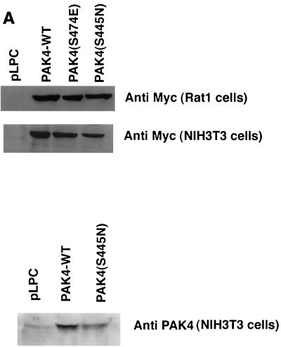
The serine/threonine kinase PAK4 is an effector molecule for the Rho GTPase Cdc42. PAK4 differs from other members of the PAK family in both sequence and function. Previously we have shown that an important function of this kinase is to mediate the induction of filopodia in response to activated Cdc42. Since previous characterization of PAK4 was carried out only with the wild-type kinase, we have generated a constitutively active mutant of the kinase to determine whether it has other functions. Expression of activated PAK4 in fibroblasts led to a transient induction of filopodia, which is consistent with its role as an effector for Cdc42. In addition, use of the activated mutant revealed a number of other important functions of this kinase that were not revealed by studying the wild-type kinase. For example, activated PAK4 led to the dissolution of stress fibers and loss of focal adhesions. Consequently, cells expressing activated PAK4 had a defect in cell spreading onto fibronectin-coated surfaces. Most importantly, fibroblasts expressing activated PAK4 had a morphology that was characteristic of oncogenic transformation. These cells were anchorage independent and formed colonies in soft agar, similar to what has been observed previously in cells expressing activated Cdc42. Consistent with this, dominant-negative PAK4 mutants inhibited focus formation by oncogenic Dbl, an exchange factor for Rho family GTPases. These results provide the first demonstration that a PAK family member can transform cells and indicate that PAK4 may play an essential role in oncogenic transformation by the GTPases. We propose that the morphological changes and changes in cell adhesion induced by PAK4 may play a direct role in oncogenic transformation by Rho family GTPases and their exchange factors.


Figures

FIG. 1
FIG. 1
PAK4(S445N) has increased kinase activity compared with wild-type PAK4 but is not enhanced in its activation of the JNK pathway. (A) NIH 3T3 cells were transiently transfected with either empty vector (Con) or vectors containing HA-tagged wild-type PAK4 (PAK4 WT) or HA-tagged PAK4(S474E) (left panel), or they were transiently transfected with empty vector or vectors containing Myc-tagged wild-type PAK4 or Myc-tagged PAK4(S445N) (right panel). After transient expression, PAK4 was immunopurified from cell lysates with anti-HA antibody (left) or anti-Myc antibody (right). Immunopurified PAK4 was incubated with HH4 or without any substrate in the presence of [γ-32P]ATP and kinase buffer. Substrate phosphorylation or autophosphorylation was analyzed after sodium dodecyl sulfate-polyacrylamide gel electrophoresis (SDS-PAGE) and autoradiography. Aliquots of each lysate were subjected to Western blot analysis with anti-HA (left) or anti-Myc (right) antibody to show that PAK4 was expressed at approximately equivalent levels in each transfection. (B) NIH 3T3 cells were transiently transfected with 1 μg of empty vector, Myc-tagged PAK4(S445N) (0.5 or 1.0 μg), or 1 μg of Myc-tagged wild-type PAK4 together with 1 μg of HA-tagged JNK expression vector. After transient transfection, HA-JNK was immunopurified from cell lysates using anti-HA antibody and then incubated with glutathione S-transferase–c-Jun in the presence of [γ-32P]ATP and kinase buffer. Substrate phosphorylation was analyzed after SDS-PAGE and autoradiography. As a positive control, cells were transfected with Rac2L61 expression vector and HA-JNK.
FIG. 2
FIG. 2
Transient expression of PAK4(S445N) results in cell rounding. NIH 3T3 cells were transfected with 1 μg of either EGFP expression vector (top), PAK4-IRES2-EGFP (middle), or PAK4(S445N)-IRES2-EGFP (bottom). Twenty-four hours after transfection, cells expressing the EGFP vectors were visualized by fluorescence microscopy under a 40× objective.
FIG. 3
FIG. 3
Morphological changes in fibroblasts stably expressing PAK4(S445N). (A) Representative Western blot analysis of Myc-tagged PAK4 expression in the different stable cell lines. Cell lysates (25 μg) from Rat1 cells or NIH 3T3 cells stably expressing the indicated plasmids were used in each blot. Blots were probed with anti-Myc antibody (top and middle panels) or anti-PAK4 antibody (bottom panel). (B) Rat1 cells expressing PAK4(S445N) produce filopodia after plating onto fibronectin. Cells expressing empty vector (pLPC) or PAK4(S445N) were plated onto fibronectin-coated coverslips and visualized by Zeiss Axiovert phase-contrast microscopy 10 min and 1 h after plating under a 100× objective. Cell morphology was visualized by time lapse photography and individual frames are shown in the figure. Panels i, ii, and iii show a PAK4(S445N) cell that was photographed at 10-s intervals 10 min after plating. Panel iv shows an example of a PAK4(S445N) cell that had already begun to spread onto the fibronectin-coated surface 1 h after plating. Panels v and vi show control cells 10 min and 1 h, respectively, after plating. (C) Morphological changes in Rat1 cells overexpressing PAK(S445N). Top panels, cells containing either PAK4(S445N) or empty vector (pLPC) were visualized by phase-contrast microscopy under a 10× objective 24 h after plating; middle panels, cells were fixed 24 h after plating onto fibronectin-coated coverslips, and polymerized actin was visualized under a 60× oil lens after staining with FITC-conjugated phalloidin; bottom panels, cells were fixed 24 h after plating, and focal adhesions were visualized by immunofluorescence microscopy under a 60× oil lens after staining with anti-vinculin antibody and rhodamine-conjugated secondary antibody. (D) Morphological changes in NIH 3T3 cells expressing PAK4(S445N). NIH 3T3 cells were analyzed as described for panel C.
FIG. 3
FIG. 3
Morphological changes in fibroblasts stably expressing PAK4(S445N). (A) Representative Western blot analysis of Myc-tagged PAK4 expression in the different stable cell lines. Cell lysates (25 μg) from Rat1 cells or NIH 3T3 cells stably expressing the indicated plasmids were used in each blot. Blots were probed with anti-Myc antibody (top and middle panels) or anti-PAK4 antibody (bottom panel). (B) Rat1 cells expressing PAK4(S445N) produce filopodia after plating onto fibronectin. Cells expressing empty vector (pLPC) or PAK4(S445N) were plated onto fibronectin-coated coverslips and visualized by Zeiss Axiovert phase-contrast microscopy 10 min and 1 h after plating under a 100× objective. Cell morphology was visualized by time lapse photography and individual frames are shown in the figure. Panels i, ii, and iii show a PAK4(S445N) cell that was photographed at 10-s intervals 10 min after plating. Panel iv shows an example of a PAK4(S445N) cell that had already begun to spread onto the fibronectin-coated surface 1 h after plating. Panels v and vi show control cells 10 min and 1 h, respectively, after plating. (C) Morphological changes in Rat1 cells overexpressing PAK(S445N). Top panels, cells containing either PAK4(S445N) or empty vector (pLPC) were visualized by phase-contrast microscopy under a 10× objective 24 h after plating; middle panels, cells were fixed 24 h after plating onto fibronectin-coated coverslips, and polymerized actin was visualized under a 60× oil lens after staining with FITC-conjugated phalloidin; bottom panels, cells were fixed 24 h after plating, and focal adhesions were visualized by immunofluorescence microscopy under a 60× oil lens after staining with anti-vinculin antibody and rhodamine-conjugated secondary antibody. (D) Morphological changes in NIH 3T3 cells expressing PAK4(S445N). NIH 3T3 cells were analyzed as described for panel C.
FIG. 4
FIG. 4
PAK4 does not phosphorylate MLCK or regulate MLC phosphorylation. (A) Wild-type PAK4, PAK4(S474E), or PAK4(S445N) were immunopurified from NIH 3T3 stable cell lines using anti-Myc antibody. Empty pLPC vector cells were used as a control. Immunoprecipitates were incubated with purified MLCK (41) and an in vitro kinase assay was carried out as described previously (41). Phosphorylated MLCK and PAK4 autophosphorylation were visualized after SDS-PAGE and autoradiography. (B) PAK4(S445N) does not lead to a decrease in MLC phosphorylation. Equal volumes of lysates from NIH 3T3 cells expressing pLPC or PAK4(S445N) were analyzed by Western blotting. Filters were probed with anti-phospho-MLC antibody, which recognizes phospho-serine 19 (top panel), or anti-MLC antibody (bottom panel).
FIG. 5
FIG. 5
Inhibition of spreading in cells expressing PAK4(S445N). Equal numbers of stable Rat1 cells expressing empty vector (pLPC) (diamonds) or PAK4(S445N) (squares) were plated onto dishes that were coated with fibronectin or collagen. At the indicated time intervals, cells were fixed and stained as described in Materials and Methods and visualized by phase-contrast microscopy. The number of spread cells was counted at each time point and expressed as a percentage of the total cell number.
FIG. 6
FIG. 6
PAK4(S445N) induces anchorage-independent growth, and PAK4 is required for oncogenic Dbl-induced focus formation. (A and B) PAK4(S445N) induces anchorage-independent growth. Soft agar growth assays were carried out as described in Materials and Methods. PAK4(S445N)-expressing cells (NIH 3T3 cells and Rat1 cells) produced colonies on soft agar similar to those of Ras-transformed cells. Pictures of the plates containing each indicated cell line were taken 2 weeks after plating. The larger colonies are visible by eye, as shown in panel A (no magnification). Representative foci of each indicated cell line were visualized by phase-contrast microscopy under a 10× objective and are shown in panel B. (C) PAK4 is required for focus formation by oncogenic Db1. NIH 3T3 cells were transfected with oncogenic Dbl or Ras (200 ng) together with either empty vector (CMV) or the indicated concentrations of PAK4(K350M), PAK4R, PAK4RΔGBD, or Cdc42N17. Cells were grown for 2 weeks following transfection and foci were counted. The number of foci is indicated as the percent of foci induced by Db1 or Ras plus empty vector. Db1 and Ras plus empty vector each produced approximately 500 foci per μg of transfected Db1 or Ras DNA. The results are the averages of two independent experiments.
FIG. 6
FIG. 6
PAK4(S445N) induces anchorage-independent growth, and PAK4 is required for oncogenic Dbl-induced focus formation. (A and B) PAK4(S445N) induces anchorage-independent growth. Soft agar growth assays were carried out as described in Materials and Methods. PAK4(S445N)-expressing cells (NIH 3T3 cells and Rat1 cells) produced colonies on soft agar similar to those of Ras-transformed cells. Pictures of the plates containing each indicated cell line were taken 2 weeks after plating. The larger colonies are visible by eye, as shown in panel A (no magnification). Representative foci of each indicated cell line were visualized by phase-contrast microscopy under a 10× objective and are shown in panel B. (C) PAK4 is required for focus formation by oncogenic Db1. NIH 3T3 cells were transfected with oncogenic Dbl or Ras (200 ng) together with either empty vector (CMV) or the indicated concentrations of PAK4(K350M), PAK4R, PAK4RΔGBD, or Cdc42N17. Cells were grown for 2 weeks following transfection and foci were counted. The number of foci is indicated as the percent of foci induced by Db1 or Ras plus empty vector. Db1 and Ras plus empty vector each produced approximately 500 foci per μg of transfected Db1 or Ras DNA. The results are the averages of two independent experiments.

References

    1. Abo A, Qu J, Cammarano M S, Dan C, Fritsch A, Baud V, Belisle B, Minden A. PAK4, a novel effector for Cdc42Hs, is implicated in the reorganization of the actin cytoskeleton and in the formation of filopodia. EMBO J. 1998;17:6527–6540. - PMC - PubMed
    1. Bagrodia S, Cerione R A. PAK to the future. Trends Cell Biol. 1999;9:350–355. - PubMed
    1. Bagrodia S, Derijard B, Davis R J, Cerione R A. Cdc42 and PAK-mediated signaling leads to Jun kinase and p38 mitogen-activated protein kinase activation. J Biol Chem. 1995;270:27995–27998. - PubMed
    1. Bagrodia S, Taylor S J, Creasy C L, Chernoff J, Cerione R A. Identification of a mouse p21 Cdc42/Rac activated kinase. J Biol Chem. 1995;270:22731–22737. - PubMed
    1. Brown J L, Stowers L, Baer M, Trejo J, Coughlin S, Chant J. Human Ste20 homologue hPAK1 links GTPases to the JNK MAP kinase pathway. Curr Biol. 1996;6:598–605. - PubMed

Publication types

Substances