. 2001 May;21(10):3534-46.
doi: 10.1128/MCB.21.10.3534-3546.2001.

Inhibition of nuclear import by protein kinase B (Akt) regulates the subcellular distribution and activity of the forkhead transcription factor AFX

Affiliations

Inhibition of nuclear import by protein kinase B (Akt) regulates the subcellular distribution and activity of the forkhead transcription factor AFX

A M Brownawell et al. Mol Cell Biol. 2001 May.

Abstract

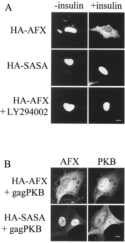
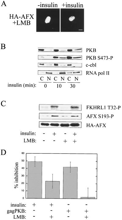
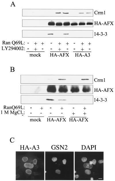
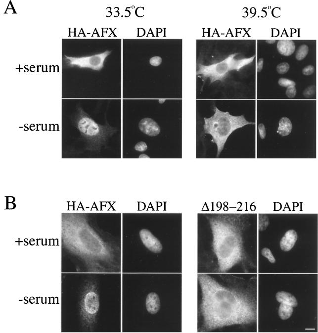
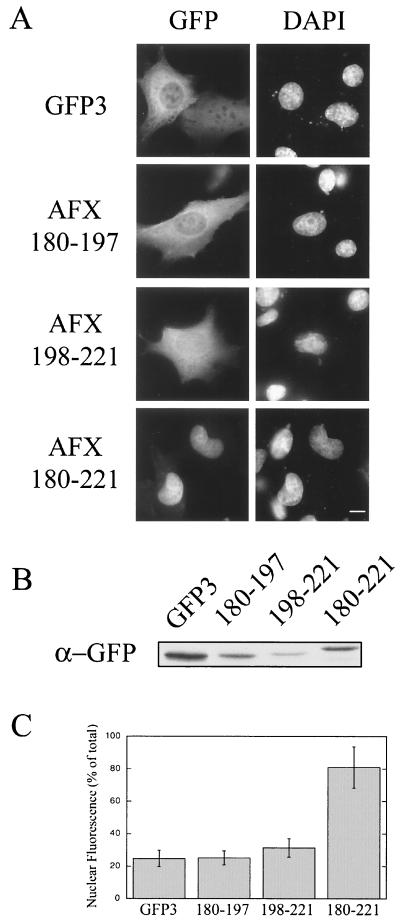
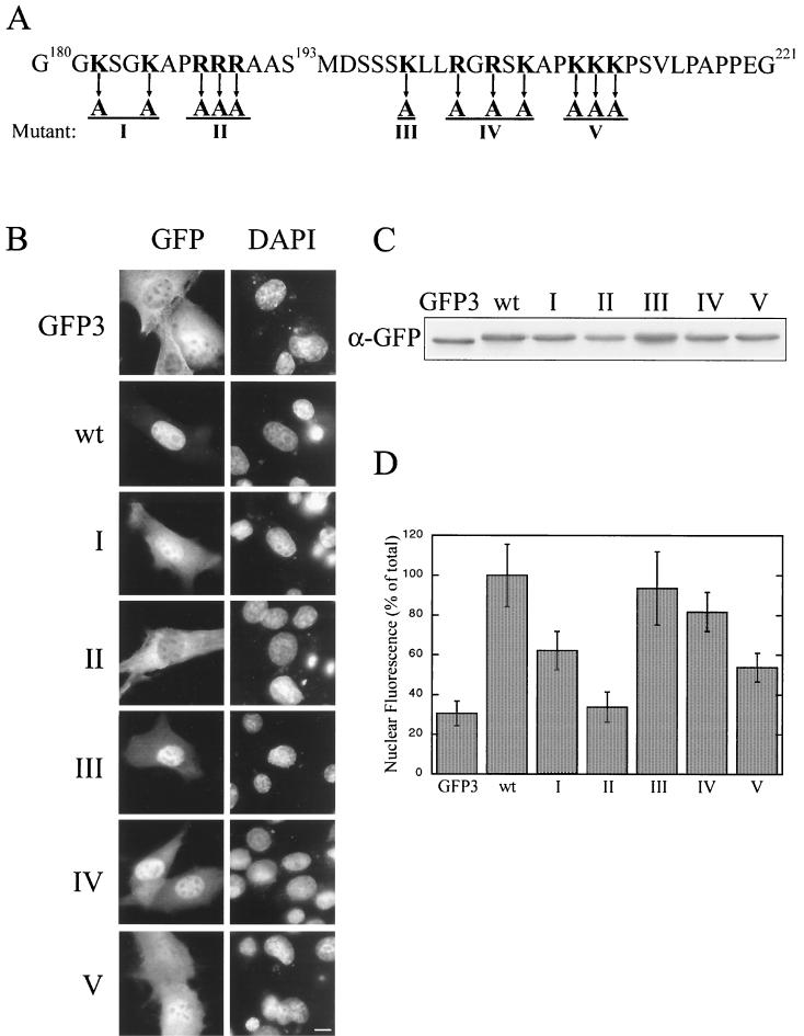
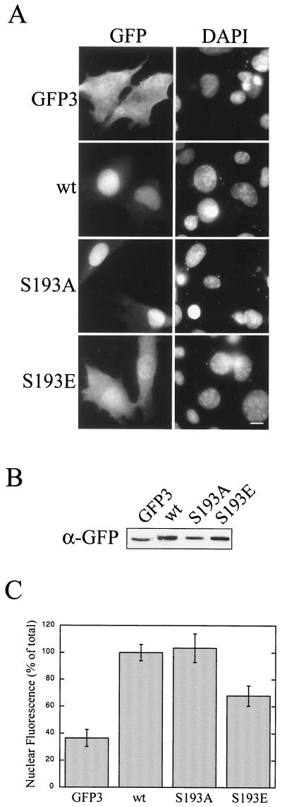
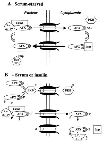
AFX belongs to a subfamily of Forkhead transcription factors that are phosphorylated by protein kinase B (PKB), also known as Akt. Phosphorylation inhibits the transcriptional activity of AFX and changes the steady-state localization of the protein from the nucleus to the cytoplasm. Our goal was threefold: to identify the cellular compartment in which PKB phosphorylates AFX, to determine whether the nuclear localization of AFX plays a role in regulating its transcriptional activity, and to elucidate the mechanism by which phosphorylation alters the localization of AFX. We show that phosphorylation of AFX by PKB occurs in the nucleus. In addition, nuclear export mediated by the export receptor, Crm1, is required for the inhibition of AFX transcriptional activity. Both phosphorylated and unphosphorylated AFX, however, bind Crm1 and can be exported from the nucleus. These results suggest that export is unregulated and that phosphorylation by PKB is not required for the nuclear export of AFX. We show that AFX enters the nucleus by an active, Ran-dependent mechanism. Amino acids 180 to 221 of AFX comprise a nonclassical nuclear localization signal (NLS). S193, contained within this atypical NLS, is a PKB-dependent phosphorylation site on AFX. Addition of a negative charge at S193 by mutating the residue to glutamate reduces nuclear accumulation. PKB-mediated phosphorylation of AFX, therefore, attenuates the import of the transcription factor, which shifts the localization of the protein from the nucleus to the cytoplasm and results in the inhibition of AFX transcriptional activity.


Figures

FIG. 1
FIG. 1
PKB-dependent phosphorylation of AFX triggers relocalization of AFX from the nucleus to the cytoplasm. (A) A14 cells were transfected with pMT2-HA-AFX or pMT2-HA-SASA. At 24 h posttransfection, serum was withdrawn for 18 to 24 h. Insulin (1 μg/ml) was then added as indicated, and these cells were incubated for 30 min. Cells treated with LY294002 (10 μM) to inhibit PIP3K were preincubated for 10 min prior to the addition of insulin. Cells were fixed, and then HA-AFX and HA-SASA were stained with 12CA5 and Texas red-conjugated secondary antibody. (B) A14 cells were transfected with pMT2-HA-AFX or pMT2-HA-SASA and with constitutively active PKB (pSG5-gagPKB). At 48 h posttransfection, the cells were fixed and HA-AFX and HA-SASA were stained as described for panel A. gagPKB was stained with anti-gag antiserum and fluorescein isothiocyanate-conjugated secondary antibody. Bars, 10 μm.
FIG. 2
FIG. 2
Nuclear export subsequent to phosphorylation by PKB in the nucleus is required for the inhibition of AFX transcriptional activity. (A) A14 cells were transfected with pMT2-HA-AFX. At 24 h posttransfection, serum was withdrawn for 18 to 24 h. The cells were treated with LMB (2 ng/ml) for 30 min to inhibit Crm1-dependent export. Then insulin (1 μg/ml) was added as indicated, and the cells were incubated for an additional 30 min. Cells were fixed, and then HA-AFX was stained as described previously. Bar, 10 μm. (B) Serum-starved A14 cells were treated with insulin (1 μg/ml) and then fractionated at the indicated times. Equal amounts of nuclear (N) and cytoplasmic (C) lysates (50 μg) were analyzed by SDS-PAGE and Western blotting. An anti-PKB antibody was used to detect endogenous PKB protein levels. An anti-PKB S473-P antibody was used to detect activated PKB. c-cb1 and RNA pol II represent cytoplasmic and nuclear protein markers, respectively. (C) A14 cells were transfected and treated as described for panel A prior to cell lysis. Equal amounts of cellular lysate (50 μg) were analyzed by SDS-PAGE and Western blotting. An anti-pT32-specific antibody was used to detect endogenous FKHRL1 phosphorylated by PKB (14). Since A14 cells do not express endogenous AFX, an anti-pS193-specific antibody was used to detect HA-AFX phosphorylated by PKB. 12CA5 was used to visualize HA-AFX expression. (D) A14 cells were cotransfected with the 1205-luc reporter, pCMV-LacZ, pMT2-HA-AFX, and where indicated, pSG5-gagPKB. Cells were preincubated with LMB where noted for 30 min prior to the addition of insulin. Cells were then treated with insulin for 16 h in the absence or presence of LMB. Luciferase activity was measured 48 h after transfection, and luciferase levels were corrected for β-galactosidase expression. pMT2-HA-AFX-transfected control cells were serum starved, and the inhibition of AFX activity was normalized to 0% (i.e., 100% relative promoter activity). Data were obtained from five separate experiments.
FIG. 3
FIG. 3
Both phosphorylated and unphosphorylated AFXs bind Crm1 and are exported from the nucleus. (A) HEK293 cells were transfected with either pMT2-HA-AFX or pMT2-HA-A3 (T28A, S193A, S258A), incubated in the presence of serum for 48 h, and where indicated, incubated with LY294002 (10 μM) for 2 h prior to cell lysis. The HA-tagged proteins were immunoprecipitated with 12CA5 and immobilized on protein A-Sepharose. The beads were then incubated with recombinant Crm1 (500 nM) in the absence or presence of RanQ69L (3 μM), a Ran mutant locked in the GTP bound conformation. Proteins that bound to AFX were analyzed by SDS-PAGE and Western blotting. An anti-His6 antibody was used to visualize Crm1, which possesses a carboxy-terminal His6 tag. Directly conjugated HRP-12CA5 was used to assess HA-AFX and HA-A3 immunoprecipitation. An anti-14-3-3 β antibody was used to detect 14-3-3 binding. (B) HEK293 cells were transfected with pMT2-HA-AFX and were treated as described for panel A. Prior to incubation with Crm1 and RanQ69L, immobilized HA-AFX was washed with 1 M MgCl2 where indicated to remove bound 14-3-3 proteins. Proteins that bound AFX were analyzed as described for panel A. (C) Heterotypic cell fusions were performed between BHK21 cells transiently transfected with pMT2-HA-A3 and a stably transfected HeLa cell line expressing GFP-streptavidin-NLS (GSN2). Cell cultures were trypsinized 24 h after transfection, mixed, allowed to adhere to coverslips overnight, and then fused by using polyethylene glycol. After incubation for 1 h with cycloheximide (50 μM), cells were fixed and stained for HA-A3 with 12CA5 and a Texas red-conjugated secondary antibody. Nuclei were visualized by staining the DNA with DAPI. Bar, 10 μm.
FIG. 4
FIG. 4
AFX import into the nucleus requires a Ran gradient and basic residues downstream from S193. (A) tsBN2 cells were transfected with pMT2-HA-AFX and were incubated at 33.5°C in the presence of serum for 48 h. Cycloheximide (50 μM) was then added to the medium to prevent new protein synthesis, and the cells were incubated at either 33.5°C (right) or 39.5°C (left) for 3 h. Where indicated, the serum was then withdrawn and the cells were returned to 33.5°C or 39.5°C for 1.5 h. Cells were fixed and HA-A3 was stained as described previously. (B) HeLa cells were transfected with pMT2-HA-AFX or pMT2-HA-AFX(Δ198–216) and were incubated in the presence of serum for 48 h. Serum was then withdrawn for 1.5 h. Cells were fixed, and HA-AFX and HA-AFX(Δ198–216) were stained as before. Nuclei were visualized by staining the DNA with DAPI. Bar, 10 μm.
FIG. 5
FIG. 5
Basic residues on both sides of S193 are sufficient for nuclear import. Basic residues on either side of PKB phosphorylation site S193 were expressed as N-terminal fusions to GFP-GFP-GFP (GFP3). pKGFP3, pKGFP3-AFX(180–197), pKGFP3-AFX(198–221), and pKGFP3-AFX(180–221) were transfected into BHK21 cells. At 18 h posttransfection, the cells were fixed and the DNA was stained with DAPI (A) or the cells were lysed and analyzed by SDS-PAGE (10 μl of lysate/lane) and immunoblotting using an anti-GFP antibody (B). (C) The relative nuclear and cytoplasmic fluorescence levels of the constructs were obtained using Openlab (Improvision). Nuclear fluorescence was calculated as a percentage of the total cellular fluorescence corrected for the background fluorescence. Each data point represents the mean fluorescence obtained from 12 randomly chosen cells. Error is expressed as standard deviations of the means. Bar, 10 μm.
FIG. 6
FIG. 6
Basic residues on both sides of S193 are required for nuclear import, and the sequence is not a classical NLS motif. (A) There are 12 lysine and arginine residues that surround S193. Five constructs with mutations of basic residues within AFX(180–221)-GFP were made (I through V). pKGFP3, pKGFP-AFX(180–221), pKGFP3-AFX(180–221)-I, pKGFP3(180–221)-II, pKGFP3-AFX(180–221)-III, pKGFP3-AFX(180–221)-IV, and pKGFP-AFX(180–221)-V were transfected into BHK21 cells. At 18 h posttransfection, the cells were either fixed (B) or lysed (C) and analyzed as described for Fig. 5 (D). Bar, 10 μm. wt, wild type.
FIG. 7
FIG. 7
Phosphorylation of S193 reduces the rate of nuclear import. Two AFX(180–221)-GFP3 mutants were created, S193A and S193E. pKGFP3, pKGFP3-AFX(180–221), pKGFP3-AFX(180–221)S193A, and pKGFP3-AFX(180–221)S193E were transfected into BHK21 cells. At 18 h posttransfection, the cells were either fixed (A) or lysed (B) and analyzed as described for Fig. 5 (C). Bar, 10 μm. wt, wild type.
FIG. 8
FIG. 8
Nuclear import, not export, of AFX is regulated by PKB. (A) Unphosphorylated AFX appears nuclear at steady state but is actually shuttling rapidly between the nucleus and the cytoplasm. Therefore, the rate of import of unphosphorylated AFX exceeds its rate of export. Nuclear export of both phosphorylated and unphosphorylated AFX is likely mediated by the exportin, Crm1, since both HA-AFX and HA-A3 bind Crm1 in the presence of RanGTP. Imp, importin. (B) The addition of insulin to cells activates PKB and causes it to translocate into the nucleus, where it phosphorylates Forkhead family members. Phosphorylated AFX exits the nucleus by binding Crm1. Phosphorylation of AFX at S193 attenuates nuclear import, perhaps by reducing the affinity of AFX for its nuclear import receptor. Therefore, phosphorylation by PKB decreases the import rate constant without altering the export rate constant. Since the steady-state localization of a protein is determined by its relative rates of import and export, the localization of AFX would shift from the nucleus to the cytoplasm as observed. This alteration in transport rates in conjunction with proposed cytoplasmic retention by binding 14-3-3 proteins would result in exclusion of AFX from the nucleus. Since AFX requires nuclear localization to carry out transcription, this mechanism of regulated transport would inhibit the activity of AFX in response to phosphorylation by PKB.

References

    1. Ahmed N N, Franke T F, Bellacosa A, Datta K, Gonzalez-Portal M E, Taguchi T, Testa J R, Tsichlis P N. The proteins encoded by c-akt and v-akt differ in post-translational modification, subcellular localization and oncogenic potential. Oncogene. 1993;8:1957–1963. - PubMed
    1. Alessi D R, James S R, Downes C P, Holmes A B, Gaffney P R, Reese C B, Cohen P. Characterization of a 3-phosphoinositide-dependent protein kinase which phosphorylates and activates protein kinase B α. Curr Biol. 1997;7:261–269. - PubMed
    1. Anderson M J, Viars C S, Czekay S, Cavenee W K, Arden K C. Cloning and characterization of three human forkhead genes that comprise an FKHR-like gene subfamily. Genomics. 1998;47:187–199. - PubMed
    1. Andjelkovic M, Alessi D R, Meier R, Fernandez A, Lamb N J, Frech M, Cron P, Cohen P, Lucocq J M, Hemmings B A. Role of translocation in the activation and function of protein kinase B. J Biol Chem. 1997;272:31515–31524. - PubMed
    1. Arts G J, Fornerod M, Mattaj I W. Identification of a nuclear export receptor for tRNA. Curr Biol. 1998;8:305–314. - PubMed

Publication types