. 2001 May;21(10):3604-8.
doi: 10.1128/MCB.21.10.3604-3608.2001.

Characterization of XIAP-deficient mice

Affiliations

Characterization of XIAP-deficient mice

H Harlin et al. Mol Cell Biol. 2001 May.

Abstract

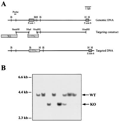
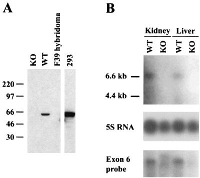
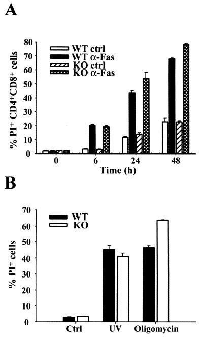
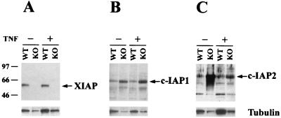
The inhibitor of apoptosis protein (IAP) family consists of a number of evolutionarily conserved proteins that function to inhibit programmed cell death. X-linked IAP (XIAP) was cloned due to its sequence homology with other family members and has previously been shown to prevent apoptosis by binding to active caspases 3, 7, and 9 in vitro. XIAP transcripts can be found in a variety of tissues, and the protein levels are regulated both transcriptionally and posttranscriptionally. To better understand the function of XIAP in normal cells, we generated mice deficient in XIAP through homologous gene targeting. The resulting mice were viable, and histopathological analysis did not reveal any differences between XIAP-deficient and wild-type mice. We were unable to detect any defects in induction of caspase-dependent or -independent apoptosis in cells from the gene-targeted mice. One change was observed in cells derived from XIAP-deficient mice: the levels of c-IAP1 and c-IAP2 protein were increased. This suggests that there exists a compensatory mechanism that leads to upregulation of other family members when XIAP expression is lost. The changes in c-IAP1 and c-IAP2 expression may provide functional compensation for loss of XIAP during development or in the induction of apoptosis.


Figures

FIG. 1
FIG. 1
Generation of XIAP-deficient mice by gene targeting. (A) Schematic representation of the targeting strategy used to disrupt the XIAP gene. The locations of exons 1 and 6 are shown. Exons 2 to 5 are contained within the 8.3-kb HindIII-HindIII fragment (not shown). The targeting construct replaces the coding sequence of exon 1 with the neomycin resistance gene, in the opposite transcriptional orientation. The sequence used as a probe to screen for homologously recombined DNA is shown. B (BglII) and H (HindIII) sites are shown for the genomic and targeted DNA. (B) Southern blot analysis of BglII-digested tail DNA from targeted mice. The WT allele is 4.0 kb, and the targeted allele is 3.2 kb. The blots were probed with the 0.2-kb BamHI-EcoRI fragment shown in panel A.
FIG. 2
FIG. 2
Targeted mice have no detectable XIAP protein or mRNA. (A) Western blot analysis of lysate from WT or XIAP-deficient (KO) embryonic fibroblasts. Lysate from a hybridoma lacking XIAP expression (F39) was used as a negative control. Lysate from 293 cells served as a positive control. Molecular mass standards, in kilodaltons, are indicated on the left. (B) Northern blot analysis of RNA from WT or XIAP-deficient (KO) tissues. The top blot was probed with a fragment from exon 1, which is deleted in the KO tissues. The bottom blot shows a duplicate blot probed with a fragment from exon 6, which is not affected by XIAP gene targeting. The loading of RNA was verified using 5S rRNA.
FIG. 3
FIG. 3
Apoptosis occurs normally in XIAP-deficient mice. (A) Fas-mediated apoptosis of double-positive (CD4+ CD8+) thymocytes. WT or XIAP-deficient thymocytes were treated with either isotype control (ctrl) or anti-Fas (α-Fas) antibody. The percentage of apoptotic (PI+) cells was determined at the indicated time points. Cells from one WT and one KO mouse were plated in triplicate, and average values are shown along with the standard deviation for each triplicate sample. The data are representative of three separate experiments. (B) Caspase-dependent or -independent apoptosis. Embryonic fibroblasts were left untreated (ctrl) or treated with either UV irradiation or oligomycin. The percentage of apoptotic cells was determined at 24 h after treatment. WT and KO cells were plated in triplicate, and average values are shown along with the standard deviation for each triplicate sample. The data are representative of two separate experiments.
FIG. 4
FIG. 4
Levels of c-IAP1 and c-IAP2 proteins are upregulated in XIAP-deficient cells. Western blot analysis of lysate from WT or XIAP-deficient (KO) embryonic fibroblasts. The cells were either untreated or stimulated with 200 U of human TNF-α/ml for 4 h. Gels were loaded in parallel, and the resulting blots were probed with antibodies directed against XIAP (A), c-IAP1 (B), or c-IAP2 (C). The blots were stripped and reprobed with an antibody against β-tubulin, shown at the bottom of each panel. Molecular mass standards, in kilodaltons, are indicated on the left.

References

    1. Ambrosini G, Adida C, Altieri D C. A novel anti-apoptosis gene, survivin, expressed in cancer and lymphoma. Nat Med. 1997;3:917–921. - PubMed
    1. Datta R, Oki E, Endo K, Biedermann V, Ren J, Kufe D. XIAP regulates DNA damage-induced apoptosis downstream of caspase-9 cleavage. J Biol Chem. 2000;275:31733–31738. - PubMed
    1. Deveraux Q L, Leo E, Stennicke H R, Welsh K, Salvesen G S, Reed J C. Cleavage of human inhibitor of apoptosis protein XIAP results in fragments with distinct specificities for caspases. EMBO J. 1999;18:5242–5251. - PMC - PubMed
    1. Deveraux Q L, Takahashi R, Salvesen G S, Reed J C. X-linked IAP is a direct inhibitor of cell-death proteases. Nature. 1997;388:300–304. - PubMed
    1. Duckett C S, Li F, Wang Y, Tomaselli K J, Thompson C B, Armstrong R C. Human IAP-like protein regulates programmed cell death downstream of Bcl-xl and cytochrome c. Mol Cell Biol. 1998;18:608–615. - PMC - PubMed

Publication types

Substances