. 2001 May;85(5):613-8.
doi: 10.1136/bjo.85.5.613.

Apoptosis mediates decrease in cellularity during the regression of Arthus reaction in cornea

Affiliations

Apoptosis mediates decrease in cellularity during the regression of Arthus reaction in cornea

N Ozaki et al. Br J Ophthalmol. 2001 May.

Abstract

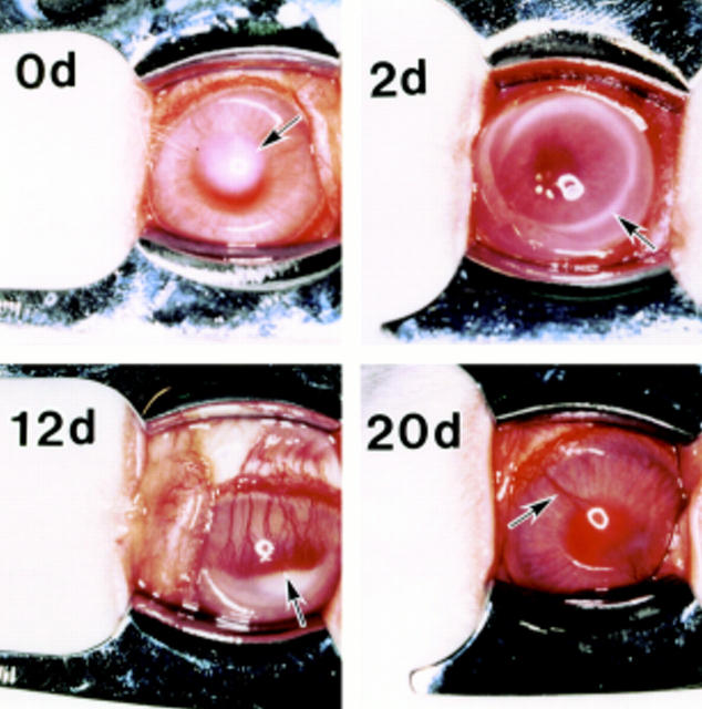
Background/aims: The Arthus type allergic reaction is characterised by inflammatory cell infiltration and marked neovascularisation in the cornea. During the healing stages, inflammatory cells and newly formed microvessels gradually disappear. The aim was to establish whether apoptosis affected the regression of inflammatory cells and newly formed microvessels, in order to define more clearly the cellular mechanisms involved in the pathobiology of corneal diseases.

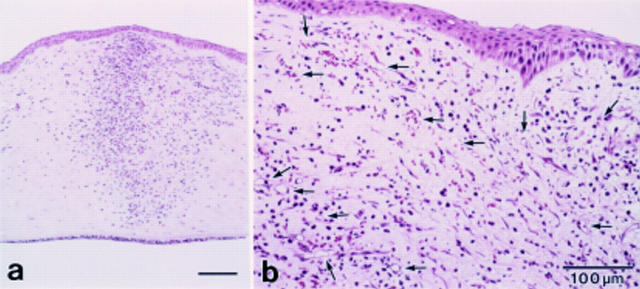
Methods: Albino male rabbits were injected subcutaneously with 5 mg/ml bovine serum albumin (BSA) incorporated in Freund's complete adjuvant twice weekly. Under the anaesthesia, 30 microl of a 0.5 mg/ml BSA solution was injected into the central corneal stroma to induce an Arthus type allergic reaction. The injured corneas were collected at various time points ranging from 3 to 20 days. Apoptotic cells were identified by both light microscopy using in situ TdT-dUTP nick end labelling (TUNEL) method and electron microscopy.

Results: With increasing time after induction of the Arthus reaction, marked neovascularisation and infiltrated inflammatory cells such as polymorphonuclear cells (PMNs) and plasma cells were observed in the cornea. Thereafter, the inflammatory cells and newly formed microvessels gradually disappeared. Coincidently, the numbers of microvessel endothelial cells and infiltrated inflammatory cells undergoing apoptosis were increased. Apoptotic bodies were taken up by macrophages, PMNs, as well as myofibroblasts derived presumably from transformation of migrated keratocytes.

Conclusions: These data demonstrate that regression of the cellular infiltrates and microvessel endothelial cells associated with the Arthus reaction in the cornea occurs via apoptosis. This finding adds insights into the cellular mechanisms regulating the pathobiology of corneal diseases.


Figures

Figure 1
Figure 1
Sensitised rabbit corneas showing opacity as a result of injection (arrow) at day 0 (0d), immuno-ring (arrow) at 2 days (2d) after injection and induction of neovascular response. Newly formed microvessels (arrow) reached to the centre of the cornea at 12 days (12d). These microvessels (arrow) gradually diminished at the healing stage on day 20 (20d).
Figure 2
Figure 2
Light microscopic observation (haematoxylin eosin staining) at 3 days. (a) The infiltration of inflammatory cells mainly polymorphonuclear neutrophils (PMNs) was noted. (b) At the limbus newly formed dense capillaries were observed (arrows).
Figure 3
Figure 3
Double staining for apoptotic cells, and microvessels in the same section at 3 days after injection of BSA. TUNEL positive cells (green, arrowheads) were present in the stroma. However, there were no positive cells in the microvessels as depicted by positive reaction for TM (red, arrows).
Figure 4
Figure 4
Double staining for apoptotic cells, and microvessels in the same section at 12 days after injection of BSA. Co-localisation of TM expression (red, arrows) and TUNEL positivity (green, arrowheads)is seen in the endothelium.
Figure 5
Figure 5
Various types of apoptotic cells and bodies in the stroma are shown (arrows).
Figure 6
Figure 6
A plasma cell in the early stage of apoptosis at 9 days after injection of BSA.
Figure 7
Figure 7
Apoptotic cell in microvessel at 12 days after injection of BSA showing an endothelial cell in the early stage of apoptosis. Note condensation of heterochromatin to form "crescent" in the lobes of the nucleus (*). Inset shows pinocytic vesicle of endothelial cells (arrows).
Figure 8
Figure 8
Phagocytosis of apoptotic cells at 12 days after injection of BSA. (A) A macrophage containing apparently a PMN with granules and fragments of chromatin condensation typical of apoptosis (arrows). (B) A PMN containing apparently a neutrophil with granules and fragments of chromatin condensation typical of apoptosis (arrows). (C) Apoptotic body (large arrows) was taken up by fibroblastic cells. This fibroblastic cell has extracellular microtendon associated with the fibronexis (small arrows), electron dense stress fibre (arrowheads), and abundant rough ER, suggesting a myofibroblast.

References

    1. Science. 1995 Nov 17;270(5239):1158-9 - PubMed
    1. J Biochem. 1994 Oct;116(4):794-800 - PubMed
    1. Br J Haematol. 1996 Feb;92(2):302-7 - PubMed
    1. J Exp Med. 1996 Aug 1;184(2):429-40 - PubMed
    1. Am J Pathol. 1996 Sep;149(3):953-61 - PubMed

Publication types