

Editorial
doi: 10.1073/pnas.011099798.
How activated receptors couple to G proteins
- PMID: 11320227
- PMCID: PMC33117
- DOI: 10.1073/pnas.011099798
Item in Clipboard
Editorial
How activated receptors couple to G proteins
Proc Natl Acad Sci U S A.
.
No abstract available
Figures
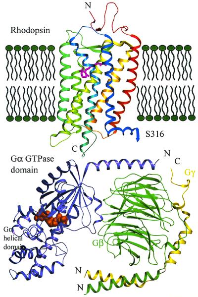
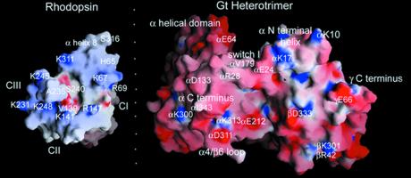
Orientation of rhodopsin, transducin, and the membrane. Refined
rhodopsin structure is from ref. , and Gt is from ref. . Models are
based on the crystal structures and are to scale. The carboxyl-terminal
residues after S316 are not shown. The orientation of Gt with respect
to rhodopsin and the membrane is based on the charge and hydrophobicity
of the surface, the known rhodopsin-binding sites on Gt, and the sites
of lipidation of Gα and Gβγ (23).
Comment on
-
Structure and function in rhodopsin: Mass spectrometric identification of the abnormal intradiscal disulfide bond in misfolded retinitis pigmentosa mutants.Proc Natl Acad Sci U S A. 2001 Apr 24;98(9):4872-6. doi: 10.1073/pnas.061632798. Proc Natl Acad Sci U S A. 2001. PMID: 11320236 Free PMC article.
-
Mapping of contact sites in complex formation between transducin and light-activated rhodopsin by covalent crosslinking: use of a photoactivatable reagent.Proc Natl Acad Sci U S A. 2001 Apr 24;98(9):4877-82. doi: 10.1073/pnas.051632898. Proc Natl Acad Sci U S A. 2001. PMID: 11320237 Free PMC article.
-
Mapping of contact sites in complex formation between light-activated rhodopsin and transducin by covalent crosslinking: use of a chemically preactivated reagent.Proc Natl Acad Sci U S A. 2001 Apr 24;98(9):4883-7. doi: 10.1073/pnas.051632998. Proc Natl Acad Sci U S A. 2001. PMID: 11320238 Free PMC article.
-
Solution 19F nuclear Overhauser effects in structural studies of the cytoplasmic domain of mammalian rhodopsin.Proc Natl Acad Sci U S A. 2001 Apr 24;98(9):4888-92. doi: 10.1073/pnas.051633098. Proc Natl Acad Sci U S A. 2001. PMID: 11320239 Free PMC article.
References
Publication types
MeSH terms
Substances
LinkOut - more resources
Full Text Sources
