. 2001 Apr 24;98(9):4872-6.
doi: 10.1073/pnas.061632798.

Structure and function in rhodopsin: Mass spectrometric identification of the abnormal intradiscal disulfide bond in misfolded retinitis pigmentosa mutants

Affiliations

Structure and function in rhodopsin: Mass spectrometric identification of the abnormal intradiscal disulfide bond in misfolded retinitis pigmentosa mutants

J Hwa et al. Proc Natl Acad Sci U S A. .

Abstract

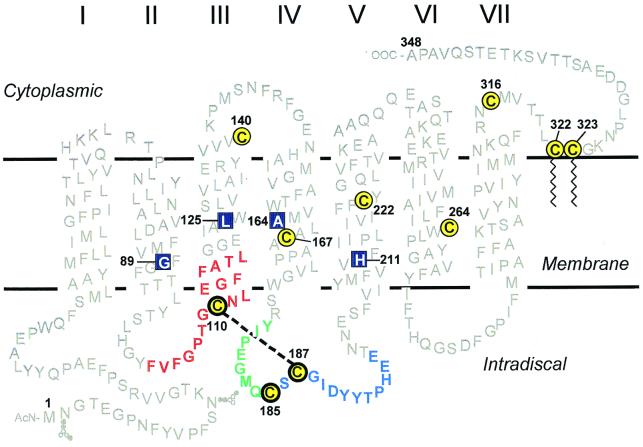
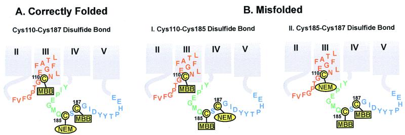
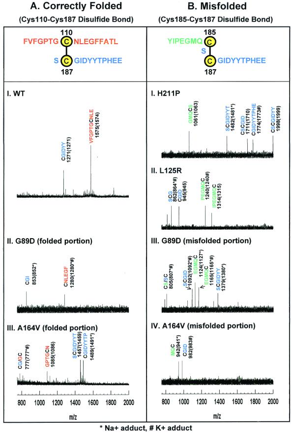
Retinitis pigmentosa (RP) point mutations in both the intradiscal (ID) and transmembrane domains of rhodopsin cause partial or complete misfolding of rhodopsin, resulting in loss of 11-cis-retinal binding. Previous work has shown that misfolding is caused by the formation of a disulfide bond in the ID domain different from the native Cys-110-Cys-187 disulfide bond in native rhodopsin. Here we report on direct identification of the abnormal disulfide bond in misfolded RP mutants in the transmembrane domain by mass spectrometric analysis. This disulfide bond is between Cys-185 and Cys-187, the same as previously identified in misfolded RP mutations in the ID domain. The strategy described here should be generally applicable to identification of disulfide bonds in other integral membrane proteins.


Figures

Figure 1
Figure 1
A secondary structure model of rhodopsin showing the cytoplasmic, TM, and the ID domains. The 10 cysteines are shown in yellow with black circles; the three ID cysteines are highlighted by bolder circles. Amino acid sequences adjoining the three cysteines of interest (for identification of the disulfide bonds) are color coded for each cysteine: red for Cys-110, green for Cys-185, and blue for Cys-187. Native rhodopsin contains a disulfide bond between Cys-110 and Cys-187 (indicated by dashed line). The RP mutants studied here are located in the TM domain (shown in blue boxes): G89D (helix II), L125R (helix III), A164V (helix IV), and H211P (helix V).
Figure 2
Figure 2
Structure of maleimido-butyryl-biocytin (MBB).
Figure 3
Figure 3
Derivatizations as expected for ID cysteines in correctly folded (A) and misfolded (B) rhodopsins. The disulfide linkage in correctly folded A rhodopsin is between Cys-110 and Cys-187, as identified by mass spectrometry (Fig. 4). Thus, steps 1 and 2 of the strategy in the identification of disulfide bonds (see above) produced rhodopsin NEM-derivatized at Cys-185, whereas Cys-110 and Cys-187 were derivatized with MBB. In misfolded rhodopsin B, the disulfide bond could either be between Cys-110 and Cys-185 (I) or between Cys-185 and Cys-187 (II). Experimentally, the disulfide bond was found between Cys-185 and Cys-187 (Fig. 4). Thus, Cys-110 was NEM-derivatized, whereas Cys-185 and Cys-187 were MBB-derivatized (II). The color code for the amino acid residues adjacent to each of the cysteines is the same as in Fig. 1. The derivatized products were then subjected to steps 3–5 (above), yielding the MBB-derivatized peptides that were analyzed by MALDI-TOF (Fig. 4).
Figure 4
Figure 4
MALDI-TOF Analysis of peptide fragments generated from the derivatized correctly folded (A) and misfolded (B) rhodopsins. The adjoining peptide sequences expected for the disulfide bonds are highlighted at the top in A and B. Rhodopsins were derivatized at cysteines as in the strategy outlined above. Because step 4 of the strategy selects only MBB-labeled peptides, all peptide assignments shown here include the mass for the MBB adducts of cysteines. The y axis shows intensity in arbitrary units. The x axis shows m/z in the range of 750–2,000. (A) Mass spectrometric results for WT rhodopsin and correctly folded portions of the A164V and G89D mutants. The observed signals in the mass spectra correspond to MBB-labeled Cys-110 peptides (red) and Cys-187 peptides (blue). (B) Mass spectrometric results for the misfolded portion of A164V and G89D (III and IV) and of completely misfolded H211P and L125R (I and II). Only peptides corresponding to a Cys-185–Cys-187 disulfide bond were observed. MBB-labeled Cys-185 containing peptides are shown in green, and MBB-labeled Cys-187 peptides are shown in blue.

Comment in

  • How activated receptors couple to G proteins.
    Hamm HE. Hamm HE. Proc Natl Acad Sci U S A. 2001 Apr 24;98(9):4819-21. doi: 10.1073/pnas.011099798. Proc Natl Acad Sci U S A. 2001. PMID: 11320227 Free PMC article. No abstract available.

References

    1. Berson E L. Invest Opthalmol Visual Sci. 1993;34:1659–1676. - PubMed
    1. Dryja T P, Berson E L. Invest Opthalmol Visual Sci. 1995;36:1197–1200. - PubMed
    1. Sung C H, Davenport C M, Hennessey J C, Maumenee I H, Jacobson S G, Heckenlively J R, Nowakowski R, Fishman G, Gouras P, Nathans J. Proc Natl Acad Sci USA. 1991;88:6481–6485. - PMC - PubMed
    1. Macke J P, Davenport C M, Jacobson S G, Hennessey J C, Gonzalez-Fernandez F, Conway B P, Heckenlively J, Palmer R, Maumenee I H, Sieving P, et al. Am J Hum Genet. 1993;53:80–89. - PMC - PubMed
    1. Inglehearn C F, Keen T J, Bashir R, Jay M, Fitzke F, Bird A C, Crombie A, Bhattacharya S S. Hum Mol Genet. 1992;1:41–45. - PubMed

Publication types

MeSH terms

LinkOut - more resources