. 2001 Apr 24;98(9):4883-7.
doi: 10.1073/pnas.051632998.

Mapping of contact sites in complex formation between light-activated rhodopsin and transducin by covalent crosslinking: use of a chemically preactivated reagent

Affiliations

Mapping of contact sites in complex formation between light-activated rhodopsin and transducin by covalent crosslinking: use of a chemically preactivated reagent

Y Itoh et al. Proc Natl Acad Sci U S A. .

Abstract

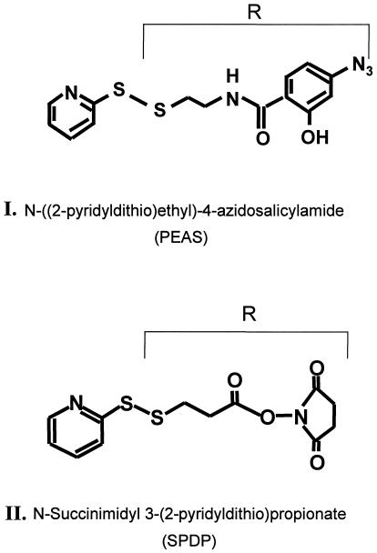
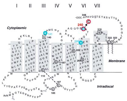
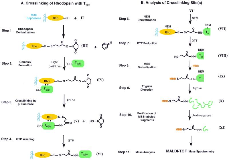
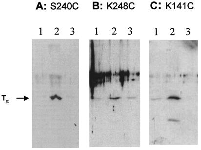
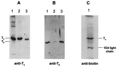
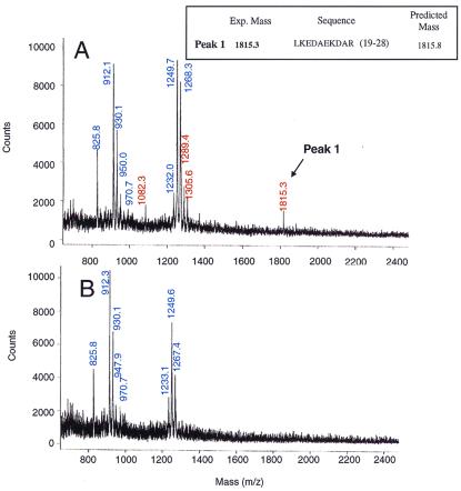
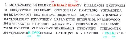
Contact sites in interaction between light-activated rhodopsin and transducin (T) have been investigated by using a chemically preactivated crosslinking reagent, N-succinimidyl 3-(2-pyridyldithio)propionate. The 3 propionyl-N-succinimidyl group in the reagent was attached by a disulfide exchange reaction to rhodopsin mutants containing single reactive cysteine groups in the cytoplasmic loops. Complex formation between the derivatized rhodopsin mutants and T was carried out by illumination at lambda > 495 nm. Subsequent increase in pH (from 6 to 7.5 or higher) of the complex resulted in crosslinking of rhodopsin to the T(alpha) subunit. Crosslinking to T(alpha) was demonstrated for the rhodopsin mutants K141C, S240C, and K248C, and the crosslinked sites in T(alpha) were identified for the rhodopsin mutant S240C. The peptides carrying the crosslinking moiety were isolated from the trypsin-digested peptide mixture, and their identification was carried out by matrix-assisted laser desorption ionization-time of flight mass spectrometry. The main site of crosslinking is within the peptide sequence, Leu-19-Arg-28 at the N-terminal region of T(alpha). The total results show that both the N and the C termini of T(alpha) are in close vicinity to the third cytoplasmic loop of rhodopsin in the complex between rhodopsin and T.


Figures

Figure 1
Figure 1
Structure of the two crosslinking reagents, I and II, used in the work described here and in ref. . In both reagents, the moiety R is transferred by a disulfide exchange reaction to the sulfhydryl group of a monocysteine rhodopsin mutant. R in I contains the photoactivatable azidophenyl group whereas R in II contains the activated carboxyl group, by derivatization with the N-oxysuccinimido group.
Figure 2
Figure 2
A secondary structure model of rhodopsin based on the crystal structure highlighting (in red) the position of the single cysteines introduced in the cytoplasmic domain. The reactive cysteines, Cys-140 and Cys-316, in native rhodopsin were replaced by serine residues (square in black). Cysteines 167, 185, 222, and 264 are not reactive. Cys-110 and Cys-187 form a disulfide bond whereas Cys-322 and Cys-323 carry palmitoyl groups (wiggly lines).
Figure 3
Figure 3
(A) Steps in the strategy for the attachment of R moiety in reagent II to rhodopsin cysteine mutants bound to 1D4-Sepharose beads, complex formation with GDP-Tαβγ on illumination, crosslinking by increasing pH to 7.5 or higher. (B) Steps in the identification of crosslinking site, carrying the HS-CH2-CH2-CO-NH group to a peptide sequence in Tα.
Figure 4
Figure 4
SDS/PAGE followed by immunoblotting in analysis of crosslinking of rhodopsin mutants, S240C, K248C, andK141C with the R group in reagent II. Crosslinking depends both on light and increase in pH. Lanes 1, eluate from 1D4-Sepharose after reduction with DTT in the dark (Fig. 3A, step 1); lanes 2, sample after illumination (Fig. 3A, step 2) and pH increase (Fig. 3A, step 3); and lanes 3, sample after illumination (Fig. 3A, step 2) only. Immunoblotting was with anti-Tα antibody.
Figure 5
Figure 5
SDS/PAGE analysis of product formed in crosslinking of rhodopsin S240C-R with T. Protein bands were detected by immunoblotting with antibodies against Tα, Tβ, and biotin. (A and B) Lanes 1, control Tαβγ; lanes 2, eluate from 1D4-Sepharose after reduction with DTT (Fig. 3B, IX); and lanes 3, eluate from 1D4-Sepharose with GTP after reduction with DTT (Fig. 3A, step 4). (C) Eluate from 1D4-Sepharose after reduction with DTT (Fig. 3B, IX).
Figure 6
Figure 6
(A) MALDI-TOF mass spectrum of peptide fragments containing MBB-β-thio-propionyloxyamido-Tα obtained after purification on avidin-agarose of trypsin-digested peptide mixture. (B) Mass spectrum of a control sample in which trypsin alone was subjected to the trypsin digestion conditions used in A. Mass (m/z) lines (blue) originate from trypsin autolysis whereas the mass (m/z) values (red) originated crosslinked Tα.
Figure 7
Figure 7
Amino acid sequence of Tα. The fragment identified by mass spectrum (Fig. 6) is underlined. This fragment (Leu-19–Arg-28, red) identified is near N-terminal region of the Tα. Also shown in blue are the two crosslinked peptide sequences at the C terminus of Tα identified in ref. .

Comment in

  • How activated receptors couple to G proteins.
    Hamm HE. Hamm HE. Proc Natl Acad Sci U S A. 2001 Apr 24;98(9):4819-21. doi: 10.1073/pnas.011099798. Proc Natl Acad Sci U S A. 2001. PMID: 11320227 Free PMC article. No abstract available.

References

    1. Khorana H G. J Biol Chem. 1992;267:1–4. - PubMed
    1. Cai K, Itoh Y, Khorana H G. Proc Natl Acad Sci USA. 2001;98:4877–4882. - PMC - PubMed
    1. Findlay J B C, Barclay P L, Bett M, Davidson M, Pappin D S L, Thompson P. Vision Res. 1984;24:1301–1308. - PubMed
    1. Kuhn H, Hargrave P A. Biochemistry. 1981;20:2410–2417. - PubMed
    1. Konig B, Arendt A, McDowell J H, Kahelrt M, Hargrave P A, Hofmann K P. Proc Natl Acad Sci USA. 1989;86:6878–6882. - PMC - PubMed

Publication types

MeSH terms

LinkOut - more resources