. 2001 Apr 24;98(9):5134-9.
doi: 10.1073/pnas.081068398.

Cancer-predisposing mutations within the RING domain of BRCA1: loss of ubiquitin protein ligase activity and protection from radiation hypersensitivity

Affiliations

Cancer-predisposing mutations within the RING domain of BRCA1: loss of ubiquitin protein ligase activity and protection from radiation hypersensitivity

H Ruffner et al. Proc Natl Acad Sci U S A. .

Abstract

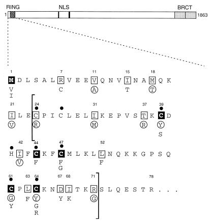
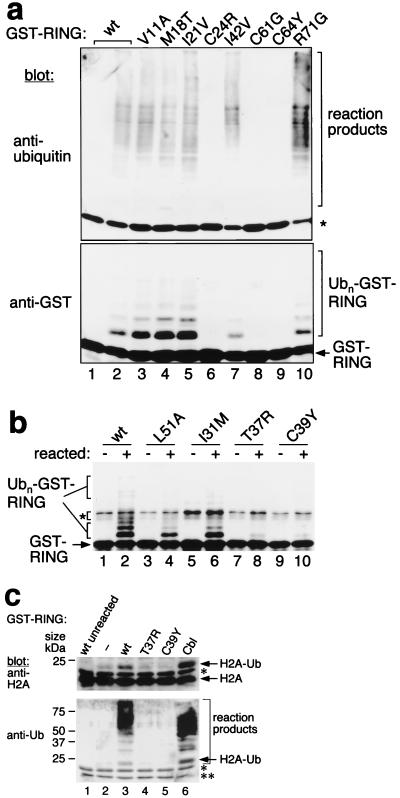
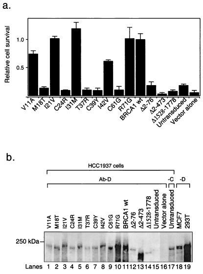
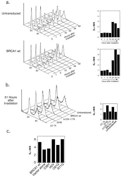
BRCA1 is a breast and ovarian cancer-specific tumor suppressor that seems to be involved in transcription and DNA repair. Here we report that BRCA1 exhibits a bona fide ubiquitin (Ub) protein ligase (E3) activity, and that cancer-predisposing mutations within the BRCA1 RING domain abolish its Ub ligase activity. Furthermore, these mutants are unable to reverse gamma-radiation hypersensitivity of BRCA1-null human breast cancer cells, HCC1937. Additionally, these mutations within the BRCA1 RING domain are not capable of restoring a G(2) + M checkpoint in HCC1937 cells. These results establish a link between Ub protein ligase activity and gamma-radiation protection function of BRCA1, and provide an explanation for why mutations within the BRCA1 RING domain predispose to cancer. Furthermore, we propose that the analysis of the Ub ligase activity of RING-domain mutations identified in patients may constitute an assay to predict predisposition to cancer.


Figures

Figure 1
Figure 1
BRCA1 N-terminal protein sequence (amino acids 1–78). A line diagram showing the domains of the BRCA1 protein is depicted at the top. The N-terminal sequence (amino acids 1–78) is represented below, as indicated by the dashed lines. The RING domain [residues 24–71 defined by comparison to the c-Cbl RING finger structure (11)] is bracketed. Numbers above individual amino acids denote the position of each residue. Filled circles indicate putative zinc-binding residues, filled squares represent cancer-predisposing mutations, and open squares represent unclassified variants. Amino acid substitutions found in patients are indicated below each residue. Circled residues denote the mutation analyzed (see also Table 1).
Figure 2
Figure 2
Ub-ligase activity of the BRCA1 RING finger. (a) Ubiquitination reactions were performed with GST fusion proteins containing BRCA1 wt or mutant RING fingers, E1, Ubc4(E2), Ub, and ATP for 0 min (lane 1) or 90 min (lanes 2–10). Ub-protein conjugates were resolved by reducing SDS/PAGE and successively analyzed by immunoblot analysis by using an Ab to Ub (Upper) or to GST (Lower). The positions of GST-RING and of reaction products (including Ubn-GST-RING) are indicated. The asterisk (Upper) indicates crossreaction between the Ab to Ub and the GST fusion proteins. (b) As in a, reactions were performed for 0 min (−) or 90 min (+) and developed with an Ab to GST. The asterisk indicates crossreacting bands. Unmodified and ubiquitinated GST-RINGs are indicated. (c) As a, reactions also contained histone H2A and were developed with Ab to H2A (Upper) or to Ub (Lower). The single asterisk in both panels indicates Ab crossreactivity to Ubc4. The double asterisk in the lower panel indicates Ab crossreactivity to H2A. Protein sizes according to prestained markers (Bio-Rad) are indicated on the left.
Figure 3
Figure 3
Effect of BRCA1 RING mutations on protection from radiation hypersensitivity. HCC1937 cells were stably reconstituted with either wt BRCA1 or different mutants by using retroviral vectors. (a) Relative cell survival after IR. The mean values (and SEM) of the ratio of the number of colonies with and without IR are shown (1.0 is defined arbitrarily for the mean value of wt BRCA1). (b) Steady-state levels of the BRCA1 proteins in HCC1937 cells. BRCA1 was immunoprecipitated from lysates of cultures that were harvested at comparable confluence (between 70% and 100% confluent). MCF7 cells were only about 50% and 293T cells about 70% confluent when harvested; therefore, BRCA1 from MCF7 cells is relatively underrepresented (lane 18). BRCA1 was immunoprecipitated by using Ab-D (lanes 1–16) or Ab-C (lane 17) from lysates of HCC1937 cells or Ab-D from lysates of MCF7 (lane 18) and 293T cells (lane 19). All immunoprecipitates were separated by SDS/PAGE and analyzed by Western blot analysis using Ab SD 118. Lanes 1–10, individual missense mutations within the N terminus of BRCA1, as indicated; lane 11, wt BRCA1; lanes 12–14, truncated BRCA1 species used as controls; lanes 15 and 17, untransduced HCC1937 cells; lane 16, HCC1937 cells infected with the empty vector; and lanes 18 and 19, endogenous BRCA1 from MCF7 and 293T cells, respectively. The 250-kDa protein marker is indicated on the left.
Figure 4
Figure 4
Restoration of a G2 + M checkpoint in HCC1937 cells by BRCA1. (a) Cell-cycle profiles of untransduced and BRCA1 wt-reconstituted cells at various times after IR. Cells were irradiated with 4 Gy 2 days after plating and analyzed for cell-cycle distribution at the times indicated (hours after irradiation). The 2n and 4n DNA contents are shown. To better illustrate the progression of the BRCA1-reconstituted cells through G2 + M, we expressed the ratio of cells in G2 + M to cells in S phase (G2 + M/S) for each time point (Right). The difference in G2 + M progression between untransduced (or GFP- or vector alone-transduced) and BRCA1-reconstituted HCC1937 cells was obtained reproducibly in independent experiments. (b) To test the specificity of a G2 + M checkpoint restoration by BRCA1, we analyzed the cell-cycle profiles of HCC1937 cells reconstituted with wt BRCA1 or the I31M, Δ2–76, and Δ1528–1778 mutants at similar time points after IR, as in a. Shown are the cell-cycle profiles (Left) and the G2 + M/S ratios (Right) at 51 h after IR. (c) Ratios of G2 + M/S of HCC1937 cells reconstituted with wt BRCA1 or C39Y, C61G, I42V, and R71G mutants at 46 h after IR (as in a and b, cell-cycle progression of the cell cultures was analyzed at various time points after IR). Similar results were obtained in an independent experiment.

References

    1. Rahman N, Stratton M R. Annu Rev Genet. 1998;32:95–121. - PubMed
    1. Claus E, Risch N, Thompson W. Am J Hum Genet. 1991;48:232–242. - PMC - PubMed
    1. Easton D F, Bishop D T, Ford D, Crockford G P, Consortium T B C L. Am J Hum Genet. 1993;52:678–701. - PMC - PubMed
    1. Miki Y, Swensen J, Shattuck-Eidens D, Futreal P A, Harshman K, Tavtigian S, Liu Q, Cochran C, Bennett L M, Ding W, et al. Science. 1994;266:66–71. - PubMed
    1. Wooster R, Bignell G, Lancaster J, Swift S, Seal S, Mangion J, Collins N, Gregory S, Gumbs C, Micklem G. Nature (London) 1995;378:789–792. - PubMed

Publication types

LinkOut - more resources