Review
. 2001 Apr;54(2):57-79.
doi: 10.1136/mp.54.2.57.

NOV (nephroblastoma overexpressed) and the CCN family of genes: structural and functional issues

Affiliations
Review

NOV (nephroblastoma overexpressed) and the CCN family of genes: structural and functional issues

B Perbal. Mol Pathol. 2001 Apr.

Abstract

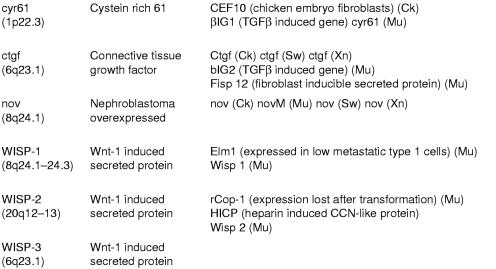
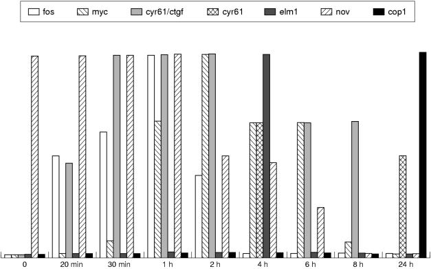
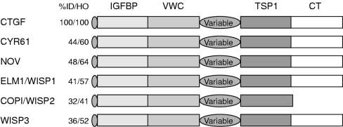
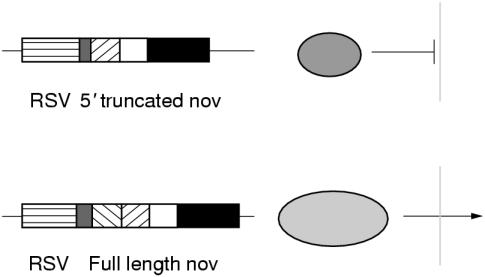
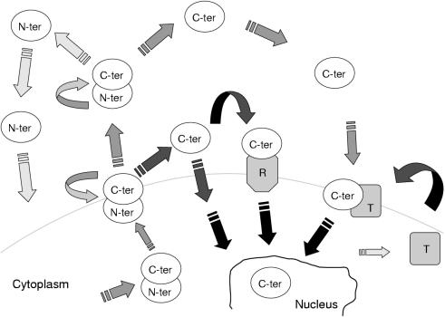
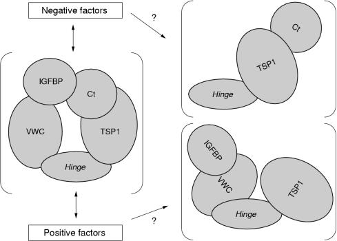
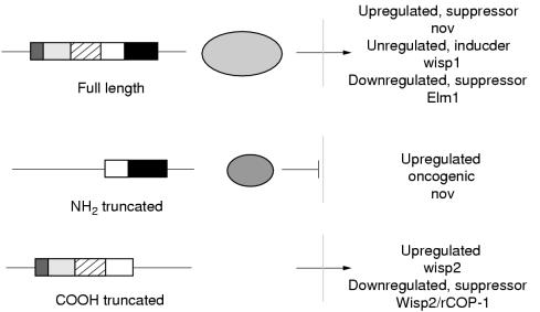
The CCN family of genes presently consists of six distinct members encoding proteins that participate in fundamental biological processes such as cell proliferation, attachment, migration, differentiation, wound healing, angiogenesis, and several pathologies including fibrosis and tumorigenesis. Whereas CYR61 and CTGF were reported to act as positive regulators of cell growth, NOV (nephroblastoma overexpressed) provided the first example of a CCN protein with negative regulatory properties and the first example of aberrant expression being associated with tumour development. The subsequent discovery of the ELM1, rCOP1, and WISP proteins has broadened the variety of functions attributed to the CCN proteins and has extended previous observations to other biological systems. This review discusses fundamental questions regarding the regulation of CCN gene expression in normal and pathological conditions, and the structural basis for their specific biological activity. After discussing the role of nov and other CCN proteins in the development of a variety of different tissues such as kidney, nervous system, muscle, cartilage, and bone, the altered expression of the CCN proteins in various pathologies is discussed, with an emphasis on the altered expression of nov in many different tumour types such as Wilms's tumour, renal cell carcinomas, prostate carcinomas, osteosarcomas, chondrosarcomas, adrenocortical carcinomas, and neuroblastomas. The possible use of nov as a tool for molecular medicine is also discussed. The variety of biological functions attributed to the CCN proteins has led to the proposal of a model in which physical interactions between the amino and carboxy portions of the CCN proteins modulate their biological activity and ensure a proper balance of positive and negative signals through interactions with other partners. In this model, disruption of the secondary structure of the CCN proteins induced by deletions of either terminus is expected to confer on the truncated polypeptide constitutive positive or negative activities.


Figures

Figure 1
Figure 1
The CCN family of genes. The names and chromosomal localisations of the human CCN genes are indicated in the left column.
Figure 2
Figure 2
Kinetics of CCN gene expression upon stimulation of cell growth.
Figure 3
Figure 3
Multimodular structure of the CCN proteins. CT, cystein knot containing family of growth regulators-like domain; IGFBP, insulin-like growth factor binding protein-like domain; TSP1, thrombospondin-like domain; and VWC, Von Willebrand factor-like domain.
Figure 4
Figure 4
Fate of the full length and truncated proteins. Rous sarcoma virus (RSV) driven expression of the truncated protein in chicken embryonic fibroblasts (CEFs) leads to the cytoplasmic accumulation (and further processing?) of the truncated NOV protein, whereas the presence of the signal peptide in full length NOV governs its secretion.
Figure 5
Figure 5
Confocal immunocytolocalisation of NOV in the nucleus of 143 osteosarcoma cells. (A) staining with anti-nov antibodies, (B) double staining with anti-nov and anti-HSV-ICP4 (herpes simplex virus infected cell protein 4) antibodies, (C) double staining with anti-nov and HSV-ICP8 antibodies. The yellow colour indicates colocalisation of ICP4 and NOV.
Figure 6
Figure 6
Schematic hypothetical pathways leading to the potential internalisation of the amino truncated NOV protein. In all cases, the secretion of a full length NOV protein is thought to be required for further processing. The full length NOV protein might be secreted and processed at the membrane site, the C-proximal moiety of the protein being internalised directly or indirectly through specific interactions with either a receptor or a transporter. Alternatively, the NOV protein could be released into the medium or the extracellular matrix where it would be submitted to proteolysis. The amino part of the protein might itself play an important role in signalling, either directly or indirectly. C-ter, amino truncated protein; N-ter, N-proximal moiety of the protein; R, receptor; T, transporter.
Figure 7
Figure 7
Schematic structure of the human (A) and chicken (B) nov promoters. The putative binding sites were identified on the basis of their consensus nucleotide sequences.
Figure 8
Figure 8
Detection of nov RNA species at sites of chondrogenesis in developing chicken. (A) Development stage (St) 3, embryonic day (E) 8.5. (B) St 31, E7. (C) St 35, E8.5. (D) St 38, E12. (E) St 35, E8.5.
Figure 9
Figure 9
Detection of the NOV protein in developing bone. (A) At stage 29 (embryonic day 6 (E6)), NOV is detected at sites of chondrogenesis in the developing chicken wing. Alcian blue staining identifies cartilage formation. (B) At stages 35–38 (E11–12) strong nov staining is seen in the perichondrium (p) and proliferating chondrocytes (pc). (C) At the same stages, flattened chondrocytes (fc) do not stain positive for nov, whereas hypertrophic chondrocytes (hc) are strongly positive for NOV staining (D).
Figure 10
Figure 10
Expression of nov RNA species and collagen II (COL II) in chicken micromass cell cultures. Staining of ribosomal RNA species allows relative quantification of the hybridisation signals obtained in two independent experiments. J0–J3, days of culture; GAPDH, glyceraldehyde-3-phosphate dehydrogenase.
Figure 11
Figure 11
The expression of nov RNA depends upon chondrogenesis. (A) 106 mesenchymal cells were seeded for micromass cell cultures. (B) 107 mesenchymal cells were seeded for micromass cell cultures. Samples were taken at the day of seeding (J0) and after 24 hours culture (J1). COL II, collagen II; GAPDH, glyceraldehyde-3-phosphate dehydrogenase.
Figure 12
Figure 12
Detection of the NOV protein in normal (A) and tumoral adrenal tissues (B and C). The normal area was isolated from a patient with adenoma. Note the uneven nov staining in the adenoma (B). An example of NOV negative adrenal carcinoma is shown (C). Photographs were kindly provided by H Yeger.
Figure 13
Figure 13
Expression of nov in renal cell carcinomas (RCCs). Increased amounts of nov were correlated with increased tumorigenicity. Nov, levels of nov RNA estimated from northern blots; Latency, time in days required to reach a tumour of a given size.
Figure 14
Figure 14
Model for the structural organisation of the CCN proteins. Potential interactions with positive and negative regulators are thought to be disrupted upon truncation of the N-proximal and C-proximal modules of CCN proteins. Ct, cystein knot containing family of growth regulators-like domain; IGFBP, insulin-like growth factor binding protein-like domain; TSP1, thrombospondin-like domain; and VWC, Von Willebrand factor-like domain.
Figure 15
Figure 15
CCN proteins and cancer. This schematic drawing summarises the diversity of situations encountered for CCN proteins in tumours. Whether the subcellular localisation of the CCN proteins other than NOV can result in opposing biological properties remains to be established.

References

    1. Bork P. The modular architecture of a new family of growth regulators related to connective tissue growth factor. FEBS Lett 1993;327:125–30. - PubMed
    1. Lau LF, Lam SC. The CCN family of angiogenic regulators: the connection. Exp Cell Res 1999; 248:44–57. - PubMed
    1. Brigstock DR. The connective tissue growth factor/cysteine-rich 61/nephroblastoma overexpressed (CCN) family. Endocr Rev 1999;20:189–206. - PubMed
    1. Essam ED, Moussad A, Brigstock D. Connective tissue growth factor: what's in a name. Mol Genet Metab 2000;71:276–92. - PubMed
    1. Lau LF, Nathans D. Expression of a set of growth-related immediate early genes in BALB/c 3T3 cells: coordinate regulation with c-fos or c-myc. Proc Natl Acad Sci USA 1987;84:1182–6. - PMC - PubMed

Publication types

MeSH terms