. 2001 Apr 27;292(5517):727-30.
doi: 10.1126/science.1059108.

Proapoptotic BAX and BAK: a requisite gateway to mitochondrial dysfunction and death

Affiliations

Proapoptotic BAX and BAK: a requisite gateway to mitochondrial dysfunction and death

M C Wei et al. Science. .

Abstract

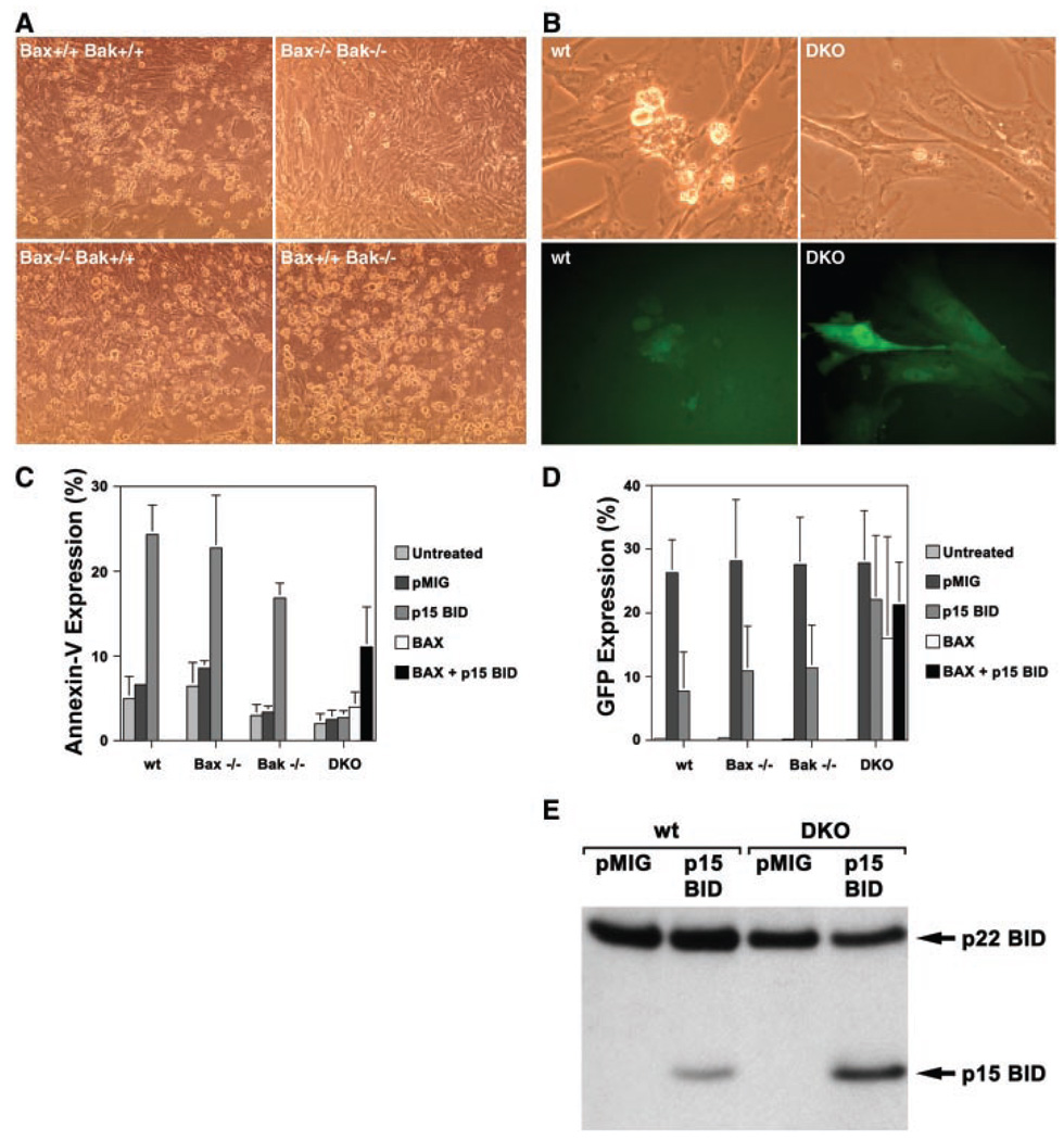
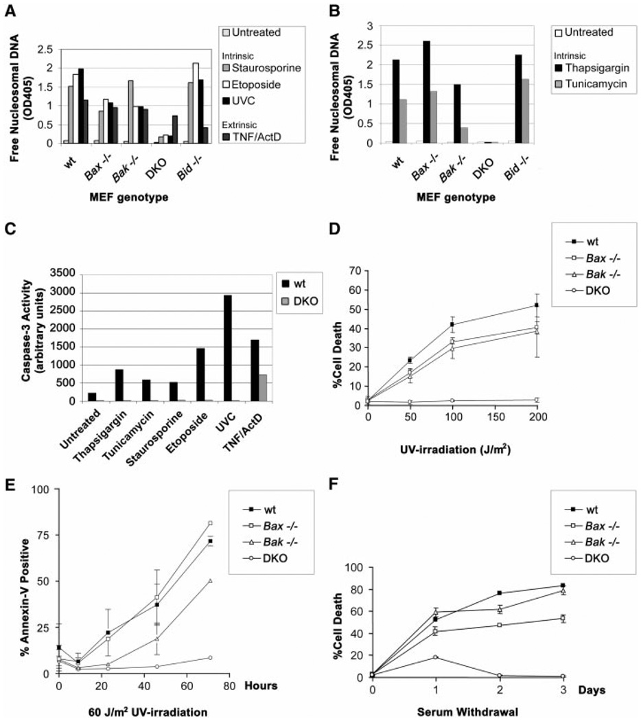
Multiple death signals influence mitochondria during apoptosis, yet the critical initiating event for mitochondrial dysfunction in vivo has been unclear. tBID, the caspase-activated form of a "BH3-domain-only" BCL-2 family member, triggers the homooligomerization of "multidomain" conserved proapoptotic family members BAK or BAX, resulting in the release of cytochrome c from mitochondria. We find that cells lacking both Bax and Bak, but not cells lacking only one of these components, are completely resistant to tBID-induced cytochrome c release and apoptosis. Moreover, doubly deficient cells are resistant to multiple apoptotic stimuli that act through disruption of mitochondrial function: staurosporine, ultraviolet radiation, growth factor deprivation, etoposide, and the endoplasmic reticulum stress stimuli thapsigargin and tunicamycin. Thus, activation of a "multidomain" proapoptotic member, BAX or BAK, appears to be an essential gateway to mitochondrial dysfunction required for cell death in response to diverse stimuli.


Figures

Fig. 1
Fig. 1
Resistance of Bax, Bak doubly deficient murine embryonic fibroblasts (MEFs) to tBID-induced apoptosis. (A) Bright-field microscopy (×20 magnification) of wild-type, Bax−/− Bak−/−, Bax−/− Bak+/+, and Bax+/+ Bak−/− primary embryonic day 13.5 MEFs 24 hours after infection with a tBID-expressing vector. Murine p15 BID and BAX were cloned into the retroviral expression vector MSCV-IRES-GFP (pMIG) (11). Retroviruses were generated by transfecting 293GPG packaging cell line as described (33). Retrovirus-containing supernatant was collected 3 and 5 days after transfection and used to infect MEFs in the presence of polybrene (8 µg/ml). (B) Bright-field and GFP fluorescence of the same field (×40 magnification) of wild-type (wt) and DKO MEFs 24 hours after infection with the tBID vector. (C) Quantitation of apoptosis in MEFs shown at the 24-hour time point after infection with pMIG (empty vector control), tBID, and/or BAX vectors. Cell death was quantitated by flow cytometric detection of Annexin-V staining (Bio-vision). Values shown are mean ± 1 SD from three independent experiments. (D) Quantitation of GFP-positive cells 24 hours after infection with pMIG, tBID, and/or BAX vectors. GFP-positive cells were detected by flow cytometry. Values shown are mean ± 1 SD from three independent experiments. (E) Immunoblot with antibody to Bid of whole-cell lysates from SV40-transformed (34) wild-type and DKO MEFs at 17 hours after infection with pMIG or tBID vectors.
Fig. 2
Fig. 2
Function of BAX and BAK downstream of tBID and upstream of cytochrome c release. (A) tBID-induced BAK oligomerization independent of BAX. Mitochondria-enriched fractions from pMIG or tBID vector-infected MEFs were prepared as described (14) and treated with dimethyl sulfoxide (DMSO) containing control buffer or 10 mM BMH cross linker (Pierce). Cross-linked BAK species were detected by antibody to BAK (Upstate Biotechnology) immunoblot. * indicates BAK complexes consistent with dimers or trimers; ** indicates inactive BAK conformer. (B) Cytochrome c release in DKO MEFs. Three-color fluorescence microscopy (35) of wild-type and DKO MEFs at 24 hours after infection with tBID and GFP-expressing vector. Red indicates cytochrome c immunostaining. Blue is Hoechst staining of DNA. Green is GFP expression in infected cells. Overlap of GFP and cytochrome c staining is denoted by yellow. Results are representative of three independent experiments. (C) Apoptosis of DKO MEFs after microinjection of cytochrome c. Primary MEFs were plated on 1% gelatin-coated gridded cover slips. Puri fied cytochrome c (1.6 mg/ml) dissolved in phosphate-buffered saline (PBS) or PBS control was mixed 1:1 with 0.5% fluorescein isothiocyanate–conjugated dextran and microinjected into cells with an Eppendorf 5246 Transjector at a pressure of 150 hPa and an injection time of 0.5 s. Apoptotic cells detach from the cover slip and were counted as a measure of apoptosis, enabling a time course of the mean percentages of microinjected cells that detach to be plotted from three independent experiments.
Fig. 3
Fig. 3
Function of BAX and BAK downstream of BID in Fas-induced hepatocellular apoptosis. (A) Hematoxylin and eosin-stained liver sections from Bak−/−, Bax−/−, and DKO mice treated with antibody to Fas (anti-Fas). Bar represents 20 µm. (B) Fas-induced BAX translocation from cytosol to mitochondria in hepatocytes is downstream of BID but independent of BAK. BAX immunoblot of mitochondrial (HM) and cytosolic (S100) fractions of hepatocytes prepared as previously described (8) from wild-type, Bid−/−, Bak+/−, and Bak−/− mice treated with saline or anti-Fas. (C) Three-color immunohistochemistry of livers from anti-Fas–treated Bak−/− and DKO mice. Cytochrome c staining is in green, activated Caspase-3/7 (CM1 antibody) in red, and nuclei stained with Hoechst in blue. Bar represents 20 µm. Histology and immunohistochemical staining of livers from anti-Fas–treated animals were done as described (14).
Fig. 4
Fig. 4
Resistance of Bax, Bak doubly deficient MEFs to multiple intrinsic death signals. (A) Susceptibility of MEFs to apoptotic death by mitochondria-dependent intrinsic signals. Wild-type, Bax−/−, Bak−/−, DKO, and Bid−/− primary MEFs were treated with 1 µM staurosporine, 100 µM etoposide, UVC (60 J/m2), or TNF-α (1 ng/ml) + actinomycin D (2 µg/ml), and a 48-hour time point is shown. Average values from duplicate samples of an enzyme-linked immunosorbent assay of apoptotic DNA fragmentation (Roche) are plotted as representative of three independent experiments. (B) Susceptibility of MEFs to apoptotic death by ER stimuli. As in (A), genotyped MEFs were treated with 2 µM thapsigargin or tunicamycin (1 µg/ml), and average values from duplicate samples at a 48-hour time point of apoptotic DNA fragmentation were plotted. DKO MEFs also demonstrated long-term survival when assessed by Annexin-V staining 4 days after the stimuli (12). (C) Quantitation of effector caspase activity (e.g., Caspase-3) using DEVD-AFC fluorogenic substrate (Clontech). Wild-type and DKO MEFs were treated with the following death signals and harvested at indicated time points: 2 µM thapsigargin (36 hours), tunicamycin (1 µg/ml; 36 hours), 1 µM staurosporine (16 hours), 100 µM etoposide (24 hours), UVC (60 J/m2; 24 hours), and TNF-α (1 ng/ml) + actinomycin D (2 µg/ml; 16 hours). Results are representative of three independent experiments. (D) Dose response of MEFs to UVC irradiation. Cell death was quantitated by trypan blue exclusion, and a 24-hour time point was plotted. (E) Time course of MEF apoptosis after exposure to UVC (60 J/m2). Cell death was quantitated by flow cytometric detection of Annexin-V staining. Long-term survival of DKO MEFs was also noted 4 days after staurosporine and etoposide, as well as UVC treatments (12). (F) Time course of MEF apoptosis in response to serum withdrawal. Cell death was quantitated by trypan blue exclusion.

Comment in

Similar articles

Cited by

References

    1. Green DR. Cell. 2000;102:1. - PubMed
    1. Kroemer G, Reed JC. Nature Med. 2000;5:513. - PubMed
    1. Adams JM, Cory S. Science. 1998;281:1322. - PubMed
    1. Gross A, McDonnell JM, Korsmeyer SJ. Genes Dev. 1999;13:1899. - PubMed
    1. Huang DC, Strasser A. Cell. 2000;103:839. - PubMed

Publication types

MeSH terms

LinkOut - more resources