Cisplatinum and taxol induce different patterns of p53 phosphorylation
- PMID: 11326311
- PMCID: PMC1505020
- DOI: 10.1038/sj.neo.7900122
Cisplatinum and taxol induce different patterns of p53 phosphorylation
Abstract
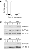
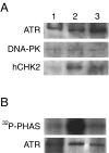
Posttranslational modifications of p53 induced by two widely used anticancer agents, cisplatinum (DDP) and taxol were investigated in two human cancer cell lines. Although both drugs were able to induce phosphorylation at serine 20 (Ser20), only DDP treatment induced p53 phosphorylation at serine 15 (Ser15). Moreover, both drug treatments were able to increase p53 levels and consequently the transcription of waf1 and mdm-2 genes, although DDP treatment resulted in a stronger inducer of both genes. Using two ataxia telangiectasia mutated (ATM) cell lines, the role of ATM in drug-induced p53 phosphorylations was investigated. No differences in drug-induced p53 phosphorylation could be observed, indicating that ATM is not the kinase involved in these phosphorylation events. In addition, inhibition of DNA-dependent protein kinase activity by wortmannin did not abolish p53 phosphorylation at Ser15 and Ser20, again indicating that DNA-PK is unlikely to be the kinase involved. After both taxol and DDP treatments, an activation of hCHK2 was found and this is likely to be responsible for phosphorylation at Ser20. In contrast, only DDP was able to activate ATR, which is the candidate kinase for phosphorylation of Ser15 by this drug. This data clearly suggests that differential mechanisms are involved in phosphorylation and activation of p53 depending on the drug type.
Figures


Similar articles
-
Hyperoxia activates the ATR-Chk1 pathway and phosphorylates p53 at multiple sites.Am J Physiol Lung Cell Mol Physiol. 2004 Jan;286(1):L87-97. doi: 10.1152/ajplung.00203.2002. Epub 2003 Sep 5. Am J Physiol Lung Cell Mol Physiol. 2004. PMID: 12959929
-
Status of p53 phosphorylation and function in sensitive and resistant human cancer models exposed to platinum-based DNA damaging agents.J Cancer Res Clin Oncol. 2003 Dec;129(12):709-18. doi: 10.1007/s00432-003-0480-4. Epub 2003 Sep 26. J Cancer Res Clin Oncol. 2003. PMID: 14513366 Free PMC article.
-
Stabilization of p53 by CP-31398 inhibits ubiquitination without altering phosphorylation at serine 15 or 20 or MDM2 binding.Mol Cell Biol. 2003 Mar;23(6):2171-81. doi: 10.1128/MCB.23.6.2171-2181.2003. Mol Cell Biol. 2003. PMID: 12612087 Free PMC article.
-
Nitric oxide and p53 in cancer-prone chronic inflammation and oxyradical overload disease.Environ Mol Mutagen. 2004;44(1):3-9. doi: 10.1002/em.20024. Environ Mol Mutagen. 2004. PMID: 15199542 Review.
-
How to activate p53.Curr Biol. 2000 Apr 20;10(8):R315-7. doi: 10.1016/s0960-9822(00)00439-5. Curr Biol. 2000. PMID: 10801407 Review.
Cited by
-
Hypoxia-induced cytotoxic drug resistance in osteosarcoma is independent of HIF-1Alpha.PLoS One. 2013 Jun 13;8(6):e65304. doi: 10.1371/journal.pone.0065304. Print 2013. PLoS One. 2013. PMID: 23785417 Free PMC article.
-
The EDN1/EDNRA/β‑arrestin axis promotes colorectal cancer progression by regulating STAT3 phosphorylation.Int J Oncol. 2023 Jan;62(1):13. doi: 10.3892/ijo.2022.5461. Epub 2022 Dec 1. Int J Oncol. 2023. PMID: 36453252 Free PMC article.
-
Ataxia telangiectasia and rad3-related kinase contributes to cell cycle arrest and survival after cisplatin but not oxaliplatin.Mol Cancer Ther. 2009 Apr;8(4):855-63. doi: 10.1158/1535-7163.MCT-08-1135. Mol Cancer Ther. 2009. PMID: 19372558 Free PMC article.
-
Cisplatin associated with LY294002 increases cytotoxicity and induces changes in transcript profiles of glioblastoma cells.Mol Biol Rep. 2014 Jan;41(1):165-77. doi: 10.1007/s11033-013-2849-z. Epub 2013 Nov 12. Mol Biol Rep. 2014. PMID: 24218165
-
In vitro and in vivo therapeutic approach for a small cell carcinoma of the ovary hypercalcaemic type using a SCCOHT-1 cellular model.Orphanet J Rare Dis. 2014 Aug 8;9:126. doi: 10.1186/s13023-014-0126-4. Orphanet J Rare Dis. 2014. PMID: 25103190 Free PMC article.
References
-
- KoL J, Prives C. p53: puzzle and paradigm. GenesDev. 1996;56:2649–2654. - PubMed
-
- Levine AJ. p53, the cellular gatekeeper for growth and division. Cell. 1997;88:323–331. - PubMed
-
- Chen X, Ko LJ, Jayaraman L, Prives C. p53 levels, functional domains, and DNA damage determine the extent of the apoptotic response of tumor cells. Genes Dev. 1996;10:2438–2451. - PubMed
-
- Ullrich SJ, Anderson CW, Mercer WE, Appella E. The p53 tumor suppressor protein, a modulator of cell proliferation. J Biol Chem. 1992;267:15259–15262. - PubMed
-
- Wahl AF, Donaldson KL, Fairchild C, Lee FY, Foster SA, Demers GW, Galloway DA. Loss of normal p53 function confers sensitization to Taxol by increasing G2/M arrest and apoptosis. Nat Med. 1996;2:72–79. - PubMed
Publication types
MeSH terms
Substances
LinkOut - more resources
Full Text Sources
Molecular Biology Databases
Research Materials
Miscellaneous
