

Giant cell tumour of tendon sheath (localised nodular tenosynovitis): clinicopathological features of 71 cases
- PMID: 11328844
- PMCID: PMC1731411
- DOI: 10.1136/jcp.54.5.404
Giant cell tumour of tendon sheath (localised nodular tenosynovitis): clinicopathological features of 71 cases
Abstract
Aims/background: Giant cell tumour of the tendon sheath (GCTTS) is regarded as the most common neoplasm of the hand that can recur after excision. The objective of this study was to review a series of cases in our department and to determine any clinical or pathological features that might predict the likelihood of recurrence.
Methods: Clinical data, obtained from pathology request forms and in patient notes, along with the gross and microscopic appearances of 71 cases of GCTTS were evaluated.
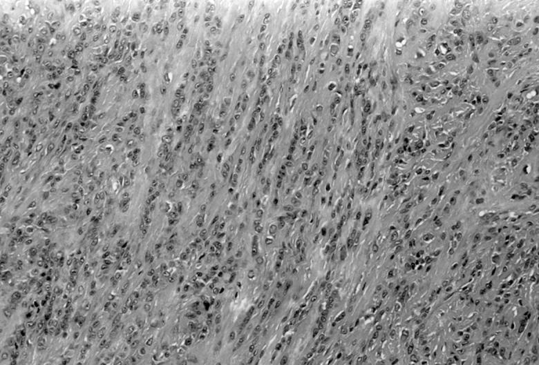
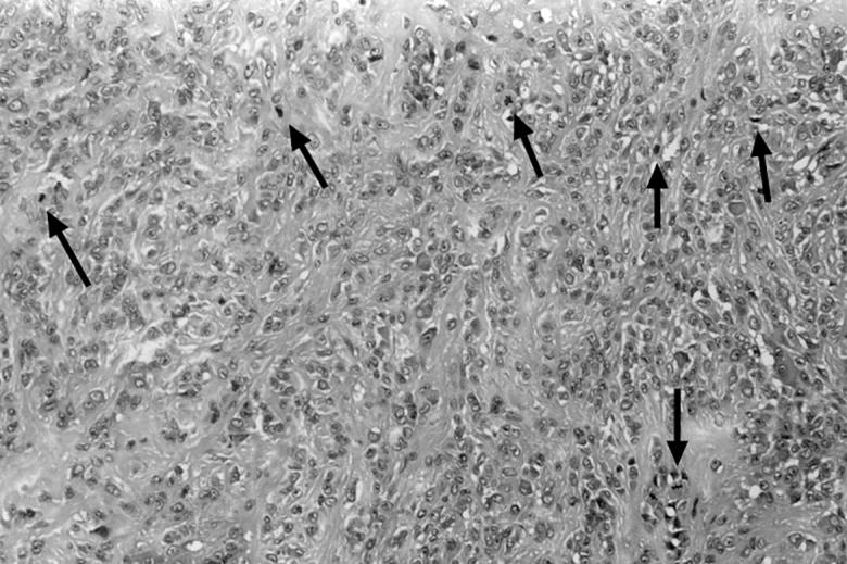
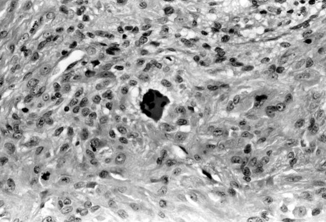
Results: Clinical features and pathological features identified were similar to those of previous studies. In comparison with previous studies a higher mitotic count (range, 1-21 mitoses/10 high power fields (HPF); mean, 5/10 HPF) was noted in all cases, irrespective of recurrence and numerous apoptotic bodies (up to 30/10 HPF), mainly formed from osteoclast-like giant cells, were present.
Conclusions: GCTTS is a relatively rare soft tissue tumour of uncertain histiogenesis. Mitotic and apoptotic figures are a common feature and do not indicate clinical behaviour. Complete local excision is the treatment of choice.
Figures
MeSH terms
LinkOut - more resources
Full Text Sources