. 2001 May 1;29(9):1852-63.
doi: 10.1093/nar/29.9.1852.

Targeting peptide nucleic acid (PNA) oligomers to mitochondria within cells by conjugation to lipophilic cations: implications for mitochondrial DNA replication, expression and disease

Affiliations

Targeting peptide nucleic acid (PNA) oligomers to mitochondria within cells by conjugation to lipophilic cations: implications for mitochondrial DNA replication, expression and disease

A Muratovska et al. Nucleic Acids Res. .

Abstract

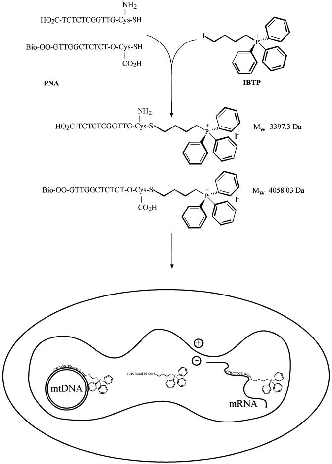
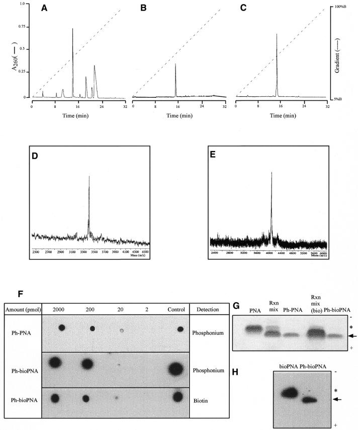
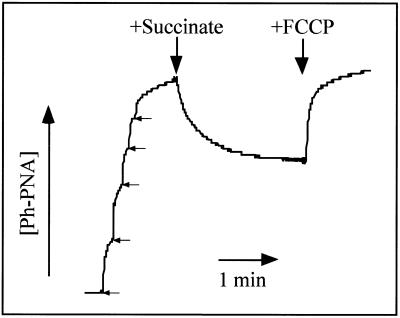
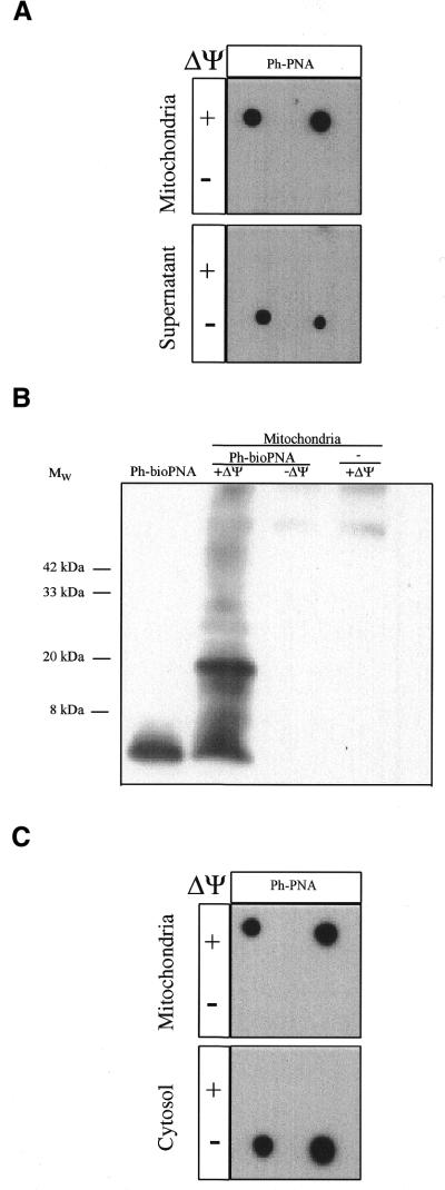
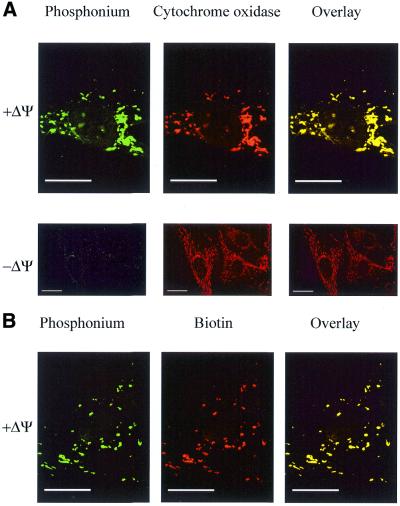
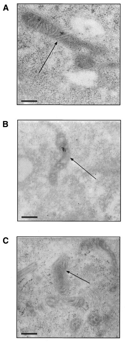
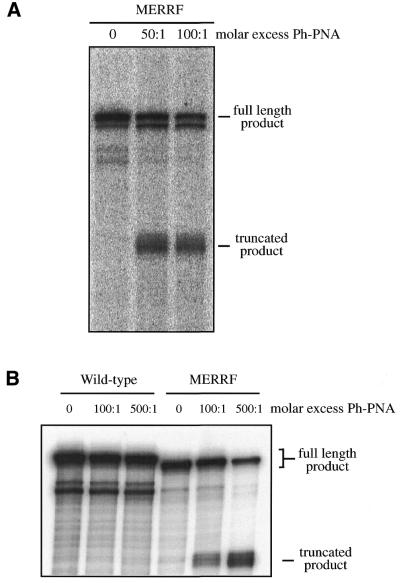
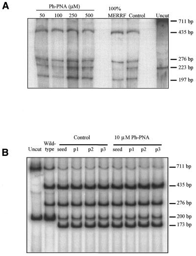
The selective manipulation of mitochondrial DNA (mtDNA) replication and expression within mammalian cells has proven difficult. One promising approach is to use peptide nucleic acid (PNA) oligomers, nucleic acid analogues that bind selectively to complementary DNA or RNA sequences inhibiting replication and translation. However, the potential of PNAs is restricted by the difficulties of delivering them to mitochondria within cells. To overcome this problem we conjugated a PNA 11mer to a lipophilic phosphonium cation. Such cations are taken up by mitochondria through the lipid bilayer driven by the membrane potential across the inner membrane. As anticipated, phosphonium-PNA (ph-PNA) conjugates of 3.4-4 kDa were imported into both isolated mitochondria and mitochondria within human cells in culture. This was confirmed by using an ion-selective electrode to measure uptake of the ph-PNA conjugates; by cell fractionation in conjunction with immunoblotting; by confocal microscopy; by immunogold-electron microscopy; and by crosslinking ph-PNA conjugates to mitochondrial matrix proteins. In all cases dissipating the mitochondrial membrane potential with an uncoupler prevented ph-PNA uptake. The ph-PNA conjugate selectively inhibited the in vitro replication of DNA containing the A8344G point mutation that causes the human mtDNA disease 'myoclonic epilepsy and ragged red fibres' (MERRF) but not the wild-type sequence that differs at a single nucleotide position. Therefore these modified PNA oligomers retain their selective binding to DNA and the lipophilic cation delivers them to mitochondria within cells. When MERRF cells were incubated with the ph-PNA conjugate the ratio of MERRF to wild-type mtDNA was unaffected, even though the ph-PNA content of the mitochondria was sufficient to inhibit MERRF mtDNA replication in a cell-free system. This unexpected finding suggests that nucleic acid derivatives cannot bind their complementary sequences during mtDNA replication. In summary, we have developed a new strategy for targeting PNA oligomers to mitochondria and used it to determine the effects of PNA on mutated mtDNA replication in cells. This work presents new approaches for the manipulation of mtDNA replication and expression, and will assist in the development of therapies for mtDNA diseases.


Figures

Figure 1
Figure 1
Synthesis of ph–PNA conjugates. The reactions of the cysteine residues of the biotinylated PNA oligomer and the PNA oligomer with IBTP are shown. The sequences and predicted MW values of the products, their uptake into mitochondria and subsequent interactions with mtDNA and mRNA are indicated.
Figure 2
Figure 2
Purification and characterisation of ph–PNA conjugates. (A) Purification of the ph–PNA reaction products by RP-HPLC. The major peak at ∼15 min, due to the ph–PNA conjugate, was collected, lyophilised, dissolved in water and a sample analysed by RP-HPLC (B). The ph–bioPNA was prepared by a similar procedure (data not shown) and purified by RP-HPLC (C). (D and E) MALDI-TOF analyses of purified ph–PNA and ph–bioPNA, respectively. The observed masses for the ph–PNA (3400 ± 4 Da) and the ph–bioPNA (4054 ± 3 Da) conjugates were within 0.1% of the calculated masses, as expected for external mass calibration. (F) Serial dilutions of the ph–PNA and ph–bioPNA conjugates were adsorbed on nitrocellulose and the triphenylphosphonium moiety detected using antitriphenylphosphonium serum. BSA conjugated to IBTP (∼1 µg protein) was used as a positive control and PNAs not conjugated to IBTP were not detected by this antibody (data not shown). Horse radish peroxidase conjugated to extravidin was used to detect biotin and the bioPNA oligomer (∼5 nmol) was used as a positive control. Unconjugated PNA oligomers were not detected by either procedure (data not shown). (G) Samples (∼5 nmol) of PNA, reaction products prior to purification, and the purified ph–PNA and ph–bioPNA conjugates were separated by Tris–tricine PAGE and stained with Coomassie blue. The precursor PNAs are marked with an asterisk, the ph–PNA conjugates with an arrow and the polarity of electrophoresis is shown. (H) bioPNA and the purified ph–bioPNA conjugate (∼5 nmol of each) were separated by Tris–tricine PAGE, transferred to nitrocellulose and probed for biotin using streptavidin.
Figure 3
Figure 3
Ph–PNA conjugate uptake measured using an ion-selective electrode. An ion-selective electrode was used to measure the concentration of a ph–PNA conjugate in a mitochondrial suspension. The arrows indicate five sequential additions of 1 µM ph–PNA to calibrate the electrode response. The mitochondria were then energised by addition of succinate (5 mM) and uncoupled with FCCP (10 µM) as indicated.
Figure 4
Figure 4
Uptake of ph–PNA conjugates by mitochondria. (A) Rat liver mitochondria were incubated with 1 µM ph–PNA in the presence or absence of ΔΨm, (±10 µM FCCP). After incubation the mitochondria were pelleted through oil and the supernatant and mitochondrial fractions were probed with antitriphenylphosphonium serum. (B) A mitochondrial matrix fraction enriched for the nucleoid was isolated from mitochondria which had been incubated ±1 µM bioPNA in the presence or absence of ΔΨm, and then crosslinked with 1% formaldehyde. Samples were separated by Tris–tricine PAGE, transferred to nitrocellulose and probed for biotin. A ph–bioPNA (∼5 nmol) control was also analysed. (C) 143B cells were incubated with 1 µM ph–PNA in the presence or absence of ΔΨm, fractionated with digitionin and separated into mitochondria- and cytoplasm-enriched fractions by centrifugation through oil. Samples from both fractions were then assayed for triphenylphosphonium as above. There was no immuno-reactivity with preimmune serum in these experiments.
Figure 5
Figure 5
Mitochondrial localisation of ph–PNA conjugates within fibroblasts by confocal immunofluorescent microscopy. Fibroblasts were incubated with 1 µM ph–PNA (A) or 1 µM ph–bioPNA (B) for 4 h at 37°C, in the presence or absence of ΔΨm. (A) Cells were fixed, incubated with antiserum against triphenylphosphonium (green) and a monoclonal antibody against cytochrome oxidase (red). In the overlaid images yellow indicates co-localisation of triphenylphosphonium and cytochrome oxidase. (B) Cells were fixed and labelled for triphenylphosphonium (green) and biotin (red). In the overlaid images yellow indicates colocalisation of the triphenylphosphonium and biotin. There was no immuno reactivity with preimmune serum in these experiments. Magnification, 1400× (B, A + ΔΨ), 600× (A – ΔΨ); scale bar, 20 µm.
Figure 6
Figure 6
Immunogold labelling of ph–PNA conjugates within the mitochondrial matrix. (A) Human fibroblasts were incubated with 1 µM ph–PNA, fixed and the intracellular localisation of triphenylphosphonium detected by immunogold electron microscopy (black dots). (B) Human fibroblasts were incubated with 1 µM ph–bioPNA and the location of the biotin determined by immunogold electron microscopy. (C) Cells were incubated with ph–PNA but the incubation with triphenylphosphonium serum was omitted, while that with the gold-linked antibody was not. Identical electron micrographs were obtained by omitting the ph–PNA conjugates from the incubation. Magnification, 52 000×; scale bar, 200 nm; arrows indicate mitochondria.
Figure 7
Figure 7
Selective inhibition of DNA replication by ph–PNA conjugates. (A) An in vitro replication run-off assay from a mtDNA template containing the MERRF mutation was performed. In the absence of ph–PNA this gave a product of 341 nt. In the presence of 50 or 100-fold molar excess of ph–PNA a truncated product of 245 nt was formed. (B) An in vitro replication run-off assay from a wild-type mtDNA template was performed. This template differed from the MERRF template in (A) at a single nucleotide position in the PNA binding region. In the absence of ph–PNA this gave a full-length product of 350 nt (the MERRF DNA template is shorter due to a 9 bp deletion at the 3′ end, downstream of the replication inhibition site). Even in the presence of a 500-fold molar excess of ph–PNA none of the truncated product of 245 nt was found, in contrast to the positive control using the MERRF template. The binding affinities of PNA and ph–PNA to the MERRF PNA templates were 11.3 ± 2.2 and 37 ± 6 nM, respectively (Paul Smith, University of Newcastle upon Tyne, personal communication).
Figure 8
Figure 8
PCR–RFLP analysis of mtDNA from MERRF fibroblasts and myoblasts incubated with ph–PNA. For all samples, after incubation the mtDNA was isolated from the cells and the region around the MERRF point mutation amplified by PCR giving a 223 bp product for fibroblasts and a 200 bp product for myoblasts. The MERRF point mutation introduces a NaeI site, leading to 197 and 26 bp fragments for fibroblasts and 173 and 27 bp fragments for myoblasts, following digestion while the wild-type PCR products are uncut. Another mtDNA region of 711 bp containing an NaeI site in both wild-type and MERRF mtDNA was amplified by PCR and digested to 435 and 276 bp fragments as an internal control for complete digestion. (A) Heteroplasmic MERRF fibroblasts were incubated with 50, 100, 250 and 500 µM ph–PNA for 30 days. In addition, control samples from untreated fibroblasts after 30 days (control), samples from homoplasmic MERRF cells (100% MERRF) and undigested samples from untreated cells (uncut) were analysed. (B) Heteroplasmic MERRF myoblasts were seeded and incubated with 10 µM ph–PNA for three passages over 3 weeks. Samples were taken after seeding or after each passage and mtDNA assayed (p1–3). In addition, control samples from untreated myoblasts (control), digested samples from wild-type myoblasts (wild-type) and undigested samples from myoblasts (uncut) were analysed. Data shown are typical results of experiments repeated several times and under a range of ph–PNA concentrations for both fibroblasts and myoblasts.

References

    1. Larsson N.G. and Clayton,D.A. (1995) Molecular genetic aspects of human mitochondrial disorders. Annu. Rev. Genet., 29, 151–178. - PubMed
    1. Taylor R.W., Chinnery,P.F., Clark,K.M., Lightowlers,R.N. and Turnbull,D.M. (1997) Treatment of mitochondrial disease. J. Bioenerg. Biomembr., 29, 195–205. - PubMed
    1. Chrzanowska-Lightowlers L.Z., Lightowlers,R.N. and Turnbull,D.M. (1995) Gene therapy for mitochondrial DNA defects: is it possible? Gene Ther., 2, 311–316. - PubMed
    1. Clayton D.A. (1991) Nuclear gadgets in mitochondrial DNA replication and transcription. Trends Biochem. Sci., 16, 107–111. - PubMed
    1. Agrawal S. (1996) Antisense therapeutics. In Walker,J.M., (ed.), Methods in Molecular Medicine. Humana Press, NJ.

Publication types

MeSH terms