. 2001 May 1;29(9):1872-83.
doi: 10.1093/nar/29.9.1872.

Human testis expresses a specific poly(A)-binding protein

Affiliations

Human testis expresses a specific poly(A)-binding protein

C Féral et al. Nucleic Acids Res. .

Abstract

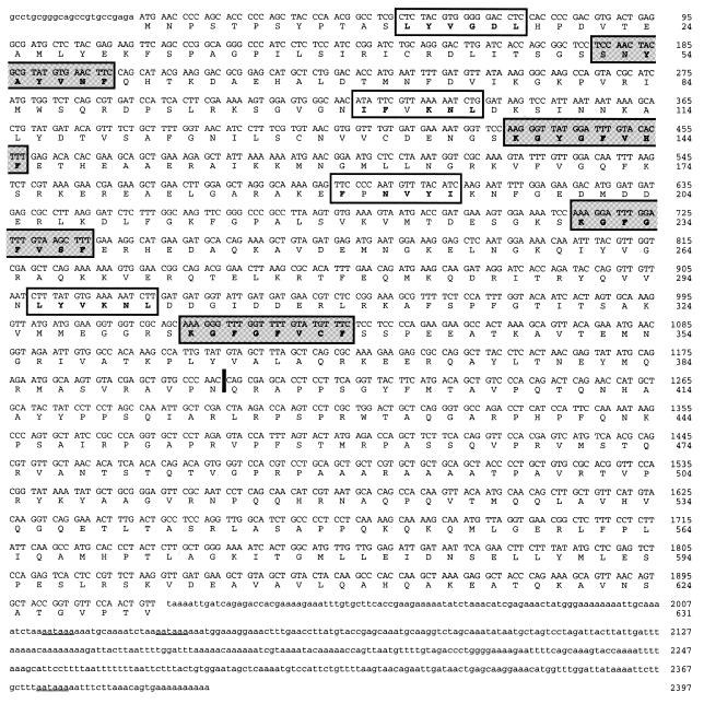
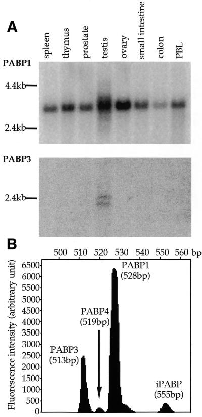
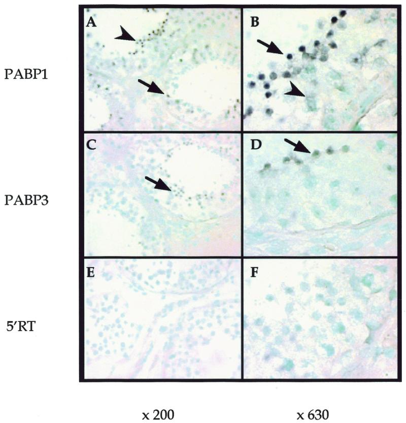
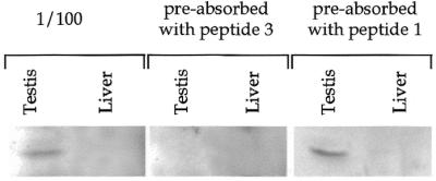
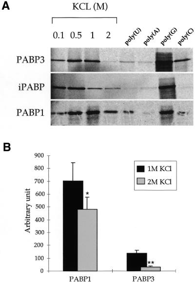
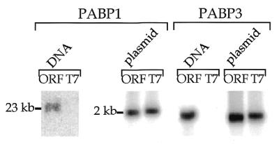
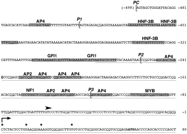
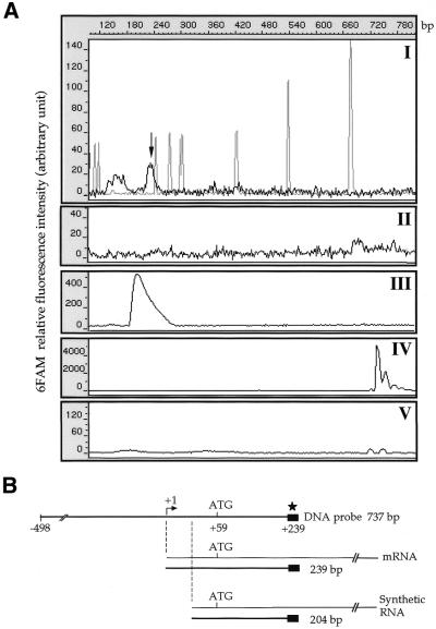
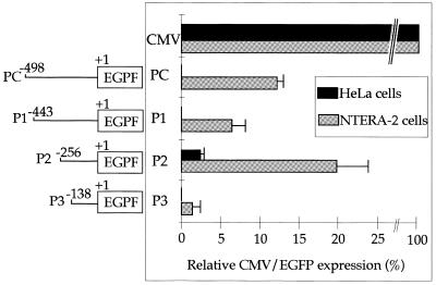
In testis mRNA stability and translation initiation are extensively under the control of poly(A)-binding proteins (PABP). Here we have cloned a new human testis-specific PABP (PABP3) of 631 amino acids (70.1 kDa) with 92.5% identical residues to the ubiquitous PABP1. A northern blot of multiple human tissues hybridised with PABP3- and PABP1-specific oligonucleotide probes revealed two PABP3 mRNAs (2.1 and 2.5 kb) detected only in testis, whereas PABP1 mRNA (3.2 kb) was present in all tested tissues. In human adult testis, PABP3 mRNA expression was restricted to round spermatids, whereas PABP1 was expressed in these cells as well as in pachytene spermatocytes. PABP3-specific antibodies identified a protein of 70 kDa in human testis extracts. This protein binds poly(A) with a slightly lower affinity as compared to PABP1. The human PABP3 gene is intronless with a transcription start site 61 nt upstream from the initiation codon. A sequence of 256 bp upstream from the transcription start site drives the promoter activity of PABP3 and its tissue-specific expression. The expression of PABP3 might be a way to bypass PABP1 translational repression and to produce the amount of PABP needed for active mRNA translation in spermatids.


Figures

Figure 1
Figure 1
cDNA and predicted amino acid sequences of PABP3. Lower case letters indicate the non-coding regions. The upper case letters correspond to the detected ORF. White and shaded boxes frame the consensus RNP2 and RNP1 sequences, respectively. The bold letters in each RNP consensus highlight the conserved amino acid residues as compared to similar regions in the PABP1 sequence. The three polyadenylation signal sequences (AATAAA) are underlined. The vertical bar represents the site of deletion of six amino acids (PVINPY) in PABP3 as compared to PABP1.
Figure 2
Figure 2
Northern blot analysis of PABP3 expression using specific oligonucleotide probes. (A) A multiple tissue blot was sequentially hybridised with oligonucleotides PABP1 and PABP3, exposed to a phosphor screen for 16 and 48 h, respectively, and analysed on a Storm 840 PhosphorImager. PBL, peripheral blood leukocytes. The size of the markers is indicated on the left. (B) Oligonucleotides 3′RT and 5′RT were used in RT–PCR on 5 ng total RNA from normal human adult testis. The amplified products were migrated on a sequencing gel and analysed using the GeneScan program as described in Materials and Methods. The surface under each peak reflects the amount of each PABP mRNA.
Figure 3
Figure 3
Localisation of PABP1 and PABP3 mRNA in human adult testis sections. Paraffin-embedded human testis sections were hybridised with the PABP1-specific antisense oligonucleotide probe (PABP1) (A and B), the PABP3-specific antisense oligonucleotide probe (PABP3) (C and D) or the common sense oligonucleotide probe (5′RT) (E and F) and counterstained with methyl green. Original magnifications: (A, C and E) 200×; B, D and F) 630×. Signals were confined to the seminiferous tubules in germinal cells (A–D). After 6 h detection the PABP1 mRNA-specific signal appeared in pachytene spermatocytes (arrowheads) and round spermatids (arrows) (A and B). The PABP3 mRNA signal appeared after 24 h reaction only in round spermatids (arrows) (C and D). No signal was observed with the control 5′RT probe after 24 h detection (E and F).
Figure 4
Figure 4
Detection of PABP3 by western blot analysis. Western blot of human testis protein extracts incubated with serum raised against a 9 amino acid specific PABP3 peptide (peptide 3). A human liver extract was used as negative control. In competition experiments the serum was pre-absorbed with peptide 1 or peptide 3.
Figure 5
Figure 5
RNA binding assays of PABP to poly(A)–Sepharose. (A) Binding of in vitro [35S]methionine-labelled PABP proteins to poly(A)–Sepharose was performed in the presence of 0.25 mg/ml yeast tRNA and 0.1 mg/ml poly(dI·dC) as non-specific competitors. In the four first lanes on the left the KCl concentration of the binding buffer varied from 0.1 to 2 M. In the four lanes on the right, binding reactions were carried out in 1 M KCl and in the presence of a 100-fold excess of the indicated competitors. (B) Analysis of five independent binding experiments of PABP1 and PABP3 to poly(A)–Sepharose in the presence of 1 or 2 M KCl. *P < 0.05; **P < 0.001.
Figure 6
Figure 6
PCR analysis of PABP1 and PABP3 genomic sequences. PCR amplification reactions of human genomic DNA (DNA) were performed using a 3′ primer (3′ORF) common for PABP1 and PABP3 and a 5′ primer (5′ORF) specific for PABP1 and PABP3 or a T7 sequence-specific primer which generates a fragment from a plasmid (plasmid). The lanes ORF contain samples amplified with oligonucleotides 3′ORF and 5′ORF, the lanes T7 contain the samples amplified with oligonucleotides 3′ORF and T7.
Figure 7
Figure 7
Sequence of the human PABP3 promoter. Grey boxes represent the location of potential binding sites for transcription factors identified using the program MatInspector (core similarity = 1; matrix similarity > 0.87). Other motifs meeting the above criteria are not shown: BRN2, CETS1P54, E47, FREAC7, IK2, LMO2COM, SRY, NF1, ROX and XFD2. The transcription start site identified by S1 mapping and corresponding to the 5′-end of the longest RACE product is indicated by an arrow at position +1. Asterisks indicate the 5′-end of cDNA sequences obtained by 5′-RACE. The translation start site is in bold and the CpG are underlined. Brackets indicate the 5′-end of the four constructs used in the transfection experiments, PC, P1, P2 and P3, respectively. The black arrowhead indicates the relative position of the transcription start site in the mouse testis-specific PABP2 gene (accession no. AF001290). The underlined sequence exhibits 91% identical nucleotide residues with the 5′-UTR of human PABP1 (accession no. AH007272).
Figure 8
Figure 8
S1 mapping of the PAPB3 promoter on human testis and liver mRNA. (A) S1 nuclease protection assay. A 737 bp gel-purified 6FAM-labeled RACE antisense probe containing potential transcription start sites identified by mRNA primer extension analysis was hybridised to 2 µg poly(A)+ RNA from either testis (I) or liver (II). As a positive control the S1 probe was also hybridised to 10 ng PABP3 synthetic RNA (accession no. AF132026) lacking nt 1–36 (see Fig. 7) (III). The undigested probe or probe digested with S1 in the absence of RNA is shown in (IV) and (V), respectively. The arrow indicates the DNA fragment protected by hybridisation to poly(A)+ mRNA. The grey peaks in (I) correspond to ROX-1000 DNA standards (Perkin Elmer, CA). The 6FAM relative fluorescence intensity (arbitrary units) is reported on the left. (B) The position of the single-stranded PABP3 DNA probe with respect to the endogenous and synthetic PABP3 mRNA, as well as the protected fragment, is diagrammed at the bottom. Numbering is relative to the transcription start site.
Figure 9
Figure 9
Expression of EGFP protein in cells transiently transfected with EGFP under the control of PABP3 promoter sequences. PABP3 genomic sequences extending from nt –498 to +30 were amplified by PCR, as described in Materials and Methods, and then inserted into a promoter-less EGFP vector (pEGFP-1). These constructs were transiently transfected into HeLa (black boxes) or NTERA-2 (grey boxes) cells. The numbers of cells counted were identical in the two transfection assays. EGFP fluorescence driven by each construct was normalised to that obtained with the CMV promoter/pEGFP-C1 vector (fluorescence of 100%). Results are the means of triplicate determinations obtained in two independent experiments. CMV corresponds to the control construct of the CMV promoter upstream from EGFP. PC, P1, P2 and P3 refer to the different PABP3 promoter constructs upstream from the EGFP reporter gene as indicated on the left.

References

    1. Yelick P.C., Kwon,Y.H., Flynn,J.F., Borzorgzadeh,A., Kleene,K.C. and Hecht,N.B. (1989) Mouse transition protein 1 is translationally regulated during the postmeiotic stages of spermatogenesis. Mol. Reprod. Dev., 1, 193–200. - PubMed
    1. Steger K., Klonisch,T., Gavenis,K., Drabent,B., Doenecke,D. and Bergmann,M. (1998) Expression of mRNA and protein of nucleoproteins during human spermiogenesis. Mol. Hum. Reprod., 4, 939–945. - PubMed
    1. Schafer M., Nayernia,K., Engel,W. and Schafer,U. (1995) Translational control in spermatogenesis. Dev. Biol., 172, 344–352. - PubMed
    1. Penttila T.L., Yuan,L., Mali,P., Hoog,C. and Parvinen,M. (1995) Haploid gene expression: temporal onset and storage patterns of 13 novel transcripts during rat and mouse spermiogenesis. Biol. Reprod., 53, 499–510. - PubMed
    1. Tafuri S.R., Familari,M. and Wolffe,A.P. (1993) A mouse Y box protein, MSY1, is associated with paternal mRNA in spermatocytes. J. Biol. Chem., 268, 12213–12220. - PubMed

Publication types

MeSH terms