. 2001 May 1;29(9):1892-7.
doi: 10.1093/nar/29.9.1892.

Modular construction for function of a ribonucleoprotein enzyme: the catalytic domain of Bacillus subtilis RNase P complexed with B. subtilis RNase P protein

Affiliations

Modular construction for function of a ribonucleoprotein enzyme: the catalytic domain of Bacillus subtilis RNase P complexed with B. subtilis RNase P protein

A Loria et al. Nucleic Acids Res. .

Abstract

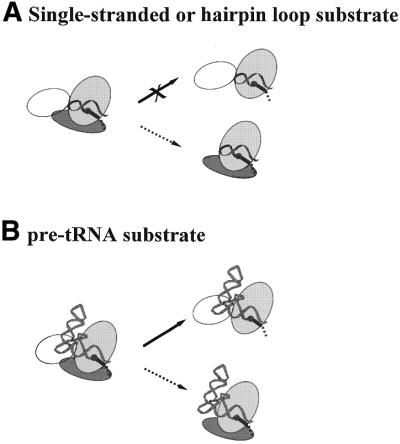
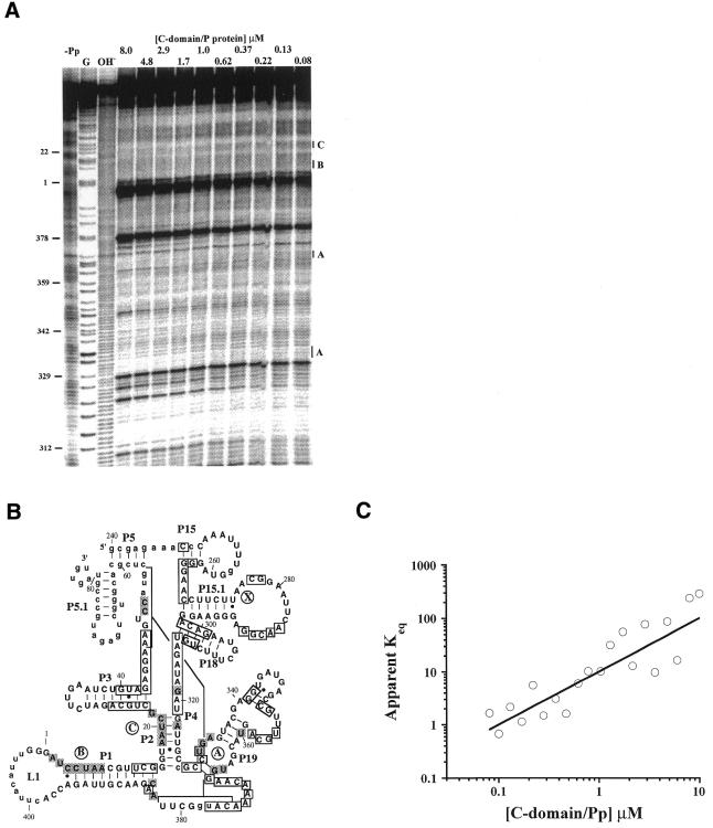
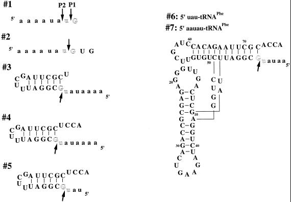
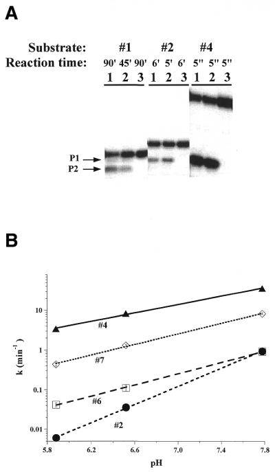
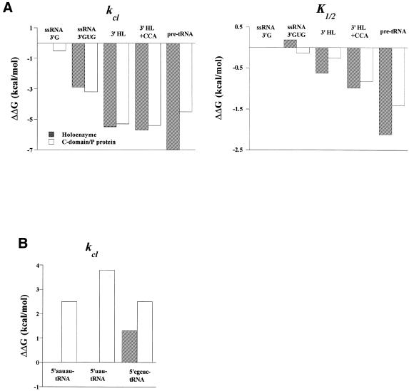
The bacterial RNase P holoenzyme catalyzes the formation of the mature 5'-end of tRNAs and is composed of an RNA and a protein subunit. Among the two folding domains of the RNase P RNA, the catalytic domain (C-domain) contains the active site of this ribozyme. We investigated specific binding of the Bacillus subtilis C-domain with the B.subtilis RNase P protein and examined the catalytic activity of this C-domain-P protein complex. The C-domain forms a specific complex with the P protein with a binding constant of approximately 0.1 microM. The C-domain-P protein complex and the holoenzyme are equally efficient in cleaving single-stranded RNA (approximately 0.9 min(-1) at pH 7.8) and substrates with a hairpin-loop 3' to the cleavage site (approximately 40 min(-1)). The holoenzyme reaction is much more efficient with a pre-tRNA substrate, binding at least 100-fold better and cleaving 10-500 times more efficiently. These results demonstrate that the RNase P holoenzyme is functionally constructed in three parts. The catalytic domain alone contains the active site, but has little specificity and affinity for most substrates. The specificity and affinity for the substrate is generated by either the specificity domain of RNase P RNA binding to a T stem-loop-like hairpin or RNase P protein binding to a single-stranded RNA. This modular construction may be exploited to obtain RNase P-based ribonucleoprotein complexes with altered substrate specificity.


Figures

Figure 1
Figure 1
Effects of P protein depletion [previous results summarized in Loria and Pan (19)] (solid arrows) or S-domain deletion (this work, dashed arrows) on catalytic activity. (A) P RNA cleaves a single-stranded or a hairpin–loop substrate at least 10 000-fold less efficiently under high salt conditions (data not shown). In contrast, the C-domain–P protein complex has the same catalytic efficiency for a hairpin–loop or a single-stranded RNA substrate as the holoenzyme. (B) P RNA effectively binds and cleaves a pre-tRNA substrate. The C-domain–P protein complex also binds and cleaves a pre-tRNA substrate, but less efficiently than a hairpin–loop substrate.
Figure 2
Figure 2
The B.subtilis C-domain forms a specific complex with the B.subtilis P protein in the absence of substrate. (A) Hydroxyl radical protection at varying concentrations of C-domain and P protein, always at a 1:1 molar ratio. All reactions contained 20 mM Tris–HCl pH 7.5, 10 mM MgCl2 and the reaction time was 30 min at 37°C. G, partial nuclease T1 digestion; OH, partial alkaline hydrolysis; –Pp, 8 µM C-domain alone. (B) The protected residues upon P protein binding superimposed on the phylogenetically derived secondary structure (33). Only residues that have protection factors of >1.5 are considered to be protected. Residues protected upon P protein binding are shaded. Residues protected upon Mg2+-induced folding are boxed (5). Region X (residues 270–280) was only protected in the holoenzyme (11). Nucleotides shown in lower case could not be analyzed either due to gel resolution or due to autolytic cleavage (around nt 405). (C) Determination of the binding constant of the C-domain–P protein complex [apparent Keq = y/(1 – y)2, curve fitted to equation 1b]. The protection factors (PF) for regions A–C are averaged. The fraction of C-domain bound with P protein corresponds to 1/PF and is plotted against the C-domain and P protein concentration, always at a 1:1 molar ratio.
Figure 3
Figure 3
Substrates used in this study with the intended cleavage site shown between the highlighted residues. The observed cleavage sites are indicated by arrows. The 5′-leader regions are shown in lower case.
Figure 4
Figure 4
(A) Cleavage of 5′-32P-labeled substrates 1, 2 and 4 by 1 µM holoenzyme (lane 1) and 1 µM C-domain–P protein complex (lane 2) under single turnover conditions. The control reaction was performed with 1 µM P protein alone (lane 3). All reactions contained 20 mM Tris–HCl pH 7.5, 10 mM MgCl2, 2% glycerol. (B) pH dependence of the cleavage rate at saturating concentrations of the C-domain–P protein complex (>10 µM) for substrates 2, 4, 6 and 7. The pH dependencies for cleavage of substrates 2 and 4 have slopes of 1.1 and 0.6 for the C-domain–P protein complex, similar to those for the holoenzyme (1.0 for substrate 2 and 0.7 for substrate 4; 8).
Figure 5
Figure 5
(A) Changes in the cleavage rate (kcl) and Michaelis constant (K1/2) upon successive addition of RNA structure/nucleotides 3′ to the cleavage site. The holoenzyme data are taken from Loria and Pan (8). The ΔΔG values [left, ΔΔG = –RT ln(k2–7/k1(P1)); right, ΔΔG = –RT ln(K2–7/K1)] are normalized to the cleavage site P1 of substrate 1 in the holoenzyme reaction. The pre-tRNA substrate data is for substrate 7, 5′-aauau-tRNAPhe. (B) Changes in the cleavage rate upon altering the 5′-leader of pre-tRNAPhe substrates. The ΔΔG values are normalized to 5′-aauau-tRNAPhe in the holoenzyme reaction. The data for the 5′-cgcuc-tRNAPhe are taken from Loria and Pan (19).

References

    1. Frank D.N. and Pace,N.R. (1998) Ribonuclease P: unity and diversity in a tRNA processing ribozyme. Annu. Rev. Biochem., 67, 153–180. - PubMed
    1. Altman S. and Kirsebom,L. (1999) Ribonuclease P. In Gesteland,R.F., Cech,T.R. and Atkins,J.F. (eds), The RNA World. Cold Spring Harbor Laboratory Press, Cold Spring Harbor, NY, pp. 351–380.
    1. Loria A. and Pan,T. (1996) Domain structure of the ribozyme from eubacterial ribonuclease P. RNA, 2, 551–563. - PMC - PubMed
    1. Massire C., Jaeger,L. and Westhof,E. (1998) Derivation of the three-dimensional architecture of bacterial ribonuclease P RNAs from comparative sequence analysis. J. Mol. Biol., 279, 773–793. - PubMed
    1. Fang X., Pan,T. and Sosnick,T.R. (1999) A thermodynamic framework and cooperativity in the tertiary folding of a Mg2+-dependent ribozyme. Biochemistry, 38, 16840–16846. - PubMed

Publication types

MeSH terms