. 2001 Apr 30;153(3):479-89.
doi: 10.1083/jcb.153.3.479.

Involvement of the lamin rod domain in heterotypic lamin interactions important for nuclear organization

Affiliations

Involvement of the lamin rod domain in heterotypic lamin interactions important for nuclear organization

E C Schirmer et al. J Cell Biol. .

Abstract

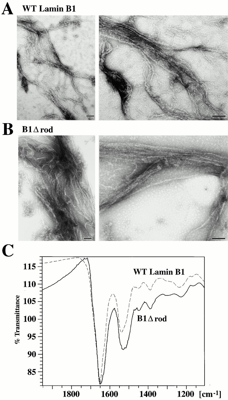
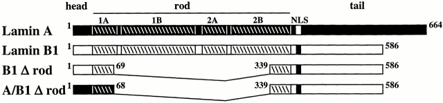
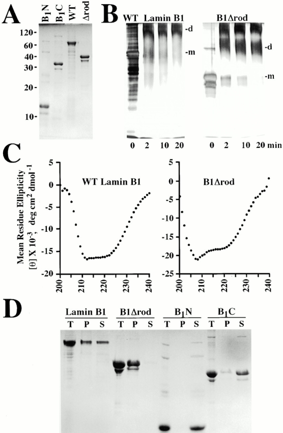
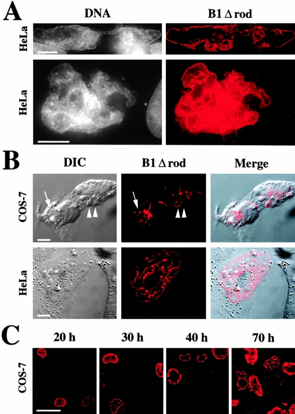
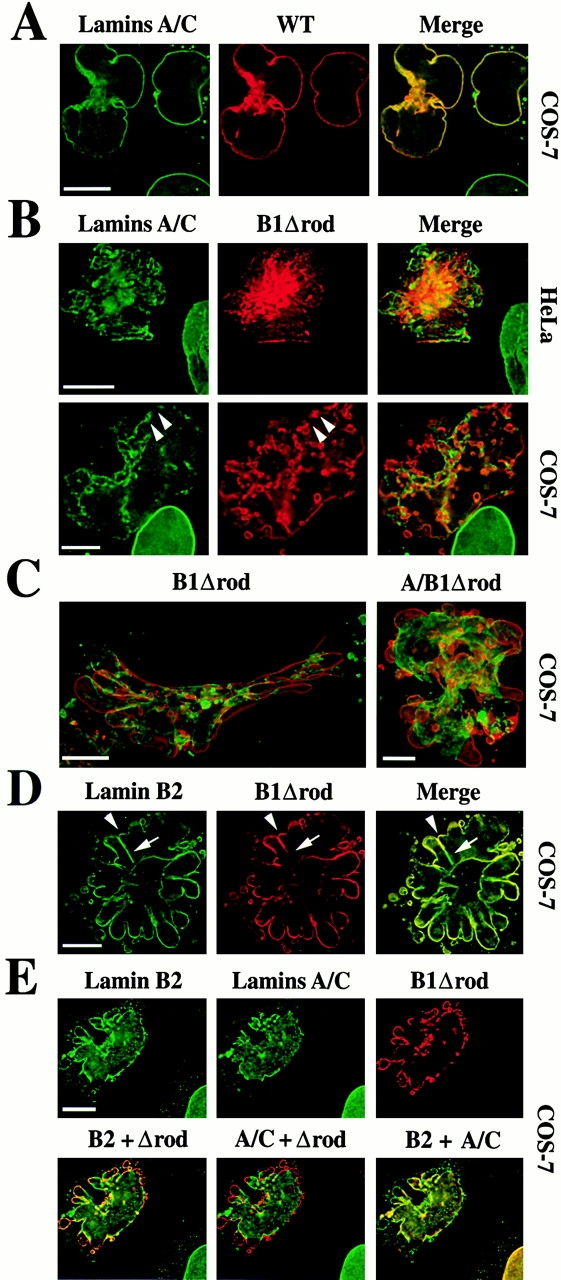
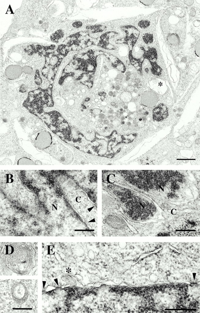
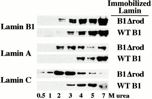
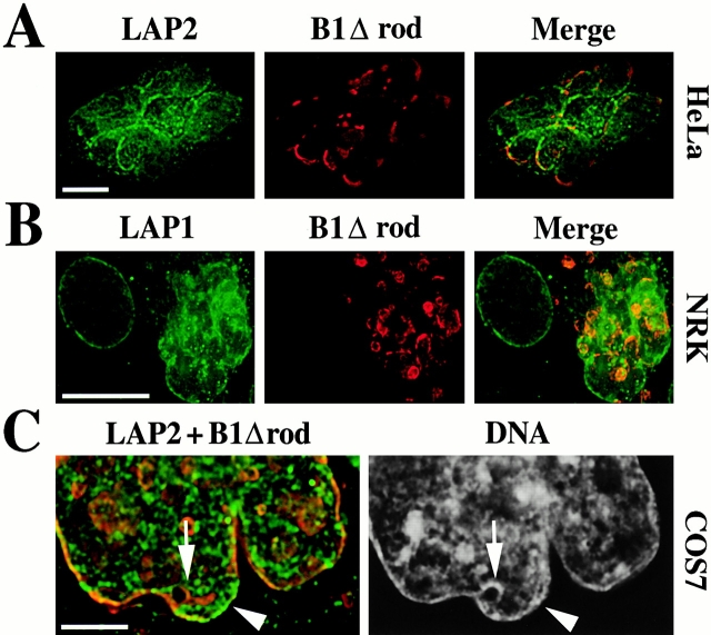
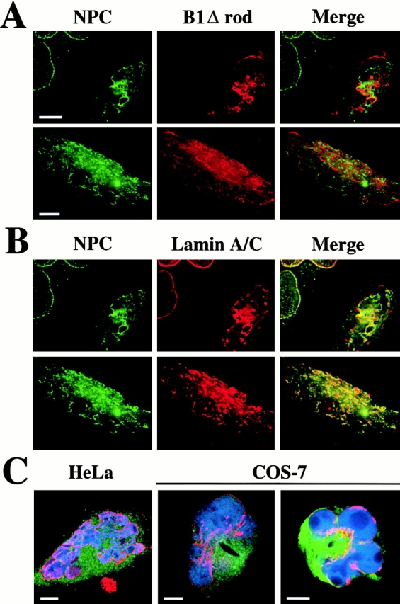
The nuclear lamina is a meshwork of intermediate-type filament proteins (lamins) that lines the inner nuclear membrane. The lamina is proposed to be an important determinant of nuclear structure, but there has been little direct testing of this idea. To investigate lamina functions, we have characterized a novel lamin B1 mutant lacking the middle approximately 4/5 of its alpha-helical rod domain. Though retaining only 10 heptads of the rod, this mutant assembles into intermediate filament-like structures in vitro. When expressed in cultured cells, it concentrates in patches at the nuclear envelope. Concurrently, endogenous lamins shift from a uniform to a patchy distribution and lose their complete colocalization, and nuclei become highly lobulated. In vitro binding studies suggest that the internal rod region is important for heterotypic associations of lamin B1, which in turn are required for proper organization of the lamina. Accompanying the changes in lamina structure induced by expression of the mutant, nuclear pore complexes and integral membrane proteins of the inner membrane cluster, principally at the patches of endogenous lamins. Considered together, these data indicate that lamins play a major role in organizing other proteins in the nuclear envelope and in determining nuclear shape.


Figures

Figure 3
Figure 3
In vitro assembly of B1Δrod. (A–B) WT lamin B1 and B1Δrod that were dialyzed out of urea-formed filaments. The structures formed by each protein were similar in appearance (bars, 100 nm). (C) FTIR indicates α-helical structures occur in the filamentous form of B1Δrod, similar to WT lamin B1. The proteins were dialyzed using conditions that had yielded filaments by EM analysis. Polymerized material was pelleted and analyzed by FTIR. The scans of B1Δrod and WT lamin B1 were very similar although the band around 1650 was wider for B1Δrod than for WT lamin B1. The position of the bands is consistent with α-helical rather than β-sheet characteristics (see text).
Figure 1
Figure 1
Design of mutant lamins. Mammalian lamins contain an NH2-terminal head domain (∼33 amino acids) followed by a coiled-coil rod domain (354 amino acids) and a COOH-terminal tail domain (200–300 amino acids). The rod is predicted to contain four separate α-helices: coil 1A, 1B, 2A, and 2B of 6, 20, 6, and 16 heptads, respectively (a slash delineates each heptad). The B1Δrod mutant lacks most of the rod, retaining only five heptads at each end that are fused in register (via an added alanine residue replacing the valine in position g of the fifth NH2-terminal heptad). The A/B1Δrod mutant contains the head and first five heptads of lamin A (black) fused to the last five heptads and tail of lamin B1 (white).
Figure 2
Figure 2
The B1Δrod mutant has characteristics of IF proteins. (A) WT lamin B1, B1Δrod, and the two lamin B1 fragments that were fused in B1Δrod (B1N and B1C) were expressed in Escherichia coli, and were analyzed by SDS PAGE and staining with Coomassie blue. (B) The B1Δrod mutant forms dimers. Soluble WT lamin B1 or B1Δrod (the same material shown in A) were cross linked with glutaraldehyde for 0–20 min and analyzed by SDS-PAGE and staining with silver. No monomeric (m) protein was observed at any time after incubation with cross linker: rather, a diffuse band at the expected Mr for the dimer species (d) was observed that did not increase over time, indicating the stability of this soluble population. An apparent cross-linked tetramer also was seen for B1Δrod, but a tetrameric form of WT lamin B1 was too large to fully migrate into the gel. Note that silver staining does not give a linear representation of the protein species (compare 0-min samples in B to Coomassie blue–stained samples in A). (C) The B1Δrod mutant has considerable α-helical structure. The dichroic spectra of soluble WT lamin B1 and B1Δrod proteins (in a similar buffer as in B) contained strong minima at 208 and 222 nm indicative of α-helical structure. (D) Relative solubility of B1Δrod and its individual segments. Proteins at 250 μg/ml were dialyzed into buffers known to promote lamin assembly. Insoluble material was pelleted (P) and compared with the supernatant (S) and the total starting material (T). This demonstrated that the B1N and B1C fragments are extremely soluble compared with their fusion product, B1Δrod, and WT lamin B1.
Figure 4
Figure 4
The B1Δrod mutant lamin grossly alters nuclear morphology. (A) Volume projections of deconvolved optical sections reveal that DNA (gray) is bounded by the mutant lamin (red). (B) Differential interference contrast (DIC) images of cells transfected with B1Δrod (left) and confocal sections of the same cells immunostained for the mutant's HA epitope tag (middle, red). The lobules observed by DIC imaging correspond to those observed with mutant lamin staining (Merge). Bars, 10 μm. (C) The lobulation phenotype increases over time. Representative fields of cells immunostained for the HA epitope from a COS7 transfection with B1Δrod are shown at 20, 30, 40, and 70 h after transfection. Initially, the mutant lamin is evenly distributed at the NE, but over time its distribution becomes patchy. The appearance of nuclear lobules correlates with the redistribution of the mutant into patches. This time course was slower for transfections with efficiencies <10%. Bar, 50 μm.
Figure 6
Figure 6
Expression of B1Δrod alters the distribution of endogenous lamins. Transfected cells were analyzed by immunofluorescent staining to detect the exogenously expressed lamin (HA epitope tag, red) and endogenous lamins (green). (A) Localization of lamins A/C in cells transfected with WT lamin B1. Both endogenous and exogenous lamins are uniformly distributed throughout the NE with complete colocalization (yellow). (B) Localization of lamins A/C in cells transfected with B1Δrod. Endogenous A/C lamins lose their normal uniform distribution and cluster at the NE, exhibiting little colocalization with B1Δrod in HeLa (top) and COS-7 (bottom) cells. Deconvolved optical sections are shown in A and B. (C) The A/B1Δrod hybrid mutant yields a similar phenotype to B1Δrod. Volume projections from deconvolved optical sections through COS-7 cells transfected with B1Δrod (left) or A/B1Δrod (right) are shown. Merged images are shown with the Δrod mutants in red and the A/C lamins in green. (D) Localization of endogenous lamin B2 in cells transfected with B1Δrod. More colocalization with the mutant was observed than for A/C lamins. (E) Lack of complete colocalization between A/C and B2 lamins. COS-7 cells triple labeled for lamin B2, lamins A/C, and B1Δrod (top) and the merges of each pair (bottom) revealed that endogenous A/C and B2 lamins become partially segregated in mutant cells. Colocalization is indicated by yellow for lamin B2 (green) and B1Δrod (red), lamins A/C (green) and B1Δrod (red), and lamins B2 (green) and lamins A/C (red). Deconvolved sections are shown in C and D. Bars, 10 μm.
Figure 5
Figure 5
Ultrastructural analysis of a HeLa cell expressing B1Δrod by thin section EM. (A) Low magnification view of a whole nucleus. Bar, 1 μm. *Concentric membrane structure. (B–E) High magnification views. Bars, 300 nm. (B and C) A double membrane encloses the dense chromatin regions within lobules. Cytoplasm (C) and nucleus (N) are indicated. Arrowheads delineate nuclear pores. (D) Views of two concentric membrane structures in the cytoplasm. (E) Arrowheads delineate nuclear pores; *concentric membrane contained within the NE.
Figure 7
Figure 7
Relative affinities of lamins for B1Δrod and WT lamin B1. WT or mutant lamin B1 were coupled with an Affi-gel matrix to which soluble WT lamins were bound. Shown are Western blots of fractions eluted with increasing concentrations of urea, probed for each specific lamin isotype.
Figure 8
Figure 8
Changes in localization of integral membrane proteins of the INM induced by B1Δrod. (A) Immunofluorescent staining to detect LAP2 (green) and B1Δrod (red). Similar results were observed when LAP2 was visualized with a polyclonal antiserum, a monoclonal antibody, and using a LAP2-YFP fusion protein (data not shown). A single deconvolved optical section is shown. (B) NRK cells transfected with B1Δrod (red) indicated that LAP1 (green) also is redistributed as a result of expressing the mutant lamin. A deconvolved volume projection is shown. Bars, 10 μm. (C) Distribution of NE proteins relative to chromatin. NE proteins LAP2 in green and B1Δrod in red are on the left and DAPI staining for DNA on the right of the deconvolved section shown. Bar, 5 μm.
Figure 9
Figure 9
Effects of B1Δrod expression on NPCs. (A) Immunofluorescent staining to detect the NPC (green) and the mutant protein (red). (B) Immunofluorescent staining of the NPC (green) and endogenous lamins A/C (red). (Merge) Colocalization is indicated by yellow. Deconvolved volume projections are shown. (C) B1Δrod cells are capable of nuclear import. HeLa cells (left) were microinjected in the cytoplasm with a fluorescently labeled NLS-containing import substrate (blue) and a fluorescent 150-kD dextran (green). COS-7 cells (right) were injected with the same import substrate (blue) and fluorescent BSA that did not carry an NLS (green). HA staining for the B1Δrod mutant (red) traces the nuclear boundary. Single confocal sections are shown. Bars, 10 μm.

Similar articles

Cited by

References

    1. Adam S.A., Marr R.S., Gerace L. Nuclear protein import in permeabilized mammalian cells requires soluble cytoplasmic factors. J. Cell Biol. 1990;111:807–816. - PMC - PubMed
    1. Aebi U., Cohn J., Buhle L., Gerace L. The nuclear lamina is a meshwork of intermediate-type filaments. Nature. 1986;323:560–564. - PubMed
    1. Benavente R., Krohne G. Involvement of nuclear lamins in postmitotic reorganization of chromatin as demonstrated by microinjection of lamin antibodies. J. Cell Biol. 1986;103:1847–1854. - PMC - PubMed
    1. Cao H., Hegele R.A. Nuclear lamin A/C R482Q mutation in Canadian kindreds with Dunnigan-type familial partial lipodystrophy. Hum. Mol. Genet. 2000;9:109–112. - PubMed
    1. Collas I., Courvalin J.C. Sorting nuclear membrane proteins at mitosis. Trends Cell Biol. 2000;10:5–8. - PubMed

Publication types

MeSH terms