. 2001 Apr 30;153(3):543-53.
doi: 10.1083/jcb.153.3.543.

Lack of pericytes leads to endothelial hyperplasia and abnormal vascular morphogenesis

Affiliations

Lack of pericytes leads to endothelial hyperplasia and abnormal vascular morphogenesis

M Hellström et al. J Cell Biol. .

Abstract

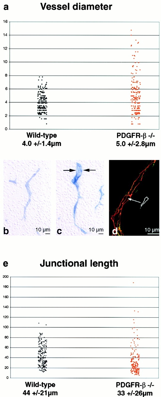
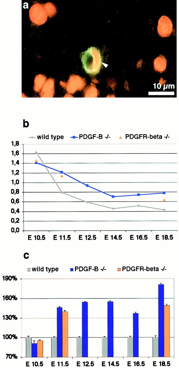
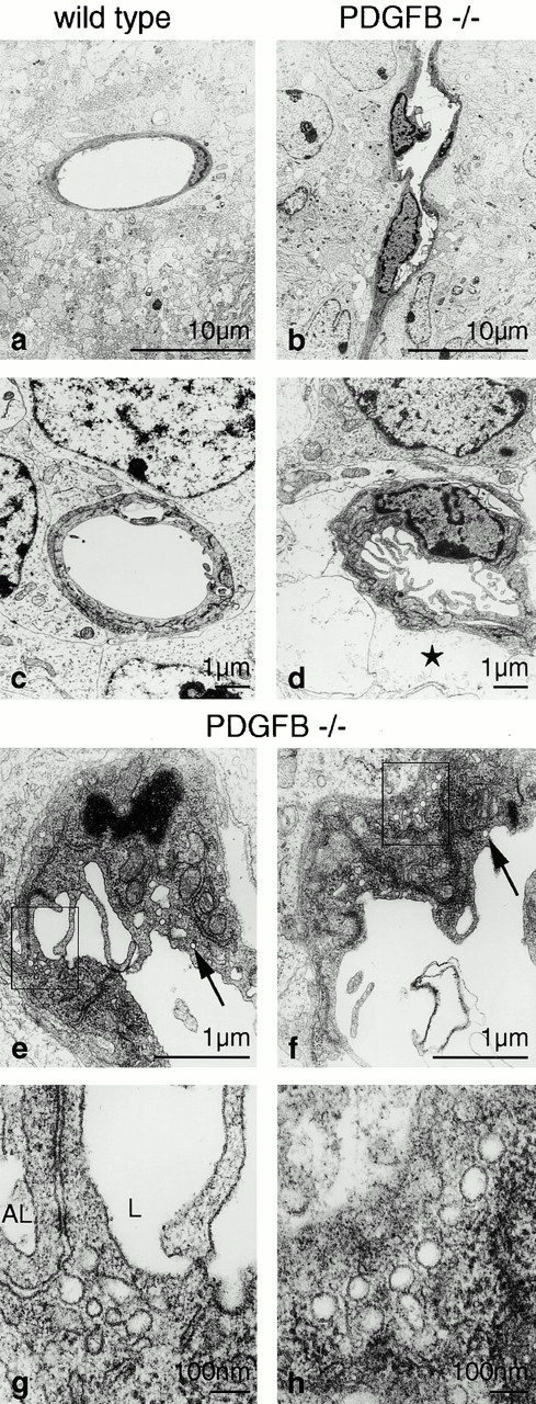
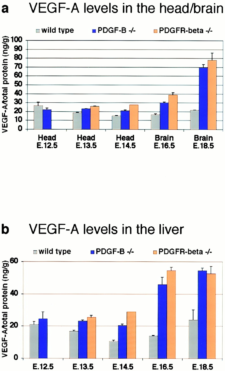
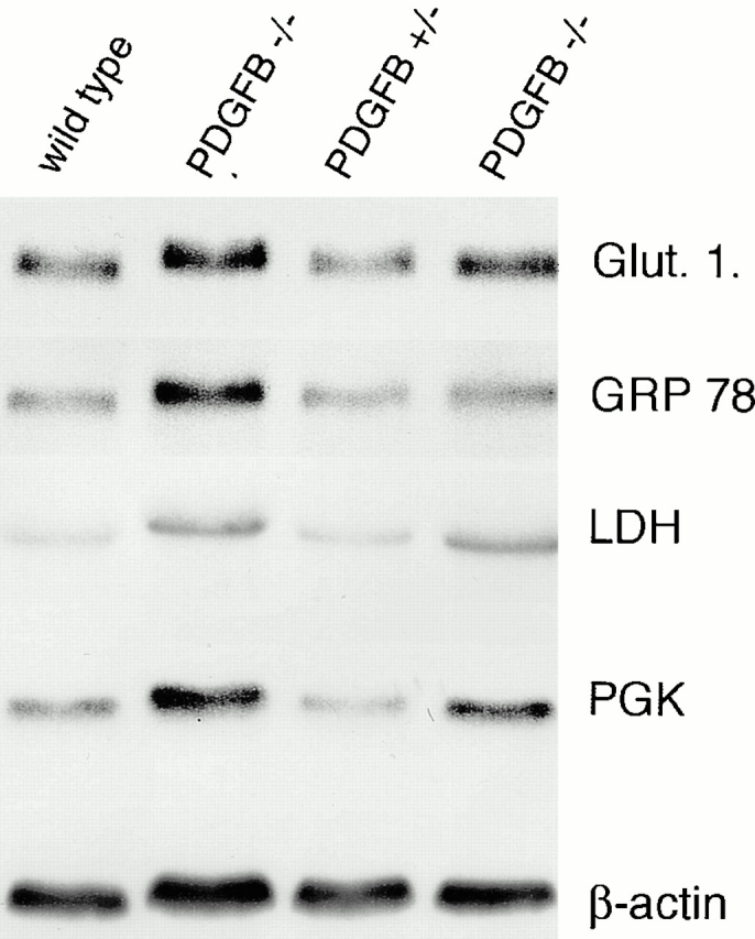
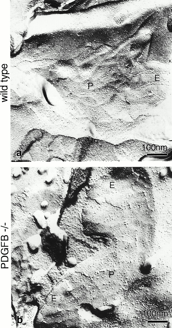
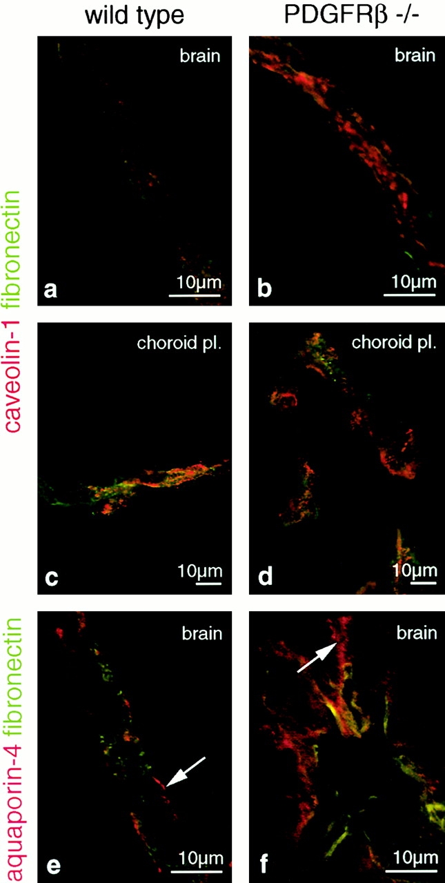
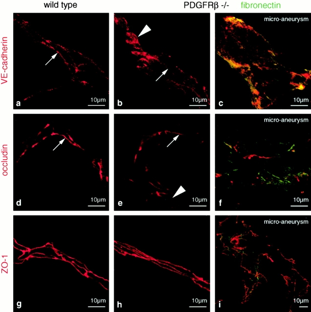
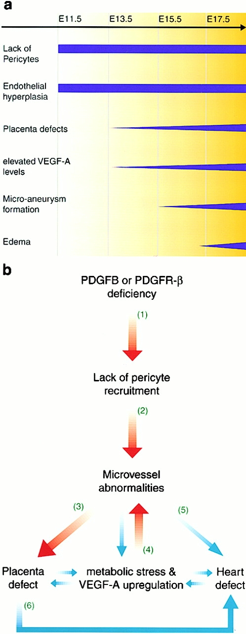
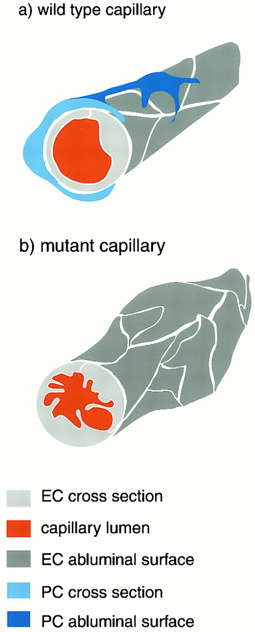
The association of pericytes (PCs) to newly formed blood vessels has been suggested to regulate endothelial cell (EC) proliferation, survival, migration, differentiation, and vascular branching. Here, we addressed these issues using PDGF-B-- and PDGF receptor-beta (PDGFR-beta)--deficient mice as in vivo models of brain angiogenesis in the absence of PCs. Quantitative morphological analysis showed that these mutants have normal microvessel density, length, and number of branch points. However, absence of PCs correlates with endothelial hyperplasia, increased capillary diameter, abnormal EC shape and ultrastructure, changed cellular distribution of certain junctional proteins, and morphological signs of increased transendothelial permeability. Brain endothelial hyperplasia was observed already at embryonic day (E) 11.5 and persisted throughout development. From E 13.5, vascular endothelial growth factor-A (VEGF-A) and other genes responsive to metabolic stress became upregulated, suggesting that the abnormal microvessel architecture has systemic metabolic consequences. VEGF-A upregulation correlated temporally with the occurrence of vascular abnormalities in the placenta and dilation of the heart. Thus, although PC deficiency appears to have direct effects on EC number before E 13.5, the subsequent increased VEGF-A levels may further abrogate microvessel architecture, promote vascular permeability, and contribute to formation of the edematous phenotype observed in late gestation PDGF-B and PDGFR-beta knock out embryos.


Figures

Figure 2
Figure 2
Vessel diameter and EC tight junction length in wild type and PDGFR-β knock out brains. Vessel diameter was measured on photographic areas from brain sections in wild type and PDGFR-β–deficient embryos at 200 locations each (a). The mean diameter was 4.0 ± 1.4 μm (SD) in the wild-type and 5.0 ± 2.8 μm in the PDGFR-β knock out embryos. There was a considerable variation of the diameter in the PDGFR-β knock out values (a), which was also seen at the microscopic level in a comparison between wild-type (b) and PDGF-B knock out (c) vessels. The vessels in the PDGF-B knock out embryos exhibited large differences in the vessel diameter with local severe dilations (arrows), whereas the wild-type vessels were more homogeneous in size. The circumferential length of 100 EC junctions (ZO-1 staining, red) were measured, as marked in d, in wild-type and PDGFR-β knock out vessels (e). The mean circumferential length was 44 ± 21 μm (SD) in the wild type and 33 ± μm 26 in the PDGFR-β knock out. Bar, 10 μm.
Figure 1
Figure 1
EC quantification in wild type, PDGF-B, and PDGFR-β knock out embryos. A brain capillary stained for isolectin BS-I (staining EC, green) and propidium iodide (staining nuclei, red) (a). The quantification of EC nuclei per vessel cross section in wild type, PDGF-B−/−, and PDFGFR-β2/− is shown in b, and the values for the mutants are shown as a percentage of the wild type in c. There is a prominent EC hyperplasia from E 11.5 on in the PDGF-B and PDGFR-β knock outs. The error bars represent the SEM. PDGF-B and PDGFR-β knock out values were significantly different (p-value < 0.001) from the wild-type values at E 11.5, 12.5, 13.5, 14.5, 16.5, and 18.5, but not at E 10.5. Bar, 10 μm.
Figure 5
Figure 5
Electron micrographs of brain capillaries in wild-type (a and c) and PDGF-B knock out (b and d–h) embryos at E 18.5. Capillaries in wild-type embryonic brains exhibited a smooth luminal and abluminal endothelial surface and a homogenous thickness of the endothelium (a and c), whereas those in PDGF-B–deficient embryos were of highly variable thickness (b and d). Luminal microfolds greatly enlarged the endothelial surface (d), and perivascular glial swelling was prominent (star in d). Caveolae were abundant in the endothelium of knock out embryos (e–h), both near the luminal (arrows) and the abluminal surface (boxed area, enlarged in g and h). In g, a series of caveolae appeared almost to connect the luminal and the abluminal compartment. Bars: (a and b) 10 μm; (c–f) 1 μm; (g and h) 100 nm.
Figure 3
Figure 3
A time course of VEGF-A ELISA measurements. VEGF-A levels were upregulated from E 13.5 on in both PDGF-B and PDGFR-β knock out head/brain (a) and liver (b). The VEGF-A concentration was normalized against the total protein in the tissue sample. There were no significant differences between PDGF-B and PDGFR-β knock out values. The SEM is shown as error bars.
Figure 4
Figure 4
Northern blot analysis of mRNAs regulated by hypoxia, hypoglycemia, and metabolic stress. Brain total RNA from wild type, PDGF-B+/−, and PDGF-B−/− were analyzed by Northern blot for Glut-1, glucose-regulated protein 78, LDH, and PGK. β-Actin was used as an internal control. The PDGF-B knock out brain showed upregulation of all these transcripts, especially LDH and PGK.
Figure 8
Figure 8
Freeze fracture replicas of brain capillary tight junctions of wild-type (a) and PDGF-B knock out (b) embryos at E 18.5. The tight junction particles were mostly associated to the external membrane leaflet (E) and only sparsely distributed on the protoplasmic membrane leaflet (P) in both the wild type and the knock out. The complexity of the junctional network as judged by the branching frequency of the strands was similar between wild type and PDGF-B knock out. Bars, 100 nm.
Figure 6
Figure 6
Confocal laser scanning micrographs of double immunolabeling for fibronectin (green) and caveolin-1 (a–d, red), and for fibronectin (green) and aquaporin-4 (e and f, red) at E 18.5. Caveolin-1 was considerably more abundant in brain capillaries of PDGFR-β knock out mice (compare a with b). The highly permeable endothelium of the choroid plexus was strongly immunopositive for caveolin-1 in both wild-type (c) and knock out embryos (d). In wild-type mice, aquaporin-4 labeling neatly outlined the blood vessels (e, arrow) whereas it was widely distributed into the surrounding brain parenchyma in knock out embryos (f, arrows). Bars, 10 μm.
Figure 7
Figure 7
Confocal laser scanning micrographs of immunolabeling for VE-cadherin (a–c), occludin (d–f), and ZO-1 (g–i) at E 18.5. (Left) Wild type. (Middle and right) PDGFR-β knock out. (Right) Show examples of brain microaneurysms with additional fibronectin staining (green) to outline the vessels. In brain capillaries of wild type mice, VE-cadherin was largely confined to the interendothelial junction (a, arrows), whereas in those of knock out embryos, VE-cadherin was more uniformly distributed (b and c). Arrow indicates weak junctional labeling; arrowhead points to cytoplasmic labeling. Occludin labeling was also more diffuse in the knock out (compare d with e; arrow, junctional labeling; arrowhead, cytoplasmic labeling). In microaneurysms, the occludin-positive junctional network was discontinuous (f). ZO-1 was equally confined to the junction in both wild-type (g) and knock out mice (h). Differences in the pattern, as shown for a microaneurysm (i), reflect the highly irregular endothelial organization at this site. Bars, 10 μm.
Figure 9
Figure 9
(a) Occurrence of phenotypes in the PDGF-B and PDGFR-β knock out embryos. The lack of PCs in PDGF-B and PDGFR-β knock out embryos coincides with the EC hyperplasia at E 11.5. At E 13.5, the first morphological signs of the placenta defects are evident, and this is also when an upregulation of VEGF-A is detected. Subsequently microaneurysms develop, and at late gestation, the embryos are edematous. (b) Hypothetical pathogenic cause–effect relationships in PDGF-B and PDGFR-β knock outs. Causal links supported by strong experimental evidence or extensive correlative data are depicted by thick red arrows. Causal links, which are plausible but lack or have only limited experimental support, are depicted with thin blue arrows. PDGF-B and PDGFR-β are required for the longitudinal recruitment of PCs to newly formed blood vessels, and lack of PCs leads to microvessel abnormalities (1 and 2) (Levéen et al. 1994; Soriano 1994; Lindahl et al. 1997; Hellstrom et al. 1999; Ohlsson et al. 1999). Deficient blood vessel development in the placenta leads to improper placenta function (3) (Ohlsson et al. 1999; Ihle 2000), which might promote hypoxia/hypoglycemia–induced VEGF-A, leading to microvessel abnormalities (4) (this report; Benjamin et al. 1998). The heart defect in the PDGF-B and PDGFR-β knock outs might stem from the abnormal heart vasculature or be due to placenta defects (5 and 6) (Barak et al. 1999; Hellstrom et al. 1999; Ihle 2000).
Figure 10
Figure 10
Schematic illustration of wild-type and knock out capillaries based on the combined data of morphological, morphometric, and immunocytochemical analysis. The absence of PCs in the PDGF-B and PDGFR-β knock outs leads to blood vessel dilation, EC hyperplasia, and microaneurysm formation. The ECs exhibit luminal membrane folding and highly variable cytoplasmic thickness and size.

References

    1. Antonelli-Orlidge A., Saunders K.B., Smith S.R., D'Amore P.A. An activated form of TGF-β is produced by co-cultures of endothelial cells and pericytes. Proc. Natl. Acad. Sci. USA. 1989;86:4544–4548. - PMC - PubMed
    1. Barak Y., Nelson M.C., Ong E.S., Jones Y.Z., Ruiz-Lozano P., Chien K.R., Koder A., Evans R.M. PPAR γ is required for placental, cardiac, and adipose tissue development. Mol. Cell. 1999;4:585–595. - PubMed
    1. Beck L.J., D'Amore P.A. Vascular developmentcellular and molecular regulation. FASEB J. 1997;11:365–373. - PubMed
    1. Benjamin L., Hemo I., Keshet E. A plasticity window for blood vessel remodelling is defined by pericyte coverage of the preformed endothelial network and is regulated by PDGF-B and VEGF. Development. 1998;125:1591–1598. - PubMed
    1. Benjamin L.E., Golijanin D., Itin A., Pode D., Keshet E. Selective ablation of immature blood vessels in established human tumors follows vascular endothelial growth factor withdrawal. J. Clin. Invest. 1999;103:159–165. - PMC - PubMed

Publication types

MeSH terms