. 2001 Apr 30;153(3):555-68.
doi: 10.1083/jcb.153.3.555.

Delta N89 beta-catenin induces precocious development, differentiation, and neoplasia in mammary gland

Affiliations

Delta N89 beta-catenin induces precocious development, differentiation, and neoplasia in mammary gland

A Imbert et al. J Cell Biol. .

Abstract

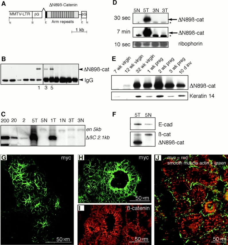
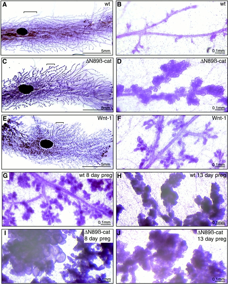
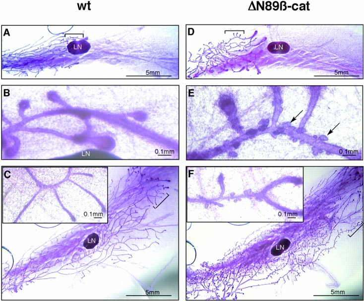
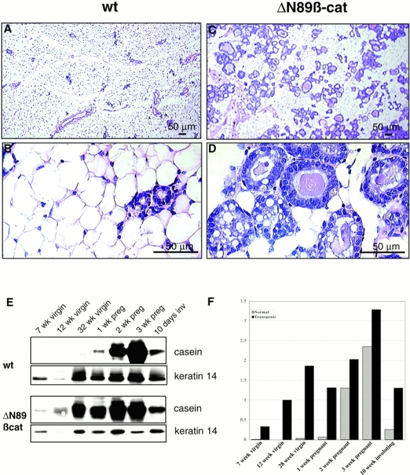
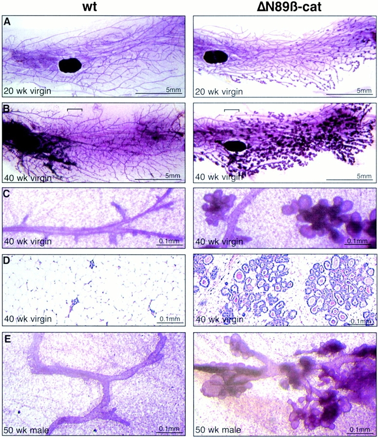
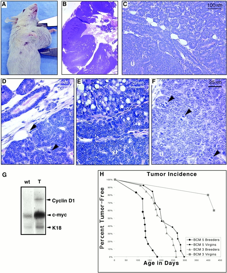
To investigate the role of beta-catenin in mammary gland development and neoplasia, we expressed a stabilized, transcriptionally active form of beta-catenin lacking the NH(2)-terminal 89 amino acids (Delta N 89 beta-catenin) under the control of the mouse mammary tumor virus long terminal repeat. Our results show that Delta N 89 beta-catenin induces precocious lobuloalveolar development and differentiation in the mammary glands of both male and female mice. Virgin Delta N 89 beta-catenin mammary glands resemble those found in wild-type (wt) pregnant mice and inappropriately express cyclin D1 mRNA. In contrast to wt mammary glands, which resume a virgin appearance after cessation of lactation, transgenic mammary glands involute to a midpregnant status. All transgenic females develop multiple aggressive adenocarcinomas early in life. Surprisingly, the Delta N89 beta-catenin phenotype differs from those elicited by overexpression of Wnt genes in this gland. In particular, Delta N 89 beta-catenin has no effect on ductal side branching. This suggests that Wnt induction of ductal branching involves additional downstream effectors or modulators.


Figures

Figure 1
Figure 1
(A) Structure of the transgene construct. DNA sequence encoding a myc epitope tag (black box), followed by sequence encoding the truncated ΔN89-terminal domain, 13 central Armadillo (Arm) repeats, and the COOH-terminal domain of Xenopus β-catenin (shaded boxes) was embedded in β-globin intron/exon/polyadenylation sequences (βG), and cloned downstream of the mouse mammary tumor virus long terminal repeat (MMTV LTR). Pertinent restriction sites are indicated: N, NotI; B, BamHI; E, EcoRI. (B) 9E10 antibody, which specifically recognizes the human myc epitope tag and does not cross-react with murine c-myc, was used to immunoprecipitate myc-tagged ΔN89β-catenin from extracts of the mammary glands from 12 individual transgenic lines. This product was detected in three lines (lines 1, 3, and 5) by Western blotting with the same antibody. Note the lower band, which migrates at M r ∼ 50,000, is the IgG band from the 9E10 immunoprecipitation detected by the HRP-rabbit anti–mouse. (C) Southern blot detection of the ΔN89β-catenin transgene in the tail genomic DNA of transgenic (T) and normal (N) littermates of F1 from lines 1, 3, and 5, as indicated. Membranes were hybridized with the 32P-labeled ΔN89β-catenin cDNA insert (ΔβC), and the mouse engrailed gene (en) fragments, which detect fragments of 2,160 bp and 5 kb, respectively, as indicated. Lanes marked 200, 20, and 2 show respective copy equivalents of ΔN89β-catenin cDNA plasmid. (D) Relative expression levels of ΔN89β-catenin protein in lines 5 and 3 as detected by Western blotting of equal amounts of total mammary proteins with anti-myc antibody. Labels on the left indicate exposure time to film after incubation in ECL reagent. Arrows point to the 75-kD ΔN89β-catenin product; asterisks indicate nonspecific band detected by the anti–mouse IgG antibody. Detection of ribophorin was used as a loading control. (E) Western blot showing expression of ΔN89β-catenin (top) in line 5 transgenic mice during mammary development and differentiation. No reaction was seen on an identical blot of wt littermates (data not shown). Immunodetection of keratin 14 on the same blot (bottom) is shown to control for epithelial content of the sample. Preg, pregnancy; inv, involution. (F) Western blot of equal amounts of total proteins from day 16 pregnant mammary glands from line 5 mice showing expression of endogenous E-cadherin (top) and the ΔN89β-catenin transgenic product and endogenous β-catenin detected with the same anti–β-catenin antibody (bottom). Note densitometry of three similar blots showed that the transgene is expressed to approximately threefold the level of endogenous β-catenin found in wt mice. The transgene downregulates endogenous β-catenin by one third, and upregulates E-cadherin twofold compared with wt levels. (G–J) Indirect immunofluorescence on frozen sections of mammary gland from 6-mo-old virgin line 5 mice (G and J) or day 16 pregnant line 3 mice (H–I). ΔN89β-catenin is detected in green (G and H) using 9E10 anti-myc antibody. Endogenous β-catenin detected with rabbit anti–β-catenin shown in red (I). Double immunolabeling to detect ΔN89β-catenin in red and smooth muscle actin, which demarcates the position of myoepithelial cells in green (J). Note the transgene is located at the intercellular borders of the luminal epithelial cells.
Figure 3
Figure 3
Comparison of the morphology of mature wt, MMTV-ΔN89β-catenin, and MMTV-Wnt-1 transgenic virgin and pregnant mammary glands. Whole mounts of inguinal mammary glands stained with carmine alum from 12-wk virgin wt (A and B) and ΔN89β-catenin transgenic littermates (C and D), and a 12-wk Wnt-1 transgenic mouse (E and F). Mammary glands from 8- and 13-d-old pregnant wt (G and H) and ΔN89β-catenin (I and J) littermates. Note that ΔN89β-catenin virgin mice show a hyperlobular phenotype, whereas Wnt-1 virgin mice show a feathery hyperbranched phenotype.
Figure 2
Figure 2
Morphology of ΔN89β-catenin mammary glands shows precocious lobular development during early puberty. Whole mounts of inguinal mammary glands stained with carmine alum from wt (A–C) and transgenic (D–F) 4-wk (A, B, D, and E) and 8-wk (C and F) line 5 littermates matched for stage of the estrous cycle. LN, lymph node. Note the extensive lobuloalveolar development on the transgenic glands (arrows in E). Images in B and E show higher magnifications of the areas bracketed in A and D. Insets in C and F are higher magnifications of bracketed areas.
Figure 5
Figure 5
Histological and biochemical evaluation shows evidence of precocious epithelial differentiation in ΔN89β-catenin virgin mice. (A–D) Hematoxylin and eosin–stained sections of mammary gland from 6-mo-old virgin wt (A and B) and ΔN89β-catenin (C and D) littermates. Note the presence of vesicles in the transgenic samples. (E) Western blot of total proteins from the inguinal mammary glands at various stages of development, pregnancy (preg), and involution (inv) with anti–β-casein antibody and anti-keratin 14 antibody on the same blot, which serves as a control of epithelial content of the sample. (F) Graph of densitometry figures derived from the casein blots shown in E after normalization for keratin 14 levels. Note β-casein is expressed in ΔN89β-catenin but not wt virgin mice, and in all pregnant mice.
Figure 4
Figure 4
Lobuloalveolar development in older virgin females of the low expressing line and in male mice. Whole mounts of inguinal mammary glands stained with carmine alum (A–C) and hematoxylin and eosin–stained sections of mammary gland (D) from 20-wk-old (A) and 40-wk-old (B and C) wt and ΔN89β-catenin transgenic littermates from the low expressing line 3. Note that low expressing line 3 ΔN89β-catenin virgin mice also show aberrant hyperlobular development with no evidence of ductal hyperbranching. (E) 1-yr-old wt and ΔN89β-catenin transgenic male littermates from line 5.
Figure 7
Figure 7
A comparison of early involution of wt and ΔN89β-catenin mammary glands. (A) Hematoxylin and eosin–stained sections of wt (top) and ΔN89β-catenin (bottom) inguinal mammary glands from littermates 1, 3, 5, and 7 d of involution (i) after removal of pups, as indicated. (B) Apoptotic changes detected by TUNEL staining of sections from wt (top) and ΔN89β-catenin (TG, bottom) inguinal mammary glands 1 and 3 d after removing pups, as indicated (1i, 3i). Note no significant differences were observed between wt and transgenic samples during this early stage of involution.
Figure 6
Figure 6
Northern analysis of cyclin D1 and c-myc mRNA levels in wt and ΔN89β-catenin mammary glands. mRNA was extracted from virgin (10 wk [10w Vir]), midpregnant (8 d [8d Preg]), and late pregnant (18 d [18d Preg]) mammary glands. Northern blots were prepared and probed with cDNAs encoding cyclin D1, c-myc, and K18 as a control to permit normalization for epithelial content of the mRNA samples. Note cyclin D1 and c-myc levels are abnormally elevated in transgenic (TG) virgin mice compared with their wt littermates.
Figure 8
Figure 8
A comparison of late involution in wt and ΔN89β-catenin mammary glands. (A) Graph of the average number of TUNEL-positive nuclei in five random frames taken from sections of wt and ΔN89β-catenin (TG) inguinal mammary glands from 1, 3, 5, and 7 d after removing pups as indicated. (B) Hematoxylin and eosin–stained sections of wt (top) and ΔN89β-catenin (bottom) inguinal mammary glands from littermates 10, 13, 16, and 21 d after removing pups. Note that in contrast to wt glands, which show continued signs of deterioration and reduction of the epithelial component, ΔN89β-catenin mice show continued lobular hyperplasia. (C) mRNA was extracted from mammary glands having undergone involution for 1, 13, and 21 d, as indicated. Northern blots were prepared and probed with cDNAs encoding cyclin D1, c-myc, casein, and K18 as a control to permit normalization for epithelial content of the mRNA samples. Note cyclin D1 becomes elevated and casein mRNA persists at day 21 of involution in transgenic mice compared with their wt littermates.
Figure 9
Figure 9
ΔN89β-catenin mice develop mammary adenocarcinomas. (A) Founder mouse line 5 with single large tumor on a thoracic mammary gland. (B) Hematoxylin and eosin–stained section of similar tumor from an F1 daughter. Tumors comprise regions of glandular hyperplasia (C and D) with central masses of disorganized, undifferentiated (U) poorly adherent cells (C, E, and F). Some showed evidence of secretory activity (E) but most showed undifferentiated cells with a high cytoplasmic to nuclear ratio and many mitotic figures (arrowheads in D and F). (G) mRNA was extracted from mammary tumors (T) and mammary glands of wt littermates. Northern blots were prepared and probed with cDNAs encoding cyclin D1, c-myc, and K18 as a control to permit normalization for epithelial content of the mRNA samples. (H) Graph of tumor incidence in ΔN89β-catenin mice. The percentage of animals in each cohort remaining free of palpable tumors was plotted as a function of age for ΔN89β-catenin line 5 breeding females (n = 12), line 3 breeding females (n = 13), and line 5 virgins (n = 13). No female wt littermates (n = 25) or wt littermate males (n = 20) developed tumors. To date, among mice that are currently > 1 yr old, three line 3 virgin females (n = 10), no line 3 males (n = 10), and only one line 5 male (n = 10) have developed tumors.

Similar articles

Cited by

References

    1. Behrens J., von Kries J.P., Kuhl M., Bruhn L., Wedlich D., Grosschedl R., Birchmeier W. Functional interaction of β-catenin with the transcription factor LEF-1. Nature. 1996;382:638–642. - PubMed
    1. Berx G., Cleton-Jansen A.M., Nollet F., de Leeuw W.J., van de Vijver M., Cornelisse C., van Roy F. E-cadherin is a tumour/invasion suppressor gene mutated in human lobular breast cancers. EMBO (Eur. Mol. Biol. Organ.) J. 1995;14:6107–6115. - PMC - PubMed
    1. Bradley R.S., Cowin P., Brown A.M.C. Expression of Wnt-1 in PC12 cells results in modulation of plakoglobin and E-cadherin and increased cellular adhesion. J. Cell Biol. 1993;123:1857–1865. - PMC - PubMed
    1. Brisken C., Heineman A., Chavarra T., Elenbaas B., Tan J., Dey S.K., McMahon A.P., Weinberg R. Essential function of Wnt-4 in mammary gland development downstream of progesterone signaling. Genes Dev. 2000;14:650–654. - PMC - PubMed
    1. Buckley A., Middleton M.C. Retinoic acid alters the keratinization of cultured rat sublingual keratinocytes in vitro. Arch. Dermatol. Res. 1987;279:257–265. - PubMed

Publication types

MeSH terms