. 2001 May 15;21(10):3322-31.
doi: 10.1523/JNEUROSCI.21-10-03322.2001.

Pyruvate released by astrocytes protects neurons from copper-catalyzed cysteine neurotoxicity

Affiliations

Pyruvate released by astrocytes protects neurons from copper-catalyzed cysteine neurotoxicity

X F Wang et al. J Neurosci. .

Abstract

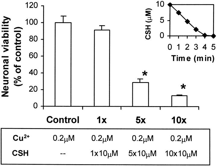
We have found previously that astrocytes can provide cysteine to neurons. However, cysteine has been reported to be neurotoxic although it plays a pivotal role in regulating intracellular levels of glutathione, the major cellular antioxidant. Here, we show that cysteine toxicity is a result of hydroxyl radicals generated during cysteine autoxidation. Transition metal ions are candidates to catalyze this process. Copper substantially accelerates the autoxidation rate of cysteine even at submicromolar levels, whereas iron and other transition metal ions, including manganese, chromium, and zinc, are less efficient. The autoxidation rate of cysteine in rat CSF is equal to that observed in the presence of approximately 0.2 microm copper. In tissue culture tests, we found that cysteine toxicity depends highly on its autoxidation rate and on the total amount of cysteine being oxidized, suggesting that the toxicity can be attributed to the free radicals produced from cysteine autoxidation, but not to cysteine itself. We have also explored the in vivo mechanisms that protect against cysteine toxicity. Catalase and pyruvate were each found to inhibit the production of hydroxyl radicals generated by cysteine autoxidation. In tissue culture, they both protected primary neurons against cysteine toxicity catalyzed by copper. This protection is attributed to their ability to react with hydrogen peroxide, preventing the formation of hydroxyl radicals. Pyruvate, but not catalase or glutathione peroxidase, was detected in astrocyte-conditioned medium and CSF. Our data therefore suggest that astrocytes can prevent cysteine toxicity by releasing pyruvate.


Figures

Fig. 1.
Fig. 1.
The factors influencing cysteine (CSH) autoxidation. The reaction conditions to assess cysteine autoxidation were 37°C and pH 7.4 in a humidified atmosphere of 5% CO2 and 95% air. The samples were taken at different time points for HPLC assays. A, Cysteine (100 μm) autoxidation in Earle's balanced salt solution (EBSS), cystine-free MEM (MEM-CSSC), and neuronal culture medium. The latter was made from MEM-CSSC, which replaced the original cystine-containing MEM in the neuronal cultures before testing. Cysteine was gradually oxidized to cystine. Cysteine concentrations in MEM-CSSC and culture medium were not significantly different from the corresponding values in EBSS (p > 0.05). B, The effects of iron on cysteine autoxidation. Ferric chloride (FeCl3), at concentrations of 0.1, 1, and 10 μm, was incubated with 100 μmcysteine in EBSS. Cysteine concentrations in the three Fe3+ solutions were not significantly different from the corresponding values in EBSS (p > 0.05). C, The effects of copper on cysteine autoxidation. Cupric sulfate (CuSO4), at concentrations of 0.1, 1, and 10 μm, was incubated with 100 μm cysteine in EBSS. Cysteine autoxidation was substantially accelerated in the presence of Cu2+ at all three concentrations. Results are normalized as the percentage of 100 μm total cysteine. Each column represents the average of three independent experiments performed in duplicate (mean ± SEM). *p < 0.01 versus corresponding values of cysteine autoxidation in EBSS.
Fig. 2.
Fig. 2.
Comparisons of cysteine autoxidation in the presence of several transition metal ions. A series of concentrations of FeSO4, FeCl3, hemin, MnSO4, CrCl3, ZnCl2, CuCl, and CuSO4 were reacted with 100 μm cysteine, respectively. The control was 100 μm cysteine without transition metal ions. The reaction mixtures were incubated in PBS at pH 7.4 and 37°C in a humidified 100% air. Cysteine concentrations were determined with Ellman's reagent after 60 min. A, Cysteine autoxidation with Fe2+, Fe3+ (1–200 μm), and hemin (1–100 μm).B, Cysteine autoxidation with Mn2+, Cr3+, and Zn2+ (1–200 μm). C, Cysteine autoxidation with Cu+ and Cu2+ (0.01–1 μm). Data are the mean ± SEM of three independent experiments in duplicate. *p < 0.01, significantly different from the control. a,bp < 0.01 versus corresponding values of Fe3+ groups.
Fig. 3.
Fig. 3.
Cysteine autoxidation rates in CSF and comparisons with those in Cu2+-supplemented solutions. CSF was taken from 3-month-old rats as described in Materials and Methods. Cysteine was added to the CSF with a final concentration of 100 μm. The CSF was incubated under conditions of pH 7.4 and 37°C in a humidified atmosphere of 5% CO2 and 95% air. Samples were taken at several time points for HPLC assays.A, Time course of the concentrations of cysteine and related compounds in the CSF during cysteine autoxidation.B, Comparison of cysteine autoxidation in CSF and in Cu2+-supplemented solutions. Cu2+was prepared in EBSS at concentrations of 0.1, 0.2, and 0.3 μm and incubated under the same conditions as the CSF. Data are the mean ± SEM of three independent experiments in duplicate. CSSC, Cystine; CSSG, cysteine-glutathione disulfide; GSH, glutathione.
Fig. 4.
Fig. 4.
Neurotoxic effects of cysteine in the presence of copper. Primary cortical neurons were cultured in serum-free MEM. Cysteine was added at concentrations as indicated in the presence of 0, 0.2, and 1.0 μm Cu2+. Neuronal viability was estimated 24 hr later using the MTT assay. Results are expressed as the percentage of surviving neurons compared with control cultures (without the addition of cysteine and Cu2+). Data represent the mean ± SEM of three independent experiments in triplicate.
Fig. 5.
Fig. 5.
Effect of total amount of cysteine being oxidized on neuronal survival. Primary cortical neurons were cultured in serum-free MEM. The concentration of Cu2+ was 0.2 μm. Cysteine (10 μm) was added each time and once or repetitively every 5 min for a total of 5 and 10 times. Cysteine (10 μm) was completely oxidized within 5 min in the presence of 0.2 μm Cu2+(inset, top right). Neuronal viability was estimated 24 hr later using the MTT assay. Results are expressed as the percentage of surviving neurons compared with control cultures (without addition of cysteine). Data represent the mean ± SEM of three independent experiments in triplicate. *p < 0.01, significantly different from the control.
Fig. 6.
Fig. 6.
Generation of hydroxyl radical (·OH) by the autoxidation of cysteine and other thiols. Cysteine and related compounds were incubated in PBS under the conditions of pH 7.4 and 37°C in a humidified atmosphere of 100% air. CCA (1 mm) was added to react with the generated ·OH, producing 7-OHCCA. Fluorescence was measured 4 hr after the reaction began.A, Cysteine, at concentrations of 0–200 μm, was incubated in the presence or absence of 0.2 μm Cu2+. B, Generation of ·OH from the autoxidation of thiols. Thiols, disulfides, and sulfur-containing amino acid (100 μm of each) were incubated with 0.2 μm Cu2+. The thiols include cysteine (CSH), glutathione (GSH), N-acetyl-cysteine (NAC), homocysteine (HSH), dithiothreitol (DTT), and 2-mercaptoethanol (2-ME). The disulfides include cystine (CSSC) and glutathione disulfide (GSSG). The sulfur-containing amino acid is cysteic acid (CA). Data represent the mean ± SEM of three independent experiments in duplicate.
Fig. 7.
Fig. 7.
The protective effects of catalase and pyruvate on cysteine neurotoxicity. Primary cortical neurons were cultured in serum-free MEM. Cysteine toxicity was induced by addition of 100 μm cysteine and 0.2 μmCu2+. Neuronal viability was estimated 24 hr later using the MTT assay. Results are expressed as the percentage of surviving neurons compared with control cultures (without addition of cysteine). A, Catalase (10 U/ml), pyruvate (1 mm), and lactate (1 mm) were added immediately before addition of cysteine and Cu2+. Data represent the mean ± SEM of three independent experiments in triplicate.B, Dose–response curve illustrating the neuroprotective effect of pyruvate. Cysteine concentrations were 0, 50, and 100 μm, respectively. Data represent the mean ± SEM of three independent experiments in duplicate. *p < 0.01, significantly different from the control.
Fig. 8.
Fig. 8.
Effects of catalase and pyruvate on the generation of ·OH from cysteine autoxidation. Cysteine (100 μm) and Cu2+ (0.2 μm) were incubated in PBS under the conditions of pH 7.4 and 37°C in a humidified atmosphere of 100% air. CCA (1 mm) was added to react with the generated ·OH, producing 7-OHCCA. The fluorescence was measured 4 hr after the reaction. Data represent the mean ± SEM of three independent experiments in duplicate. A, Catalase (10 U/ml), pyruvate (1 mm), and lactate (1 mm) were added immediately before addition of cysteine and Cu2+. B, Pyruvate, at concentrations of 0, 0.1, 1, and 10 mm, was added immediately before addition of cysteine and Cu2+. *, **p < 0.01, significantly different from the control (cysteine only).
Fig. 9.
Fig. 9.
Time course of pyruvate release by astrocytes. Confluent astrocytes in flasks were rinsed with Hank's solution. The serum-free MEM was added at a concentration of 1.33 ml per 1 × 10⁶ cells. Samples of the ACM were taken at 0, 6, 12, 24, and 48 hr and used for pyruvate assays. Data represent the mean ± SEM of three independent experiments in triplicate.
Fig. 10.
Fig. 10.
Diagram of the proposed mechanism of protection by astrocytes in preventing cysteine toxicity catalyzed by copper. Astrocytes release glutathione and indirectly produce cysteine in the extracellular fluid of the CNS. Cysteine, as well as glutathione or other thiols, will be oxidized to disulfide under the catalysis of protein-unbound or loosely bound copper. Molecular oxygen, as the oxidant, accepts electrons step by step to produce superoxide radicals, hydrogen peroxide, and hydroxyl radicals. The latter is the major damaging free radical to the cells. In parallel, astrocytes also release pyruvate, which can react with hydrogen peroxide, preventing the formation of hydroxyl radicals.

Similar articles

Cited by

References

    1. Aguilar MV, Jimenez-Jimenez FJ, Molina JA, Meseguer I, Mateos-Vega CJ, Gonzalez-Munoz MJ, de Bustos F, Gomez-Escalonilla C, Ort-Pareja M, Zurdo M, Martinez-Para MC. Cerebrospinal fluid selenium and chromium levels in patients with Parkinson's disease. J Neural Transm. 1998;105:1245–1251. - PubMed
    1. Beutler E. Nutritional and metabolic aspects of glutathione. Annu Rev Nutr. 1989;9:287–302. - PubMed
    1. Bunton CA. Oxidation of α-diketones and α-keto-acids by hydrogen peroxide. Nature. 1949;163:444. - PubMed
    1. Cavallini D, De Marco C, Dupre S, Rotilio G. The copper catalyzed oxidation of cysteine to cystine. Arch Biochem Biophys. 1969;130:354–361. - PubMed
    1. Chevion M. A site-specific mechanism for free radical induced biological damage: the essential role of redox-active transition metals. Free Radic Biol Med. 1988;5:27–37. - PubMed

Publication types

MeSH terms

LinkOut - more resources