. 2001 May 15;21(10):3360-8.
doi: 10.1523/JNEUROSCI.21-10-03360.2001.

Ezrin-dependent promotion of glioma cell clonogenicity, motility, and invasion mediated by BCL-2 and transforming growth factor-beta2

Affiliations

Ezrin-dependent promotion of glioma cell clonogenicity, motility, and invasion mediated by BCL-2 and transforming growth factor-beta2

W Wick et al. J Neurosci. .

Abstract

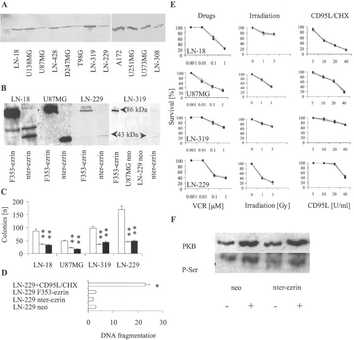
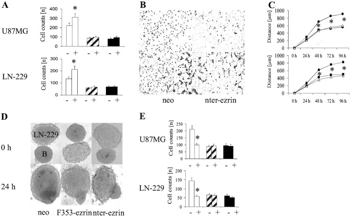
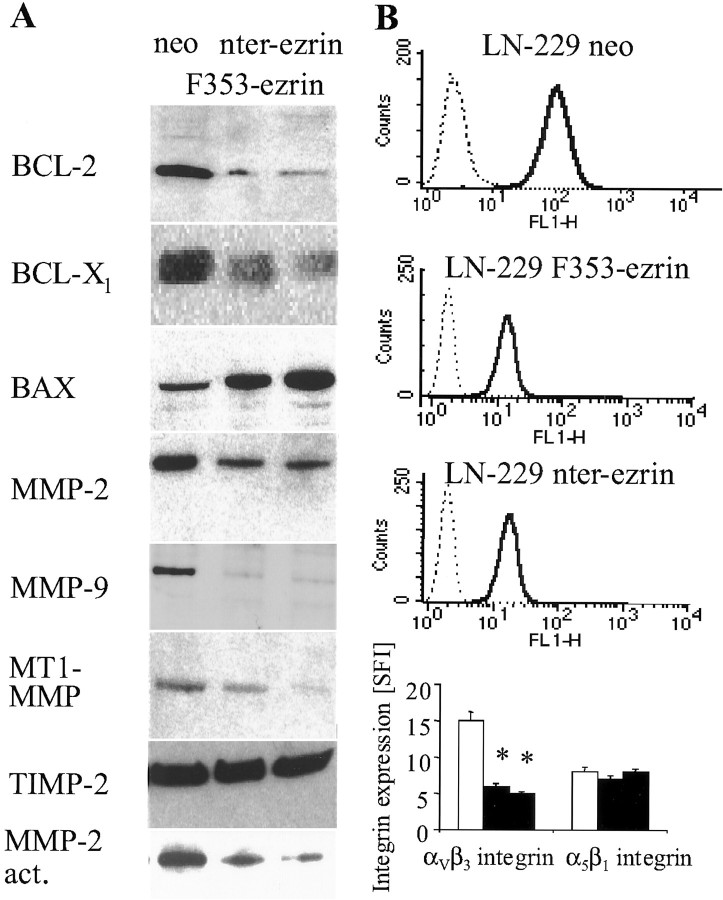
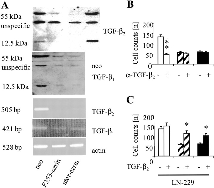
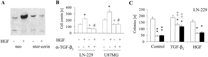
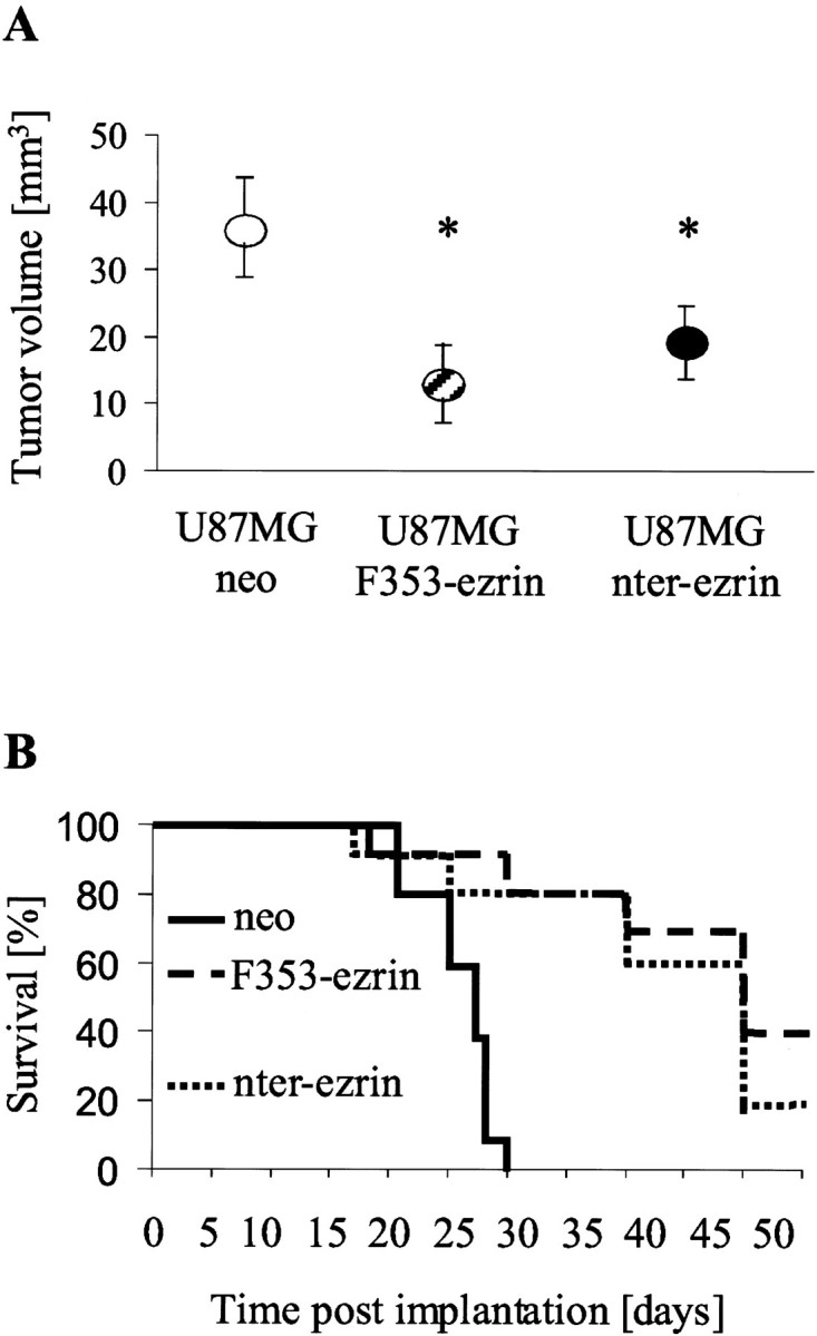
Ezrin belongs to the ezrin-radixin-moesin family proteins, which cross-link actin cytoskeleton and plasma membrane. Malignant glioma cells are paradigmatic for their strong migratory and invasive properties. Here, we report that the expression of dominant-negative ezrins inhibits clonogenicity, migration, and invasiveness of human malignant glioma cells. Furthermore, dominant-negative ezrins block hepatocyte growth factor (HGF)-mediated stimulation of clonogenicity and migration, without altering HGF-induced protein kinase B/Akt and focal adhesion kinase phosphorylation. Glioma cells expressing dominant-negative ezrins exhibit a shift of the BCL-2/BAX rheostat toward apoptosis, reduced alpha(V)beta(3) integrin expression and reduced matrix metalloproteinase (MMP) expression and activity. These changes are associated with a dramatic loss of transforming growth factor beta(2) (TGF-beta(2)) release. Exogenous supplementation of TGF-beta(2) overcomes the inhibitory effects of dominant-negative ezrins on migration and clonogenicity. A neutralizing TGF-beta(2) antibody mimics the effects of dominant-negative ezrins on clonogenicity and migration. Exogenous HGF markedly induces TGF-beta(2) protein levels, and a neutralizing TGF-beta(2) antibody abolishes the HGF-mediated increase in glioma cell motility. Finally, TGF-beta(2) does not modulate BCL-2 or BAX expression, but BCL-2 gene transfer increases the levels of latent and active TGF-beta(2). Intracranial xenografts of U87MG glioma cells transfected with the dominant-negative ezrins in athymic mice grow to significantly smaller volumes, and the median survival of these mice is 50 d compared with 28 d in the control group. These data define a novel pathway for HGF-induced glioma cell migration and invasion, which requires ezrin, changes in the BCL-2/BAX rheostat, and the induction of TGF-beta(2) expression in vitro, and underscore the important role of HGF signaling in vivo.


Figures

Fig. 1.
Fig. 1.
Dominant-negative ezrin inhibits clonogenicity but modulates neither sensitivity to apoptosis nor HGF-mediated PKB/Akt stimulation. A, Immunoblot analysis for ezrin was performed using a polyclonal ezrin antibody. B,Immunoblot analysis for the VSVG-tagged NH2-terminal domain of ezrin (nter-ezrin, 43 kDa), and the VSVG-tagged mutant (F353-ezrin, 86 kDa) was performed using a VSVG antibody. C,Clonogenicity of control transfectants (open bars,left) or F353-ezrin (open bars,middle)- or nter-ezrin-expressing cells (black bars) was assessed by colony-forming assay (mean and SEM; n = 3; **p < 0.01;t test). D, Spontaneous DNA fragmentation of neo-, F353-ezrin-, or nter-ezrin-transfected LN-229 cells was assessed by DNA fluorometry (Wick et al., 1999a). As a positive control, nontransfected parental cells were treated with CD95L (50 U/ml) and CHX (10 μg/ml) for 16 hr (mean percentages and SEM;n = 3; *p < 0.05;t test). E, Neo (filled circles)-, F353-ezrin (filled squares)-, or nter-ezrin (open triangles)-transfected sublines of the LN-18, U87MG, LN-319, or LN-229 cell lines were treated with vincristine (VCR) for 72 hr (left panel), irradiated, and assessed for colony formation (middle panel), or treated with CD95L plus CHX (10 μg/ml) for 16 hr (right panel) (mean percentages; n = 3; SEM < 10%).F, PKB was immunoprecipitated from untreated (−) or HGF (10 ng/ml, 24 hr)-treated (+) LN-229 cells transfected with the neo control plasmid or nter-ezrin. The lysates were analyzed by SDS-PAGE and immunoblot analysis for PKB levels (top panel) and serine phosphorylation (bottom panel).
Fig. 2.
Fig. 2.
Dominant-negative ezrin reduces constitutive and HGF-induced migration and invasion. A, Migration was measured in the absence (−) or presence (+) of HGF (10 ng/ml) in a chemotaxis chamber assay in glioma cells transfected with control plasmid (open bars), F353-ezrin (striped bars), or nter-ezrin (black bars). Migrated cells were counted in five random fields (mean and SEM;n = 3; *p < 0.05;t test). Representative filters demonstrating nter-ezrin-mediated inhibition of migration are shown inB (magnification, 200×). C, The transfected cell lines (neo, filled circles; F353-ezrin,filled squares; nter-ezrin, open triangles) were analyzed for migration from preformed spheroids. The distance in micrometers from the center of the spheroid minus the diameter of the spheroid was measured for 50 representative migrated cells (mean values; n = 3; *p < 0.05; t test; SEM < 10%). D, The transfected LN-229 cell lines were analyzed in confrontation assays that assess invasion of a rat brain aggregate. The images show the brain aggregate (labeledB) as smaller and the tumor spheroid (labeled LN-229) as relatively larger. E, Migration was measured in the absence (−) or presence (+) of neutralizing α-HGF-antibody (2 ng/ml) in a chemotaxis chamber assay in glioma cells transfected with control plasmid (open bars), F353-ezrin (striped bars), or nter-ezrin (black bars). Migrated cells were counted in five random fields (mean and SEM;n = 3; *p < 0.05;t test).
Fig. 3.
Fig. 3.
Dominant-negative ezrin-induced inhibition of migration and invasion: association with altered BCL-2/BAX rheostat, decreased MMP activity, and decreased αvβ3integrin expression. A, The levels of BCL-2, BCL-XL, and BAX in LN-229 whole-cell lysates and of MMP-2, MMP-9, MT1-MMP, and TIMP-2 in the supernatant were examined by immunoblot. MMP-2 activity in conditioned medium of neo, nter-ezrin-, and F353-ezrin-transfected LN-229 cells was examined by gelatin zymography. All blots are representative of experiments performed three times with similar results. B,αvβ3 and α5β1integrin expression were determined by flow cytometry. The curves for αVβ3 antibody (bold line) and control antibody (dotted line) are shown. As documented in the bottom panel, there was no change in α5β1 integrin. In the bar graph, data are expressed as mean SFI values (n = 3).
Fig. 4.
Fig. 4.
Inhibition of glioma cell clonogenicity, migration, and invasion by dominant-negative ezrin involves loss of TGF-β2. A, TGF-β2 and TGF-β1 levels in the conditioned medium of neo (lanes 1 and 4) or F353-ezrin or nter-ezrin-transfected LN-229 cells were assessed by immunoblot. In thebottom panels, the levels ofTGF-β1 andTGF-β2 mRNA were assessed by semiquantitative RT-PCR. B, C, The migration of LN-229 sublines was measured in the absence (−) or presence (+) of neutralizing TGF-β2 antibody (2 μg/ml) (B) or the absence (−) or presence (+) of TGF-β2 (2 ng/ml) (C) (mean and SEM;n = 3; *p < 0.05, **p < 0.01 for the effect of TGF-β2antibody in B or of TGF-β2 inC).
Fig. 5.
Fig. 5.
HGF-promoted glioma cell migration requires enhanced TGF-β2. A, LN-229 neo or nter-ezrin cells were untreated or exposed to HGF (10 ng/ml) for 24 hr and analyzed for TGF-β2 release as in Figure4A. B, LN-229 and U87MG cells were treated with HGF (10 ng/ml for 24 hr) or TGF-β2 antibody as in Figure 4B, as indicated, and assessed for migration (mean and SEM; n = 3; ttest; *p < 0.05 for the effect of HGF;#p < 0.05 for the effect of TGF-β2 antibody compared with control antibody-treated cells; +p < 0.05 for the effect of TGF-β2 antibody plus HGF compared with HGF alone).C, The cells were untreated or treated with TGF-β2 (2 ng/ml) or HGF (10 ng/ml) and assessed for colony formation as in Figure 1C (mean and SEM;n = 3; t test; *p < 0.05; **p < 0.01 for the effect of dominant-negative ezrin; ++p< 0.01 for the effect of TGF-β2 compared with nontreated isogenic cells).
Fig. 6.
Fig. 6.
Increased TGF-β2 release by BCL-2-transfected glioma cells. A, The levels of BCL-2 and BAX were measured in whole-cell lysates of LN-229 neo and nter-ezrin cells after no treatment (−) or exposure to TGF-β2 (2 ng/ml) for 24 hr (+) by immunoblot.B, Released TGF-β2 was detected in the conditioned medium of LN-229 neo or BCL-2 cells by immunoblot analysis as in Figure 4A.
Fig. 7.
Fig. 7.
Expression of dominant-negative ezrins prolongs survival of U87MG human glioma xenograft-bearing athymic mice. U87MG neo, U87MG F353-ezrin, or U87MG nter-ezrin human glioma cells were implanted stereotactically into the striatum of athymic mice as detailed in Materials and Methods. A, Mice (n = 3) were killed when the first animal developed symptoms (at 28 d). Brains were fixed, cut, and tumor volumes of hematoxylin–eosin-stained brains were determined with the MCID digitalization system (*p < 0.05;n = 3; t test). B, In an independent set of experiments, the animals of each group (n = 6) were observed at regular intervals until they developed neurological symptoms. Then the mice were killed.

Similar articles

Cited by

References

    1. Algrain M, Turunen O, Vaheri A, Louvard D, Arpin M. Ezrin contains cytoskeleton and membrane binding domains accounting for its proposed role as a membrane-cytoskeletal linker. J Cell Biol. 1993;120:129–139. - PMC - PubMed
    1. Belien A, Paganetti P, Schwab M. Membrane-type matrix metalloprotease (MT1-MMP) enables invasive migration of glioma cells in central nervous system white matter. J Cell Biol. 1999;144:373–384. - PMC - PubMed
    1. Beviglia L, Kramer R. HGF induces FAK activation and integrin-mediated adhesion in MTLn3 breast carcinoma cells. Int J Cancer. 1999;83:640–649. - PubMed
    1. Bourguignon LY, Zhu H, Shao L, Chen YW (2000) CD44 interaction with c-Src kinase promotes cortactin-mediated cytoskeleton function and hyaluronic acid (HA)-dependent ovarian tumor cell migration. J Biol Chem: Nov 17, epub ahead of print. - PubMed
    1. Brooks P, Stromblad S, Sanders LC, von Schalscha TL, Aimes RT, Stetler-Stevenson WG, Quigley JP, Cheresh DA. Localization of matrix metalloproteinase MMP-2 to the surface of invasive cells by interaction with integrin αVβ3. Cell. 1996;85:683–693. - PubMed

Publication types

MeSH terms