

Proliferation of NG2-positive cells and altered oligodendrocyte numbers in the contused rat spinal cord
- PMID: 11331369
- PMCID: PMC6762495
- DOI: 10.1523/JNEUROSCI.21-10-03392.2001
Proliferation of NG2-positive cells and altered oligodendrocyte numbers in the contused rat spinal cord
Abstract
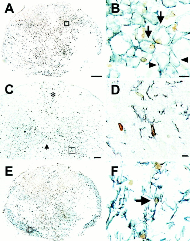
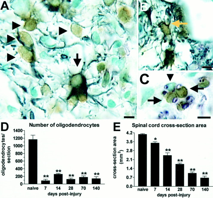
Given the numerous reparative roles glia may play after spinal cord injury (SCI), glial proliferation and cell number were examined in a model of traumatic SCI. Emphasis was placed on analysis of oligodendrocytes and NG2-positive (NG2+) cells, an endogenous cell population that may be involved in oligodendrocyte replacement. Overall, proliferation (assessed by bromodeoxyuridine incorporation) was markedly elevated during the first 2 weeks after injury and declined thereafter; a large portion of these dividing cells likely consisted of microglia-macrophages. Although the total number of NG2+ cells in the epicenter was reduced by half, we noted protracted proliferation in surviving NG2+ cells, with values sevenfold greater than in uninjured controls. Elevated proliferation of NG2+ cells persisted throughout the first 4 weeks after injury. However, the absolute number of NG2+ cells was not increased over controls, suggesting that the daughter cells either did not survive or they differentiated into other cell types. As expected, oligodendrocyte numbers were drastically altered after SCI. By 7 d after injury, the number of oligodendrocytes at the impact site was reduced by 93%. Despite ongoing tissue loss, the number of oligodendrocytes in spared tissue rose threefold at 14 d after injury. Although the function of NG2+ cells within the spinal cord is not completely understood, several studies suggest that they may differentiate into oligodendrocytes. Thus, proliferating NG2+ cells may contribute to the increased oligodendrocyte number observed at 2 weeks after injury. Future studies are required, however, to definitively determine the role NG2+ cells play in oligodendrocyte genesis, remyelination, and other post-injury events.
Figures


References
-
- Adrian EK., Jr Cell division in injured spinal cord. Am J Anat. 1968;123:501–520. - PubMed
-
- Adrian EK, Jr, Walker BE. Incorporation of thymidine-H3 by cells in normal and injured mouse spinal cord. J Neuropathol Exp Neurol. 1962;21:597–609. - PubMed
-
- Adrian EK, Jr, Williams MG, George FC. Fine structure of reactive cells in injured nervous tissue labeled with 3H-thymidine injected before injury. J Comp Neurol. 1978;180:815–839. - PubMed
-
- Amat JA, Farooq M, Ishiguro H, Norton WT. Cells of the oligodendrocyte lineage proliferate following cortical stab wounds: an in vitro analysis. Glia. 1998;22:64–71. - PubMed
-
- Balentine JD. Pathology of experimental spinal cord trauma. II. Ultrastructure of axons and myelin. Lab Invest. 1978;39:254–266. - PubMed
Publication types
MeSH terms
Substances
Grants and funding
LinkOut - more resources
Full Text Sources
Other Literature Sources
Medical
Research Materials