

Macrophages are eliminated from the injured peripheral nerve via local apoptosis and circulation to regional lymph nodes and the spleen
- PMID: 11331370
- PMCID: PMC6762479
- DOI: 10.1523/JNEUROSCI.21-10-03401.2001
Macrophages are eliminated from the injured peripheral nerve via local apoptosis and circulation to regional lymph nodes and the spleen
Abstract
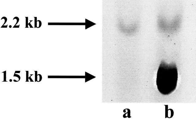
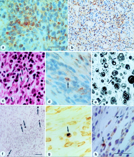
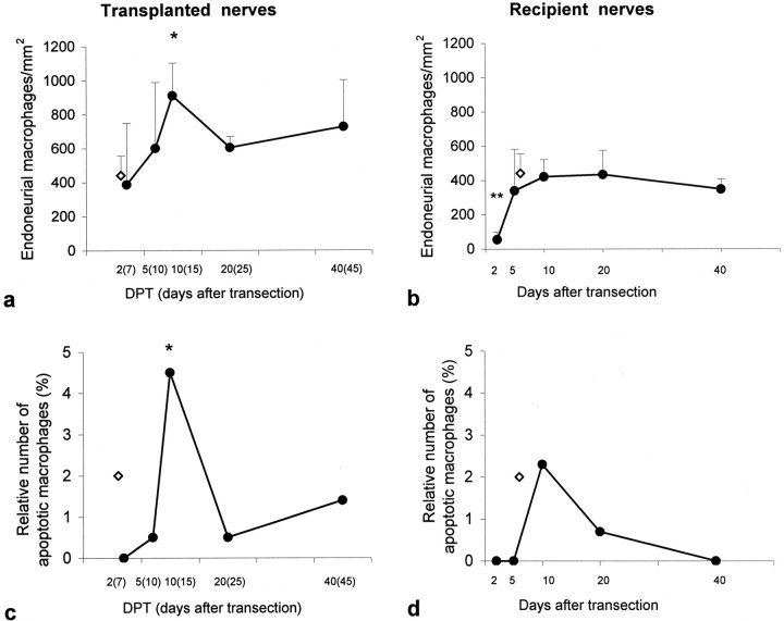
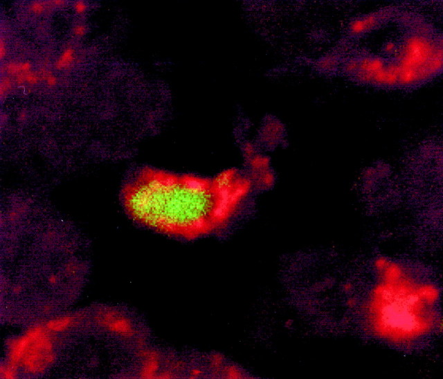
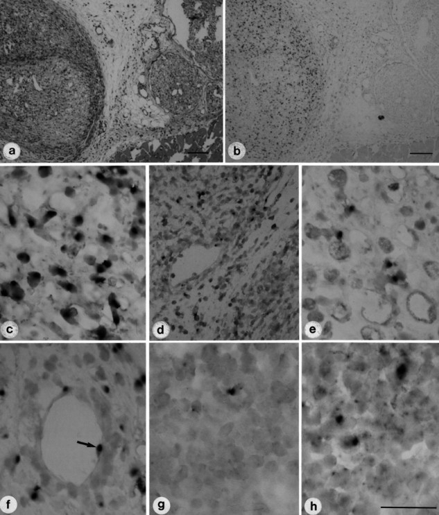
The present study investigated the fate of macrophages in peripheral nerves undergoing Wallerian degeneration, especially their disappearance from the injured nerves after phagocytosis of axonal and myelin debris. Wallerian degeneration was induced in adult male C57Bl/6 mice by transecting the right sciatic nerve. Five days after transection, the male sciatic nerves were transplanted into female recipient mice by placing them exactly parallel to the host sciatic nerves. Nerves of the female recipient mice were also transected to induce breakdown of the blood-nerve barrier in the host animal. Apoptosis was assessed by morphological, immunohistochemical (activated caspase-3), and molecular (DNA fragmentation) methods in transplanted, recipient, and in control nerves. A subpopulation of macrophages within the degenerating nerves died locally by apoptosis in each experiment. The fate of the male macrophages within the transplanted nerves and the host organism was investigated by in situ hybridization with a Y-chromosome-specific DNA probe (145SC5). In situ hybridization specifically stained cells within the transplanted male nerve. Y-chromosome-positive cells were detected not only inside the transplanted nerve, but also inside the female host nerve, the perineurial tissue, the local perineurial blood vessels, draining lymph nodes and the spleen of the female host, suggesting hematogenous as well as lymphatic elimination of macrophages from the injured nerve. These data indicate that local apoptosis and systemic elimination via circulation to the local lymph nodes and the spleen are involved in the disappearance of macrophages from the injured peripheral nervous system.
Figures
Similar articles
-
ATF3 upregulation in glia during Wallerian degeneration: differential expression in peripheral nerves and CNS white matter.BMC Neurosci. 2004 Mar 4;5:9. doi: 10.1186/1471-2202-5-9. BMC Neurosci. 2004. PMID: 15113454 Free PMC article.
-
Over-expression of alpha-synuclein in the nervous system enhances axonal degeneration after peripheral nerve lesion in a transgenic mouse strain.J Neurochem. 2010 Aug;114(4):1007-18. doi: 10.1111/j.1471-4159.2010.06832.x. Epub 2010 May 26. J Neurochem. 2010. PMID: 20524960
-
Macrophage-specific RhoA knockout delays Wallerian degeneration after peripheral nerve injury in mice.J Neuroinflammation. 2021 Oct 15;18(1):234. doi: 10.1186/s12974-021-02292-y. J Neuroinflammation. 2021. PMID: 34654444 Free PMC article.
-
The macrophage: a key player in the pathophysiology of peripheral neuropathies.J Neuroinflammation. 2022 Apr 16;19(1):97. doi: 10.1186/s12974-022-02454-6. J Neuroinflammation. 2022. PMID: 35429971 Free PMC article. Review.
-
The origin, fate and function of macrophages in the peripheral nervous system-an update.Int Immunol. 2020 Oct 20;32(11):709-717. doi: 10.1093/intimm/dxaa030. Int Immunol. 2020. PMID: 32322888 Review.
Cited by
-
Wallerian degeneration: gaining perspective on inflammatory events after peripheral nerve injury.J Neuroinflammation. 2011 Aug 30;8:110. doi: 10.1186/1742-2094-8-110. J Neuroinflammation. 2011. PMID: 21878126 Free PMC article. Review.
-
Unveiling the Role of Schwann Cell Plasticity in the Pathogenesis of Diabetic Peripheral Neuropathy.Int J Mol Sci. 2024 Oct 8;25(19):10785. doi: 10.3390/ijms251910785. Int J Mol Sci. 2024. PMID: 39409114 Free PMC article. Review.
-
Fas determines differential fates of resident and recruited macrophages during resolution of acute lung injury.Am J Respir Crit Care Med. 2011 Sep 1;184(5):547-60. doi: 10.1164/rccm.201011-1891OC. Am J Respir Crit Care Med. 2011. PMID: 21471090 Free PMC article.
-
Schwann cell influence on motor neuron regeneration accuracy.Neuroscience. 2009 Sep 29;163(1):213-21. doi: 10.1016/j.neuroscience.2009.05.073. Epub 2009 Jun 6. Neuroscience. 2009. PMID: 19505536 Free PMC article.
-
Next-generation RNA sequencing elucidates transcriptomic signatures of pathophysiologic nerve regeneration.Sci Rep. 2023 May 31;13(1):8856. doi: 10.1038/s41598-023-35606-6. Sci Rep. 2023. PMID: 37258605 Free PMC article.
References
-
- Baggiolini M, Dewald B, Moser B. Interleukin-8 and related chemotactic cytokines—CXC and CC chemokines. Adv Immunol. 1994;55:97–179. - PubMed
-
- Baggiolini M, Dewald B, Moser B. Human chemokines: an update. Annu Rev Immunol. 1997;15:675–705. - PubMed
-
- Barth MW, Hendrzak JA, Melnicoff MJ, Morahan PS. Review of the macrophage disappearance reaction. J Leukoc Biol. 1995;57:361–367. - PubMed
-
- Beuche W, Friede RL. The role of non-resident cells in Wallerian degeneration. J Neurocytol. 1984;13:767–796. - PubMed
-
- Breitschopf H, Suchanek G, Gould RM, Colman DR, Lassmann H. In situ hybridization with digoxigenin-labeled probes: sensitive and reliable detection method applied to myelinating rat brain. Acta Neuropathol. 1992;84:581–587. - PubMed
MeSH terms
Substances
LinkOut - more resources
Full Text Sources
Research Materials