. 2001 May 15;21(10):3457-75.
doi: 10.1523/JNEUROSCI.21-10-03457.2001.

Neurotrophic factors and receptors in the immature and adult spinal cord after mechanical injury or kainic acid

Affiliations

Neurotrophic factors and receptors in the immature and adult spinal cord after mechanical injury or kainic acid

J Widenfalk et al. J Neurosci. .

Abstract

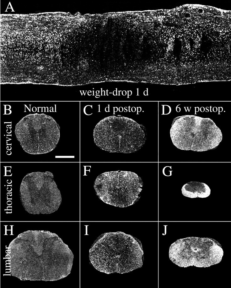
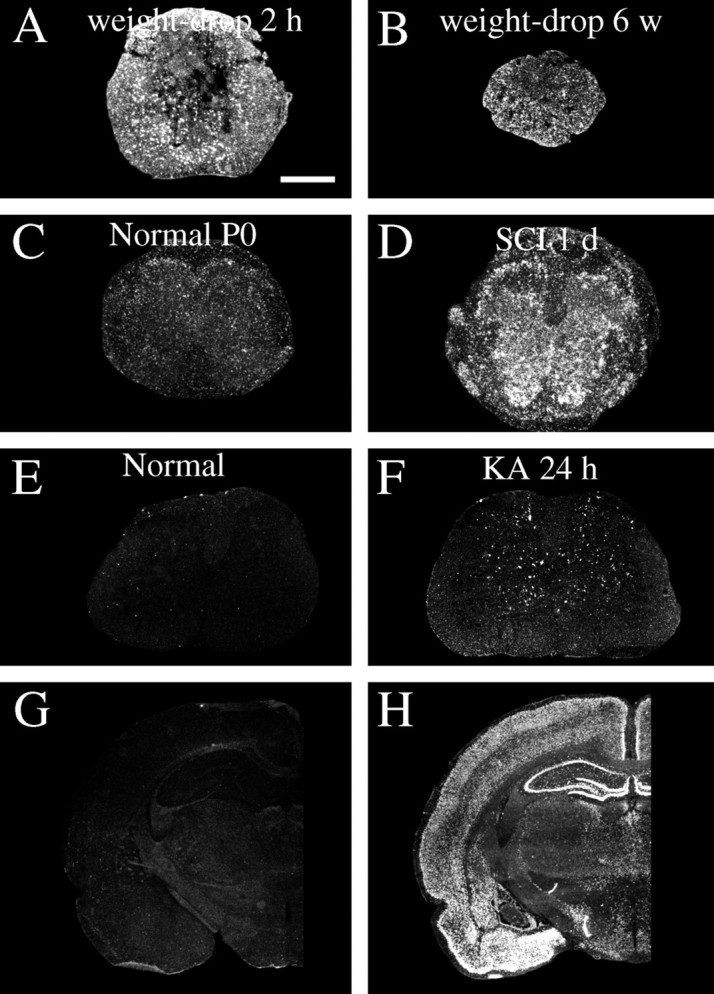
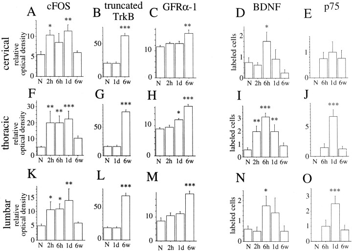
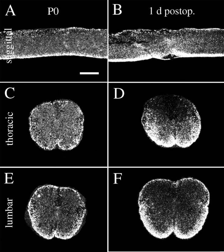
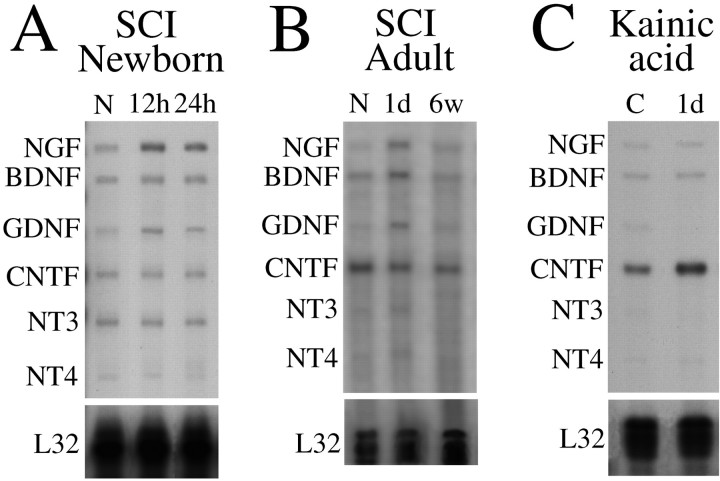
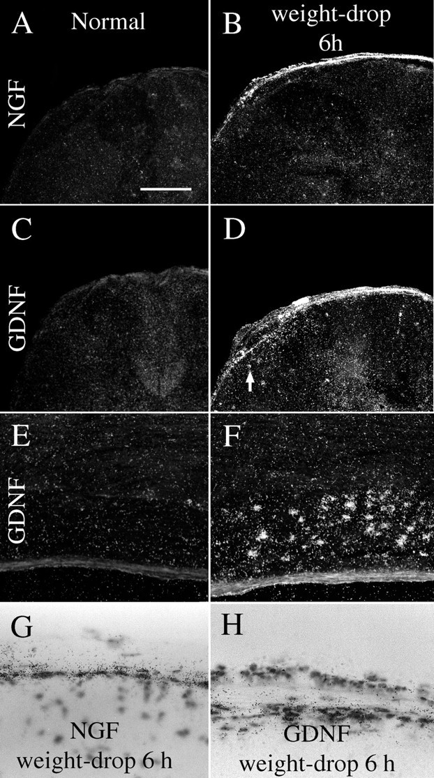
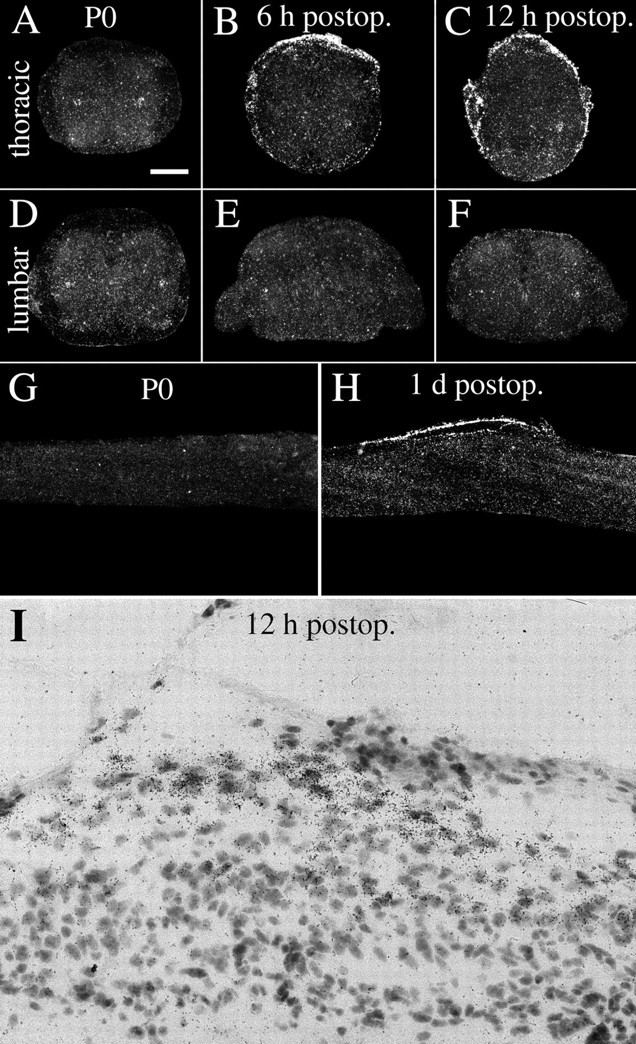
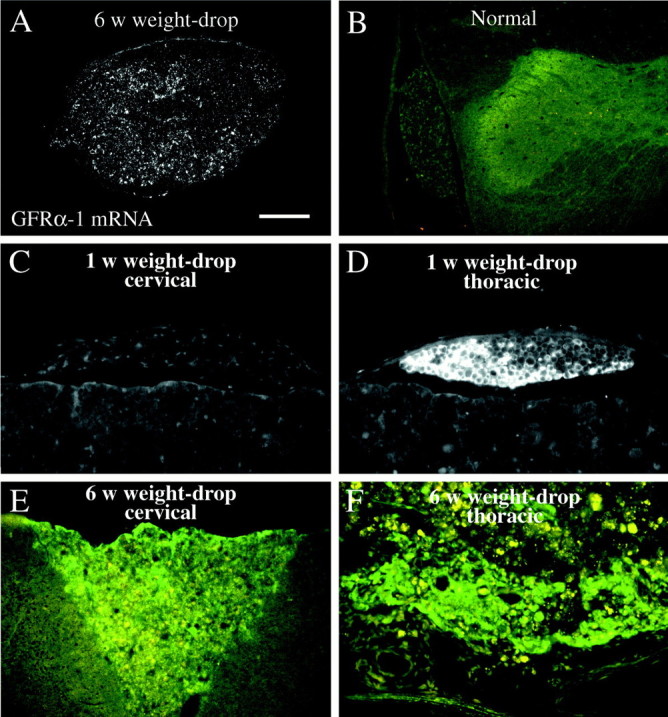
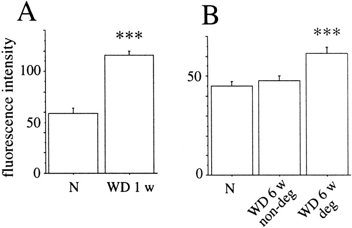
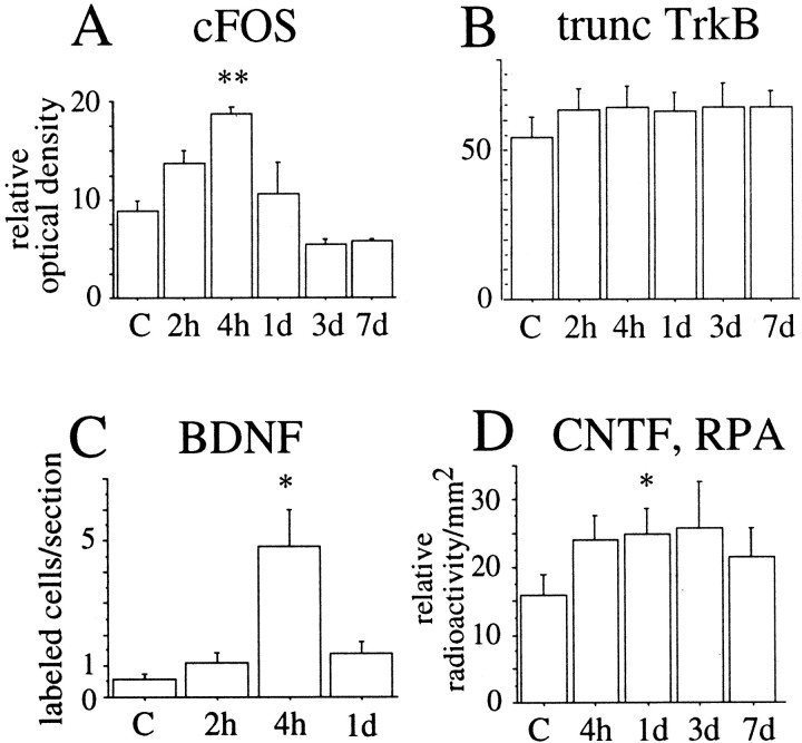
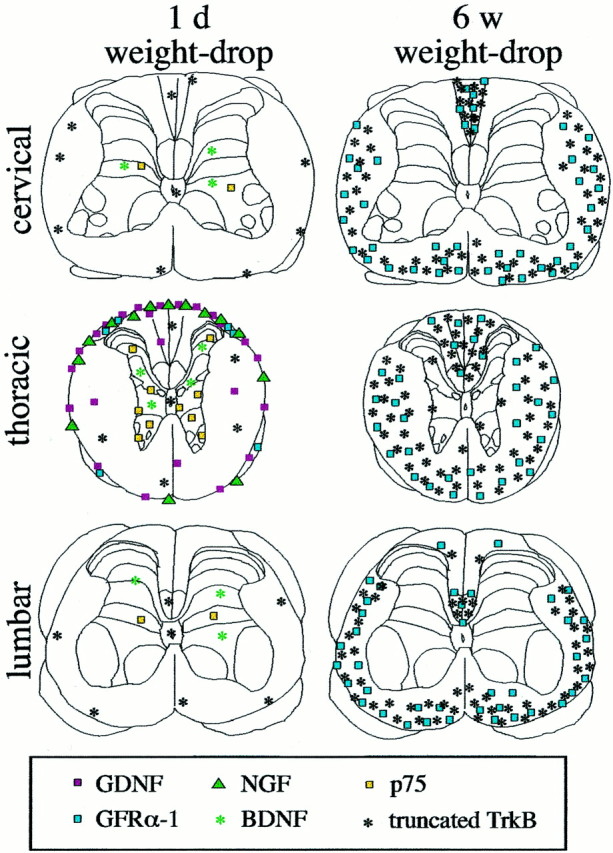
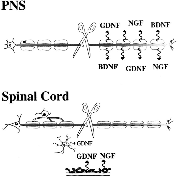
Delivery of neurotrophic factors to the injured spinal cord has been shown to stimulate neuronal survival and regeneration. This indicates that a lack of sufficient trophic support is one factor contributing to the absence of spontaneous regeneration in the mammalian spinal cord. Regulation of the expression of neurotrophic factors and receptors after spinal cord injury has not been studied in detail. We investigated levels of mRNA-encoding neurotrophins, glial cell line-derived neurotrophic factor (GDNF) family members and related receptors, ciliary neurotrophic factor (CNTF), and c-fos in normal and injured spinal cord. Injuries in adult rats included weight-drop, transection, and excitotoxic kainic acid delivery; in newborn rats, partial transection was performed. The regulation of expression patterns in the adult spinal cord was compared with that in the PNS and the neonate spinal cord. After mechanical injury of the adult rat spinal cord, upregulations of NGF and GDNF mRNA occurred in meningeal cells adjacent to the lesion. BDNF and p75 mRNA increased in neurons, GDNF mRNA increased in astrocytes close to the lesion, and GFRalpha-1 and truncated TrkB mRNA increased in astrocytes of degenerating white matter. The relatively limited upregulation of neurotrophic factors in the spinal cord contrasted with the response of affected nerve roots, in which marked increases of NGF and GDNF mRNA levels were observed in Schwann cells. The difference between the ability of the PNS and CNS to provide trophic support correlates with their different abilities to regenerate. Kainic acid delivery led to only weak upregulations of BDNF and CNTF mRNA. Compared with several brain regions, the overall response of the spinal cord tissue to kainic acid was weak. The relative sparseness of upregulations of endogenous neurotrophic factors after injury strengthens the hypothesis that lack of regeneration in the spinal cord is attributable at least partly to lack of trophic support.


Figures

Fig. 1.
Fig. 1.
Dark-field photomicrograph depicting GFAP mRNAin situ hybridization signals in normal and weight-drop-injured adult spinal cord. A, Horizontal section showing the lesion area 1 d after injury. The primary lesion zone with the shape of an ellipse is characterized by markedly reduced mRNA synthesis, whereas cells surrounding the lesion are strongly positive for the GFAP mRNA probe. B,E, H, GFAP mRNA hybridization signals in normal uninjured spinal cord at cervical, thoracic, and lumbar levels.C, F, I, One day after SCI, GFAP mRNA signals have increased at the thoracic level close to the lesion (F) but also at cervical (C) and lumbar levels (I). Note increased signals in cells around the central canal at cervical and lumbar levels.D, G, J, Six weeks after injury, strong GFAP mRNA expression is present in degenerated white matter, especially at the level of injury (G). Degenerating white matter fiber tracts, such as the ascending sensory tract in the dorsal funiculus above the lesion (D), and the descending corticospinal tract below the lesion in the dorsal funiculus (J), are also strongly labeled. Scale bar (shown in B):A, 650 μm; BJ, 1 mm.
Fig. 2.
Fig. 2.
Alterations of c-fos mRNA expression in response to SCI and kainic acid delivery. A, Two hours after weight-drop injury of the adult spinal cord, glial cells and neurons present strong c-fos mRNA signals close to the lesion.B, Six weeks after injury, glial cells and some neurons close to the injury are still positively labeled by the c-fos probe.C, A weak c-fos mRNA expression is found in newborn (P0) spinal cord. D, One day after partial transection of the newborn spinal cord (SCI 1 d), robust c-fos mRNA upregulations occur primarily in cells of the gray matter and in nerve roots. E, In normal adult rat spinal cord, very low levels of c-fos mRNA were found.F, One day after kainic acid delivery, scattered neurons in Rexed's laminas III–VII present strong c-fos mRNA signals.G, In a normal brain (the brain of the animal depicted in E), no robust c-fos mRNA signals can be found.H, One day after kainic acid delivery (the brain of the animal depicted in F), dramatic c-fos hybridization signals appear in areas such as the neocortex, the hippocampal formation, and amygdala. Scale bar (shown inA): A, B,G, H, 1 mm; C,D, 2 mm; E, F, 300 μm.
Fig. 3.
Fig. 3.
Regulation of different mRNA species in the spinal cord after weight-drop injury as monitored by in situhybridization. Quantifications of c-fos, truncated TrkB, and GFRα-1 were performed by measuring the relative optical density on x-ray films exposed to hybridized sections. Quantifications of BDNF and p75 were performed by counting the number of positively labeled cells per section. All statistical comparisons were made to normal animals (N). A, At the cervical level c-fos was significantly increased at 2 hr and 1 d after injury (F(4,20) = 4.9;p < 0.01). B, Significant increase of truncated TrkB in white matter at cervical levels was found 6 weeks after surgery (F(3,18) = 48;p < 0.001) C, At the cervical level significant upregulation of GFRα-1 mRNA in white matter was found 6 weeks after injury (F(4,21) = 3.3;p < 0.05). D, The number of BDNF mRNA-labeled cells was significantly increased 6 hr after injury (F(4,24) = 3.3;p < 0.05). E, There was an increase in the number of p75 cells 6 hr, 1 d, and 6 weeks after injury at the cervical level, but the difference compared with normal was not statistically significant. F, At the thoracic level c-fos was significantly increased at 2 hr, 6 hr, and 1 d after injury (F(4,20) = 6.6;p < 0.01). G, A significant increase of truncated TrkB in white matter at the thoracic level was found 6 weeks after surgery (F(3,18) = 108; p < 0.001). H, At the thoracic level significant upregulation of GFRα-1 mRNA in white matter was found 6 weeks after injury (F(4,21) = 28; p < 0.001). I, The number of BDNF mRNA-labeled cells was significantly increased at the thoracic level 2 hr, 6 hr, and 1 d after injury (F(4,24) = 11; p < 0.001). J, The number of p75 probe-labeled cells was elevated at 6 hr, 1 d, and 6 weeks after injury at the thoracic level. The difference was significant after 1 d (F(3,17) = 21; p < 0.001). K, c-fos mRNA expression levels were significantly increased 2 hr, 6 hr, and 1 d after injury (F(4,20) = 5.1; p< 0.01). L, A significant increase of truncated TrkB in white matter at lumbar levels was found 6 weeks after surgery (F(3,18) = 54; p < 0.001). M, At the lumbar level significant upregulation of GFRα-1 mRNA in white matter was found 6 weeks after injury (F(4,21) = 14; p < 0.001). N, The number of BDNF mRNA-labeled cells was significantly increased at the lumbar level 6 hr after injury (F(4,30) = 3.1; p< 0.05). O, The number of p75 mRNA-labeled cells was elevated at 6 hr, 1 d, and 6 weeks after injury at the thoracic level. The increase was significant after 1 d (F(3,17) = 8; p < 0.01).
Fig. 4.
Fig. 4.
Dark-field photomicrograph depicting GFAP mRNAin situ hybridization signals in normal and injured neonatal spinal cord. A, C,E, GFAP mRNA hybridization signals in normal uninjured neonatal spinal cord. B, D,F, One day after injury GFAP mRNA expression is increased in tissue close to the lesion (B,D) and slightly increased in ventral white matter at the lumbar level (F). Scale bar (shown inA): A, B, 600 μm;CF, 300 μm.
Fig. 5.
Fig. 5.
Appearance of the adult spinal cord of neonatally injured animal. Five levels, cervical to lumbar, of a spinal cord collected from an adult individual that underwent a partial spinal cord transection as newborn. A cut spanning approximately two-thirds of the width of the thoracic spinal cord beginning from the right side was carried out on the first postnatal day, a time point when long nerve fiber tracts, such as the corticospinal tract, are developing. The white matter of the cut side is reduced in size at all levels, but most prominently at thoracic levels close to the injury. This animal scored 21 (= normal) on the BBB score as an adult. Luxol fast blue was used for staining. Scale bar, 1 mm.
Fig. 6.
Fig. 6.
Expression of mRNA encoding neurotrophic factor receptors in the intact normal spinal cord of the adult rat.A, RET mRNA signals are seen in motoneurons.B, p75 mRNA signals were not detectable in spinal cord gray or white matter but were detectable in a subpopulation of dorsal root ganglia neurons, which is shown here in a horizontal section of cervical spinal cord. C, GFRα-1 mRNA signals are present in motoneurons and interneurons. D, TrkA mRNA expression was not found in spinal cord gray or white matter but was found in a subpopulation of dorsal root ganglia neurons, which is shown here in a horizontal section of cervical spinal cord (section adjacent to B). E, GFRα-2 mRNA signals are seen in the dorsal horns of gray matter and in a subpopulation of dorsal root ganglia neurons. F, TrkB mRNA synthesis occurs throughout the gray matter. Scale bar (shown inA): AF, 700 μm.
Fig. 7.
Fig. 7.
Expression of mRNA encoding neurotrophic factor receptors in the intact normal spinal cord of newborn rat.A, GFRα-1 mRNA signals are found throughout the gray matter, with motoneurons displaying particularly strong signals, and in a subpopulation of dorsal root ganglia neurons. B, TrkA mRNA expression is present in dorsal root ganglia neurons.C, GFRα-2 mRNA signals are found throughout the gray matter and in a subpopulation of dorsal root ganglia neurons.D, TrkB mRNA synthesis occurs throughout the gray matter and in some dorsal root ganglia neurons. E, RET mRNA signals were confined to motoneurons and a subpopulation of dorsal root ganglia neurons. F, The probe directed against truncated TrkB plus full-length TrkB-labeled cells in dorsal root ganglia and spinal cord gray and white matter. G, p75 mRNA signals are seen in motoneurons and many dorsal root ganglia neurons.H, TrkC mRNA expression is present throughout gray matter and in many dorsal root ganglia neurons. Scale bar (shown inA): AH, 170 μm.
Fig. 8.
Fig. 8.
Truncated TrkB and p75 mRNA are both upregulated after injury to the adult spinal cord. Hybridization was performed with a probe that recognizes both truncated and full-length TrkB. The expression of truncated TrkB was deduced from comparisons with the expression pattern of the full-length-specific TrkB probe. No robust increases in hybridization signals could be detected when using a probe only complementary to full-length TrkB. A,D, G, The expression of full-length TrkB and truncated TrkB mRNAs in normal uninjured spinal cord is found in neurons of the gray matter and glial cells, respectively.B, E, H, One day after injury glial cells near the injury upregulated truncated TrkB mRNA signals (E). The truncated TrkB mRNA expression is also increased to some extent at cervical and lumbar levels (B, H). C,F, I, Six weeks after injury, white matter tissue at the injury level is strongly positive for truncated TrkB probe (F). In addition, the truncated TrkB mRNA expression is markedly increased in cells of degenerated fiber tracts, such as the ascending sensory tract in the dorsal funiculus above the lesion (C), and the descending corticospinal tract below the lesion in the dorsal funiculus (I). J, Horizontal section of a spinal cord 6 weeks after complete transection. Cells strongly positive for the truncated TrkB probe are found in both gray and white matter tissue but not in the glial scar separating the rostral and caudal stumps. Ependymal cells of the central canal are also strongly positive, as seen in the rostral stump (left) along the midline and reaching the glial scar in the middle. K, Bright-field photomicrograph showing strong truncated TrkB mRNA signals in a subpopulation of cells in the lateral funiculus of lumbar white matter 6 weeks after the operation. L, Neurons close to the lesion 1 d after injury labeled by p75 mRNA hybridization. Scale bar (shown in A):AI, 1 mm; J, 300 μm;K, L, 25 μm.
Fig. 9.
Fig. 9.
Regulation of different mRNA species after SCI measured by RPA (A, B, E,F, I, J) andin situ hybridization (C,D, G, H, K,L). Bars in RPA graphs represent average radioactivity per area (detected by phosphoimaging) expressed in arbitrary units and displayed as percentage of the housekeeping gene L32. A, GDNF mRNA was increased at the level of injury 1 d after transection (T), but not 6 weeks after weight-drop (WD) injury (F(2,30) = 8; p < 0.01). B, GDNF mRNA expression levels were significantly elevated at the level of injury 1 d after partial transection of the neonatal spinal cord measured by RPA (F(2,15) = 4.3; p< 0.05). C, Quantification of in situhybridization demonstrated increased GDNF mRNA expression at the level of the injury, 6 hr, 12 hr, and 1 d after injury.D, No significant difference in GDNF mRNA expression could be found at the lumbar level. E, NGF mRNA probe signals were upregulated 1 d after transection, but not 6 weeks after weight-drop injury (F(2,30) = 11;p < 0.001). F, NGF mRNA expression levels were significantly elevated at the level of injury 12 hr and 1 d after partial transection of neonatal spinal cord (F(2,15) = 4.3; p< 0.05). G, NGF mRNA expression increased at the thoracic level 6 hr, 12 hr, and 1 d after injury.H, No significant difference in NGF mRNA expression could be found at the lumbar level. I, BDNF mRNA probe signals were upregulated 1 d after transection but not 6 weeks after weight-drop injury (F(2,30) = 9;p < 0.001). J, No statistically significant differences in NT3 mRNA expression were found after injury.K, No statistically significant differences were found in truncated TrkB mRNA expression at the thoracic level after injury.L, Likewise, no statistically significant differences were found in truncated TrkB mRNA expression at the lumbar level.
Fig. 10.
Fig. 10.
Neurotrophic factor expression in intact and injured adult and neonatal spinal cord. Representative autoradiograms from RPAs are shown. A riboprobe complementary to the housekeeping gene L32 was used as an internal standard. A, At the newborn stage, NGF and GDNF mRNA signals increased. B, GDNF, NGF, and BDNF signals were upregulated 1 d after transection in adults. C, CNTF mRNA levels were upregulated 1 d after kainic acid delivery.
Fig. 11.
Fig. 11.
GDNF and NGF mRNA expression is increased after injury of the adult spinal cord. A, No robust NGF mRNA signals were found in normal uninjured spinal cord. B, Six hours after weight-drop injury, strong NGF mRNA signals were found in meningeal and Schwann cells close to the lesion. C, In normal adult spinal cord, no robust GDNF mRNA expression could be detected. D, Six hours after weight-drop injury, a marked upregulation of GDNF mRNA signals was found in meningeal and Schwann cells at the level of the lesion. Weak GDNF mRNA signals were observed in glial cells, presumably astrocytes, in the immediate lesion vicinity (arrow). E, Horizontal section of a thoracic dorsal root in a normal animal. No specific GDNF mRNA labeling is observable. F, Six hours after weight-drop injury, intense GDNF mRNA labeling was found in dorsal root Schwann cells at the lesion level. G, Bright-field images depicting meningeal cells expressing NGF mRNA 6 hr after injury.H, GDNF mRNA signals in cells of the dorsal meninges 6 hr after injury. Scale bar (shown in A): A–D, 400 μm;E, F, 250 μm; G,H, 60 μm.
Fig. 12.
Fig. 12.
NGF mRNA hybridization signals increase after partial transection injury to the neonatal spinal cord.A, D, NGF mRNA expression in normal uninjured neonatal (P0) spinal cord is present in cells of the gray matter lateral horn (A, D).B, C, E, F, Six and 12 hr after injury, NGF mRNA synthesis is markedly increased in meningeal cells around the lesion. Schwann cells of dorsal roots on the transected side (left side) also exhibit strong NGF mRNA signals, whereas the dorsal roots of the opposite side are devoid of signal or only weakly positive (B, C). At lumbar levels no altered expression of NGF mRNA could be found (E, F). G, Sagittal section of normal uninjured neonatal spinal cord. No robust NGF mRNA signals are seen in meningeal cells. H, Sagittal section of neonatal spinal cord 1 d after injury exhibiting strong NGF mRNA expression in meningeal cells close to the lesion.I, Bright-field view depicting NGF probe-labeled meningeal cells close to the lesion. Scale bar (shown inA): AF, 250 μm;G, H, 400 μm; I, 40 μm.
Fig. 13.
Fig. 13.
GFRα-1 protein immunoreactivity is increased in nerve roots and degenerating white matter after SCI. A, Dark-field photomicrograph depicting GFRα-1 in situhybridization signals in degenerating white matter, close to the lesion, 6 weeks after weight-drop. B, GFRα-1 immunostaining in a dorsal horn and nerve root in a normal animal. Robust immunoreactivity is seen in lamina II of the dorsal horn. Other laminas of the gray matter are weakly positive, and in the nerve root the GFRα-1 antibody labels some nerve fibers and possibly weakly Schwann cells. C, GFRα-1 immunoreactivity in a cervical dorsal root 6 weeks after weight-drop injury. Some nerve fibers are positive, and a weak staining is possibly also present in Schwann cells. D, One week after injury a dramatic increase in GFRα-1 immunoreactivity is seen in Schwann cells of a dorsal root immediately caudal to the weight-drop site (thoracic level). Meningeal cells are also GFRα-1 positive. E, Increased GFRα-1 immunoreactivity (greenfluorescence), was found in degenerating white matter of the ascending fiber tract in the dorsal funiculus 6 weeks after weight-drop injury. Autofluorescence in infiltrating immune cells (orange-yellow fluorescence) is also seen. Note that no robust immunolabeling is seen in the white matter adjacent to the ascending portion of the dorsal funiculus. F, Six weeks after injury, robust GFRα-1 antibody immunoreactivity (green) is observed in the remaining white matter at the level of injury. Numerous immune cells/phagocytes are present, displaying an orange-yellow autofluorescence. Scale bar (shown in A): A, 200 μm;B, 400 μm; CF, 100 μm.
Fig. 14.
Fig. 14.
GFRα-1 protein levels increase in injured nerve roots and degenerating white matter of the spinal cord.A, The intensity of GFRα-1 immunoreactivity in thoracic nerve roots in normal animals (N) and animals subjected to SCI 1 week earlier (WD 1 w). A marked increase is observed in injured nerve roots (unpaired t test; df = 14;t = 8.6; p < 0.001).B, An increase in GFRα-1 protein levels was also found in degenerating white matter of the spinal cord 6 weeks after injury (ANOVA; F(2,21) = 11;p < 0.001). Measurements were performed in intact white matter in normal animals (N), in nondegenerated white matter in the dorsal funiculus at the cervical level 6 weeks after injury (WD 6 wnon-deg), and in degenerated white matter in the dorsal funiculus at the cervical level 6 weeks after injury (WD 6 w deg).
Fig. 15.
Fig. 15.
GDNF mRNA hybridization signals increase after partial transection injury to the neonatal spinal cord.A, D, Normal uninjured neonatal (P0) spinal cord exhibits strong GDNF mRNA signals in cells of Clark's column (A) and weak signals in the dorsal horns. B, C, E,F, Six and 12 hr after injury, a robust increase of GDNF mRNA signals is seen in meningeal cells around the lesion. Schwann cells of dorsal roots on the transected (left) side also exhibit strong GDNF mRNA signals (especially in C), whereas the dorsal roots of the opposite side are devoid of signal or are only weakly positive (B, C). At lumbar levels no altered expression of GDNF mRNA could be found (E, F). G, Sagittal section of normal uninjured neonatal spinal cord. GDNF mRNA expression is seen in Clark's column and in the dorsal horn. H, Sagittal section of neonatal spinal cord 1 d after injury with strong GDNF mRNA expression in meningeal cells close to the lesion.I, Bright-field photomicrograph depicting GDNF probe-labeled cells in meningeal cells close to the lesion. Scale bar (shown in A): AF, 250 μm; G, H, 400 μm; I, 40 μm.
Fig. 16.
Fig. 16.
Regulation of different mRNA species in lumbar spinal cord after kainic acid injection. Data were obtained fromin situ hybridization (AC) and RPA (D).A, Quantification of in situhybridization signals for c-fos on x-ray film at different time points after kainic acid delivery. Compared with controls (Ringer's solution injected, designated C), c-fos increased gradually, peaking at 4 hr (F(3,18) = 5.4;p < 0.01). B, No robust changes were found in truncated TrkB mRNA expression. C, The number of BDNF mRNA-labeled cells per section increased 4 hr after treatment (F(3,25) = 17.3;p < 0.001). D, CNTF mRNA levels measured by RPA were significantly increased 1 d after kainic acid delivery (F(3,30) = 4.3;p < 0.05). Bars represent average radioactivity per area (detected by phosphoimaging) expressed as percentage of L32 values.
Fig. 17.
Fig. 17.
Schematic figure summarizing the upregulations of different neurotrophic factors and related receptors 1 d and 6 weeks after injury. Only elevated levels are shown. The number of symbols shown for each gene product correlates to the level of expression found. Rexed's laminas were drawn according to Paxinos and Watson (1986).
Fig. 18.
Fig. 18.
Schematic representation of differences in non-neuronal neurotrophic support between the PNS and the spinal cord after injury. The comparison is valid, for instance, for upper and lower motoneurons. Although the Schwann cells in the PNS increase their synthesis of NGF, BDNF, and GDNF (Johnson et al., 1988; Meyer et al., 1992; Funakoshi et al., 1993; Naveilhan et al., 1997; Trupp et al., 1997), the oligodendrocytes in the CNS do not. After SCI the short-lived sources of neurotrophic support are meningeal cells and astrocytes localized to the site of injury. In comparison with GDNF mRNA upregulation in injured nerve roots, the GDNF mRNA signals in astrocytes were only minor. Higher relative mRNA levels are indicated by larger letters.

References

    1. Altar CA, DiStefano PS. Neurotrophin trafficking by anterograde transport. Trends Neurosci. 1998;21:433–437. - PubMed
    1. Baloh R, Tansey M, Golden J, Creedon D, Heuckeroth R, Keck C, Zimonjic D, Popescu N, Johnson E, Jr, Milbrandt J. TrnR2, a novel receptor that mediates neurturin and GDNF signaling through Ret. Neuron. 1997;18:793–802. - PubMed
    1. Baloh RH, Enomoto H, Johnson EM, Jr, Milbrandt J. The GDNF family ligands and receptors: implications for neural development. Curr Opin Neurobiol. 2000;10:103–110. - PubMed
    1. Basso DM, Beattie MS, Bresnahan JC. A sensitive and reliable locomotor rating scale for open field testing in rats. J Neurotrauma. 1995;12:1–21. - PubMed
    1. Bennett DL, Michael GJ, Ramachandran N, Munson JB, Averill S, Yan Q, McMahon SB, Priestley JV. A distinct subgroup of small DRG cells express GDNF receptor components and GDNF is protective for these neurons after nerve injury. J Neurosci. 1998;18:3059–3072. - PMC - PubMed

Publication types

MeSH terms