Enhancement of sensorimotor behavioral recovery in hemiparkinsonian rats with intrastriatal, intranigral, and intrasubthalamic nucleus dopaminergic transplants
- PMID: 11331381
- PMCID: PMC6762505
- DOI: 10.1523/JNEUROSCI.21-10-03521.2001
Enhancement of sensorimotor behavioral recovery in hemiparkinsonian rats with intrastriatal, intranigral, and intrasubthalamic nucleus dopaminergic transplants
Abstract
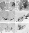
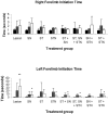
One of the critical variables that influences the efficacy of clinical neural transplantation for Parkinson's disease (PD) is optimal graft placement. The current transplantation paradigm that focuses on ectopic placement of fetal grafts in the striatum (ST) fails to reconstruct the basal ganglia circuitry or normalize neuronal activity in important basal ganglia structures, such as the substantia nigra (SN) and the subthalamic nucleus (STN). The aim of this study was to investigate a multitarget neural transplantation strategy for PD by assessing whether simultaneous dopaminergic transplants in the ST, SN, and STN induce functional recovery in hemiparkinsonian rats. Forty-six female Wistar rats with unilateral 6-hydroxydopamine lesions of the nigrostriatal pathway were randomly divided into eight groups and received lesions only or injections of 900,000 embryonic rat ventral mesencephalic cells in the (1) ST, (2) SN, (3) STN, (4) ST and SN, (5) ST, SN, and STN, (6) ST and STN, or (7) SN and STN. The number of cells transplanted was equally divided among grafting sites. Animals with two grafts received 450,000 cells in each structure, and animals with three grafts received 300,000 cells per structure. Recovery was assessed by amphetamine-induced rotations and the stepping tests. Graft survival was assessed using tyrosine hydroxylase immunohistochemistry. At 8 weeks after transplantation, simultaneous dopaminergic transplants in the ST, SN, and STN induced significant improvement in rotational behavior and stepping test scores. Intrastriatal transplants were associated with significant recovery of rotational asymmetry, whereas SN and STN transplants were associated with improved forelimb function scores. These results suggest that restoration of dopaminergic activity to multiple basal ganglia targets, such as the ST and SN, or the ST and STN, promotes a more complete functional recovery of complex sensorimotor behaviors. A multitarget transplant strategy aimed at optimizing dopaminergic reinnervation of the basal ganglia may be crucial in improving clinical outcomes in PD patients.
Figures


Similar articles
-
Transplantation of mouse embryonic stem cell-derived neurons into the striatum, subthalamic nucleus and substantia nigra, and behavioral recovery in hemiparkinsonian rats.Neurosci Lett. 2005 Oct 28;387(3):151-6. doi: 10.1016/j.neulet.2005.06.029. Neurosci Lett. 2005. PMID: 16023291
-
Intranigral transplants of GABA-rich striatal tissue induce behavioral recovery in the rat Parkinson model and promote the effects obtained by intrastriatal dopaminergic transplants.Exp Neurol. 1999 Feb;155(2):165-86. doi: 10.1006/exnr.1998.6916. Exp Neurol. 1999. PMID: 10072293
-
Simultaneous intrastriatal and intranigral dopaminergic grafts in the parkinsonian rat model: role of the intranigral graft.J Comp Neurol. 2000 Oct 9;426(1):106-16. J Comp Neurol. 2000. PMID: 10980486
-
A multiple target neural transplantation strategy for Parkinson's disease.Rev Neurosci. 2002;13(3):243-56. doi: 10.1515/revneuro.2002.13.3.243. Rev Neurosci. 2002. PMID: 12405227 Review.
-
How to improve the survival of the fetal ventral mesencephalic cell transplanted in Parkinson's disease?Neurosci Bull. 2007 Nov;23(6):377-82. doi: 10.1007/s12264-007-0056-4. Neurosci Bull. 2007. PMID: 18064069 Free PMC article. Review.
Cited by
-
Recovery from experimental parkinsonism by semaphorin-guided axonal growth of grafted dopamine neurons.Mol Ther. 2013 Aug;21(8):1579-91. doi: 10.1038/mt.2013.78. Epub 2013 Jun 4. Mol Ther. 2013. PMID: 23732989 Free PMC article.
-
Botulinum Neurotoxin-A Injected Intrastriatally into Hemiparkinsonian Rats Improves the Initiation Time for Left and Right Forelimbs in Both Forehand and Backhand Directions.Int J Mol Sci. 2019 Feb 25;20(4):992. doi: 10.3390/ijms20040992. Int J Mol Sci. 2019. PMID: 30823527 Free PMC article.
-
Dopamine Cell Therapy: From Cell Replacement to Circuitry Repair.J Parkinsons Dis. 2021;11(s2):S159-S165. doi: 10.3233/JPD-212609. J Parkinsons Dis. 2021. PMID: 33814467 Free PMC article. Review.
-
Transplantation of hypocretin neurons into the pontine reticular formation: preliminary results.Sleep. 2004 Dec 15;27(8):1465-70. doi: 10.1093/sleep/27.8.1465. Sleep. 2004. PMID: 15683135 Free PMC article.
-
Endogenous versus exogenous cell replacement for Parkinson's disease: where are we at and where are we going?Neural Regen Res. 2022 Dec;17(12):2637-2642. doi: 10.4103/1673-5374.336137. Neural Regen Res. 2022. PMID: 35662194 Free PMC article. Review.
References
-
- Abercrombie M. Estimation of nuclear population from microtome sections. Anat Rec. 1946;94:239–247. - PubMed
-
- Albin RL, Young AB, Penney JB. The functional anatomy of disorders of the basal ganglia. Trends Neurosci. 1995;18:63–64. - PubMed
-
- Baker KA, Sadi D, Hong M, Mendez I. Simultaneous intrastriatal and intranigral dopaminergic grafts in the parkinsonian rat model: role of the intranigral graft. J Comp Neurol. 2000;426:106–116. - PubMed
-
- Bergman H, Wichmann T, DeLong MR. Reversal of experimental parkinsonism by lesions of the subthalamic nucleus. Science. 1990;249:1436–1438. - PubMed
-
- Bergman H, Wichmann T, Karmon B, Delong MR. The primate subthalamic nucleus. II. Neuronal activity in the MPTP model of parkinsonism. J Neurophysiol. 1994;72:507–520. - PubMed
Publication types
MeSH terms
Substances
LinkOut - more resources
Full Text Sources
