

Status epilepticus causes necrotic damage in the mediodorsal nucleus of the thalamus in immature rats
- PMID: 11331388
- PMCID: PMC6762492
- DOI: 10.1523/JNEUROSCI.21-10-03593.2001
Status epilepticus causes necrotic damage in the mediodorsal nucleus of the thalamus in immature rats
Abstract
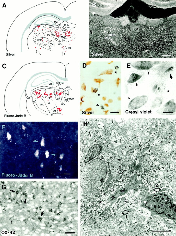
Status epilepticus (StE) in immature rats causes long-term functional impairment. Whether this is associated with structural alterations remains controversial. The present study was designed to test the hypothesis that StE at an early age results in neuronal loss. StE was induced with lithium-pilocarpine in 12-d-old rats, and the presence of neuronal damage was investigated in the brain from 12 hr up to 1 week later using silver and Fluoro-Jade B staining techniques. Analysis of the sections indicated consistent neuronal damage in the central and lateral segments of the mediodorsal nucleus of the thalamus, which was confirmed using adjacent cresyl violet-stained preparations. The mechanism of thalamic damage (necrosis vs apoptosis) was investigated further using TUNEL, immunohistochemistry for caspase-3 and cytochrome c, and electron microscopy. Activated microglia were detected using OX-42 immunohistochemistry. The presence of silver and Fluoro-Jade B-positive degenerating neurons in the mediodorsal thalamic nucleus was associated with the appearance of OX-42-immunopositive activated microglia but not with the expression of markers of programmed cell death, caspase-3, or cytochrome c. Electron microscopy revealed necrosis of the ultrastructure of damaged neurons, providing further evidence that the mechanism of StE-induced damage in the mediodorsal thalamic nucleus at postnatal day 12 is necrosis rather than apoptosis. Finally, these data together with previously described functions of the medial and lateral segments of the mediodorsal thalamic nucleus suggest that some functions, such as adaptation to novelty, might become compromised after StE early in development.
Figures
Similar articles
-
Dynamic changes of status epilepticus-induced neuronal degeneration in the mediodorsal nucleus of the thalamus during postnatal development of the rat.Epilepsia. 2002;43 Suppl 5:54-60. doi: 10.1046/j.1528-1157.43.s.5.36.x. Epilepsia. 2002. PMID: 12121296
-
Neuronal degeneration is observed in multiple regions outside the hippocampus after lithium pilocarpine-induced status epilepticus in the immature rat.Neuroscience. 2013 Nov 12;252:45-59. doi: 10.1016/j.neuroscience.2013.07.045. Epub 2013 Jul 27. Neuroscience. 2013. PMID: 23896573 Free PMC article.
-
Seizure-induced neuronal necrosis: implications for programmed cell death mechanisms.Epilepsia. 2000;41 Suppl 6:S9-13. doi: 10.1111/j.1528-1157.2000.tb01549.x. Epilepsia. 2000. PMID: 10999512
-
The mediodorsal thalamic nucleus and schizophrenia.J Psychiatry Neurosci. 2008 Nov;33(6):489-98. J Psychiatry Neurosci. 2008. PMID: 18982171 Free PMC article. Review.
-
Programmed Necrosis After Status Epilepticus.In: Noebels JL, Avoli M, Rogawski MA, Olsen RW, Delgado-Escueta AV, editors. Jasper's Basic Mechanisms of the Epilepsies [Internet]. 4th edition. Bethesda (MD): National Center for Biotechnology Information (US); 2012. In: Noebels JL, Avoli M, Rogawski MA, Olsen RW, Delgado-Escueta AV, editors. Jasper's Basic Mechanisms of the Epilepsies [Internet]. 4th edition. Bethesda (MD): National Center for Biotechnology Information (US); 2012. PMID: 22787596 Free Books & Documents. Review.
Cited by
-
The outcome of early life status epilepticus-lessons from laboratory animals.Epilepsia Open. 2023 May;8 Suppl 1(Suppl 1):S90-S109. doi: 10.1002/epi4.12664. Epub 2022 Nov 9. Epilepsia Open. 2023. PMID: 36352789 Free PMC article.
-
Brain energy metabolism during experimental neonatal seizures.Neurochem Res. 2010 Dec;35(12):2193-8. doi: 10.1007/s11064-010-0339-4. Epub 2010 Dec 7. Neurochem Res. 2010. PMID: 21136154 Free PMC article.
-
Sex-specific consequences of early life seizures.Neurobiol Dis. 2014 Dec;72 Pt B(Pt B):153-66. doi: 10.1016/j.nbd.2014.05.021. Epub 2014 May 27. Neurobiol Dis. 2014. PMID: 24874547 Free PMC article. Review.
-
fMRI shows atypical language lateralization in pediatric epilepsy patients.Epilepsia. 2006 Mar;47(3):593-600. doi: 10.1111/j.1528-1167.2006.00474.x. Epilepsia. 2006. PMID: 16529628 Free PMC article.
-
Behavioral impairments in rats with chronic epilepsy suggest comorbidity between epilepsy and attention deficit/hyperactivity disorder.Epilepsy Behav. 2014 Feb;31:267-75. doi: 10.1016/j.yebeh.2013.10.004. Epub 2013 Nov 18. Epilepsy Behav. 2014. PMID: 24262783 Free PMC article.
References
-
- Aicardi J, Chevrie JJ. Convulsive status epilepticus in infants and children: a study of 239 cases. Epilepsia. 1970;11:187–197. - PubMed
-
- Babb TL, Leite JP, Mathern GW, Pretorius JK. Kainic acid induced hippocampal seizures in rats: comparison of acute and chronic seizures using intrahippocampal versus systemic injection. Ital J Neurol Sci. 1995;16:39–44. - PubMed
Publication types
MeSH terms
Substances
LinkOut - more resources
Full Text Sources
Research Materials