

Reorganization of motor and somatosensory cortex in upper extremity amputees with phantom limb pain
- PMID: 11331390
- PMCID: PMC6762494
- DOI: 10.1523/JNEUROSCI.21-10-03609.2001
Reorganization of motor and somatosensory cortex in upper extremity amputees with phantom limb pain
Abstract
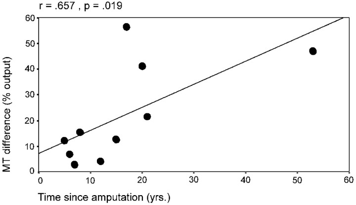
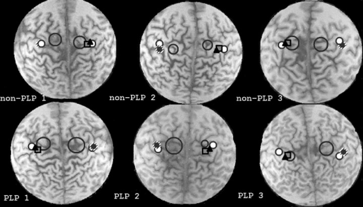
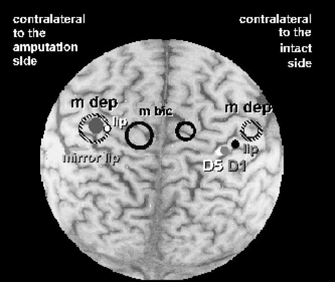
Phantom limb pain (PLP) in amputees is associated with reorganizational changes in the somatosensory system. To investigate the relationship between somatosensory and motor reorganization and phantom limb pain, we used focal transcranial magnetic stimulation (TMS) of the motor cortex and neuroelectric source imaging of the somatosensory cortex (SI) in patients with and without phantom limb pain. For transcranial magnetic stimulation, recordings were made bilaterally from the biceps brachii, zygomaticus, and depressor labii inferioris muscles. Neuroelectric source imaging of the EEG was obtained after somatosensory stimulation of the skin overlying face and hand. Patients with phantom limb pain had larger motor-evoked potentials from the biceps brachii, and the map of outputs was larger for muscles on the amputated side compared with the intact side. The optimal scalp positions for stimulation of the zygomaticus and depressor labii inferioris muscles were displaced significantly more medially (toward the missing hand representation) in patients with phantom limb pain only. Neuroelectric source imaging revealed a similar medial displacement of the dipole center for face stimulation in patients with phantom limb pain. There was a high correlation between the magnitude of the shift of the cortical representation of the mouth into the hand area in motor and somatosensory cortex and phantom limb pain. These results show enhanced plasticity in both the motor and somatosensory domains in amputees with phantom limb pain.
Figures
Similar articles
-
Remote activation of referred phantom sensation and cortical reorganization in human upper extremity amputees.Exp Brain Res. 2004 Jan;154(1):97-102. doi: 10.1007/s00221-003-1649-4. Epub 2003 Oct 14. Exp Brain Res. 2004. PMID: 14557916
-
Neuroelectric source imaging of steady-state movement-related cortical potentials in human upper extremity amputees with and without phantom limb pain.Pain. 2004 Jul;110(1-2):90-102. doi: 10.1016/j.pain.2004.03.013. Pain. 2004. PMID: 15275756
-
Mapping phantom movement representations in the motor cortex of amputees.Brain. 2006 Aug;129(Pt 8):2202-10. doi: 10.1093/brain/awl180. Epub 2006 Jul 14. Brain. 2006. PMID: 16844715
-
The motor cortex and its role in phantom limb phenomena.Neuroscientist. 2008 Apr;14(2):195-202. doi: 10.1177/1073858407309466. Epub 2007 Nov 7. Neuroscientist. 2008. PMID: 17989169 Review.
-
[Cortical reorganization and pain. Empirical findings and therapeutic implication using the example of phantom pain].Schmerz. 2001 Apr;15(2):131-7. doi: 10.1007/s004820170037. Schmerz. 2001. PMID: 11810344 Review. German.
Cited by
-
Postamputation pain: epidemiology, mechanisms, and treatment.J Pain Res. 2013;6:121-36. doi: 10.2147/JPR.S32299. Epub 2013 Feb 13. J Pain Res. 2013. PMID: 23426608 Free PMC article.
-
Neurochemical analysis of primary motor cortex in chronic low back pain.Brain Sci. 2012 Sep 1;2(3):319-31. doi: 10.3390/brainsci2030319. Brain Sci. 2012. PMID: 23766894 Free PMC article.
-
Oculomotor responses of the visual system to an artificial central scotoma may not represent genuine visuomotor adaptation.J Vis. 2022 Sep 2;22(10):17. doi: 10.1167/jov.22.10.17. J Vis. 2022. PMID: 36136045 Free PMC article.
-
Phantom limb pain, cortical reorganization and the therapeutic effect of mental imagery.Brain. 2008 Aug;131(Pt 8):2181-91. doi: 10.1093/brain/awn124. Epub 2008 Jun 20. Brain. 2008. PMID: 18567624 Free PMC article.
-
Amputation and prosthesis implantation shape body and peripersonal space representations.Sci Rep. 2013 Oct 3;3:2844. doi: 10.1038/srep02844. Sci Rep. 2013. PMID: 24088746 Free PMC article.
References
-
- Aglioti S, Cortese F, Franchini C. Rapid sensory remapping in the adult human brain as inferred from phantom breast perception. NeuroReport. 1994;5:473–476. - PubMed
-
- Aglioti S, Bonazzi A, Cortese F. Phantom lower limb as a perceptual marker of neural plasticity in the mature human brain. Proc R Soc Lond B Biol Sci. 1995;255:273–278. - PubMed
-
- Aglioti S, Smania N, Atzei BA, Berlucchi G. Spatio-temporal pattern of evoked phantom sensations in a left index amputee patient. Behav Neurosci. 1997;111:867–872. - PubMed
-
- Borsook D, Becerra L, Fishman S, Edwards A, Jennings CL, Stojanovic M. Acute plasticity in the human somatosensory cortex following amputation. NeuroReport. 1998;9:1013–1017. - PubMed
Publication types
MeSH terms
LinkOut - more resources
Full Text Sources
Medical
Research Materials