

Neurotoxic lesions of the lateral nucleus of the amygdala decrease conditioned fear but not unconditioned fear of a predator odor: comparison with electrolytic lesions
- PMID: 11331391
- PMCID: PMC6762462
- DOI: 10.1523/JNEUROSCI.21-10-03619.2001
Neurotoxic lesions of the lateral nucleus of the amygdala decrease conditioned fear but not unconditioned fear of a predator odor: comparison with electrolytic lesions
Abstract
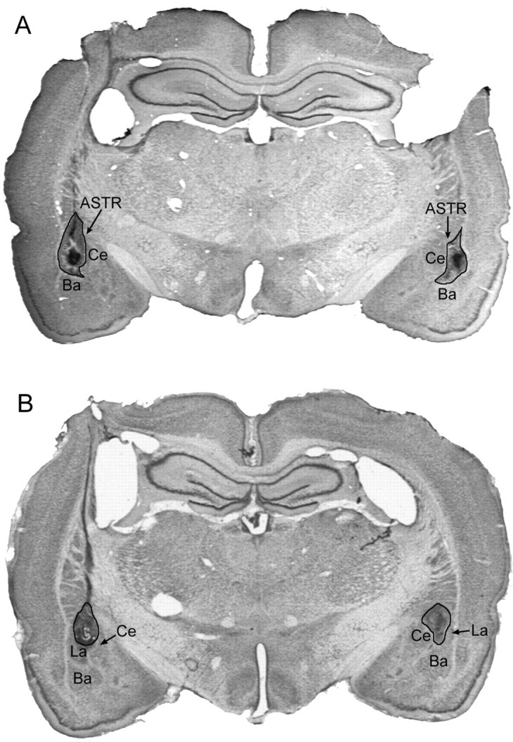
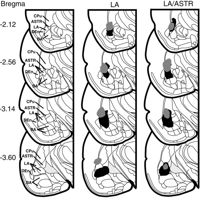
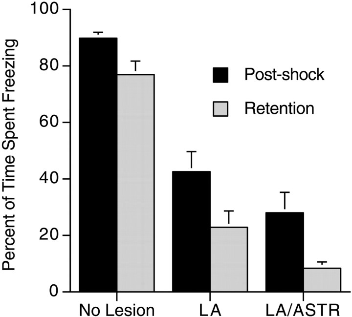
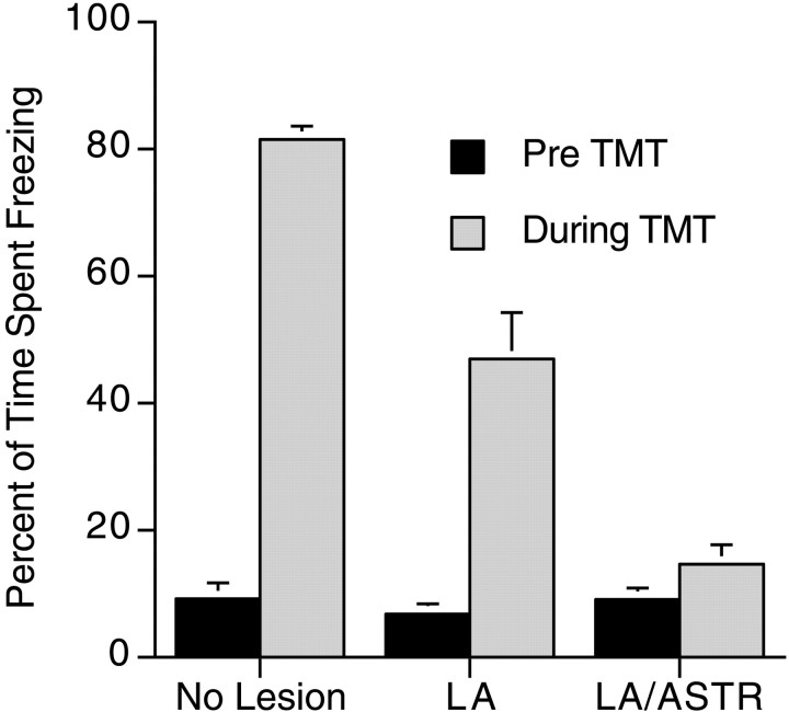
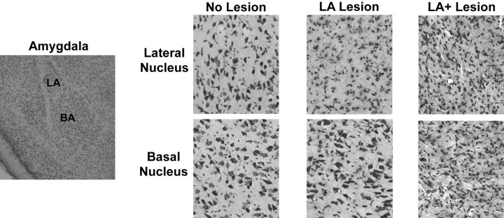
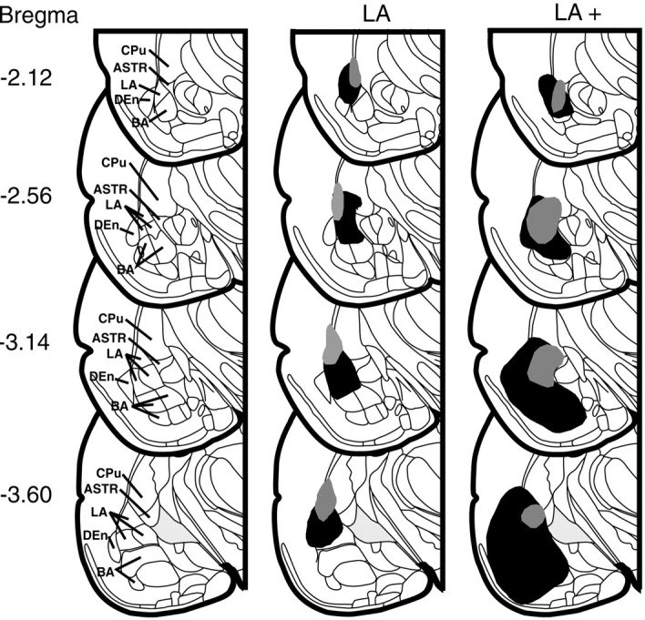
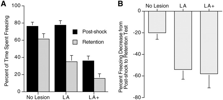
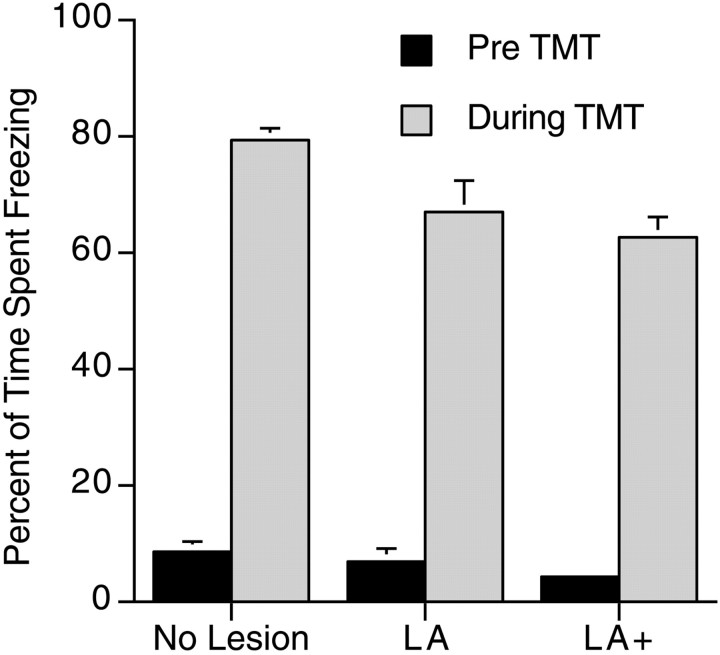
Considerable evidence suggests that the lateral (LA) and basal (BA) nuclei of the amygdala are sites of plasticity and storage of emotional memory. Recent arguments, however, have seriously challenged this view, suggesting that the effects of amygdala lesions are attributable to interference with performance of fear behavior and not learning and memory. One way to address this controversy is to measure the same behavioral response during both conditioned and unconditioned fear. This is done in the present study by measuring fear-related freezing behavior after electrolytic and neurotoxic lesions of the LA or LA/BA nuclei in rats in a contextual fear conditioning paradigm and unconditioned fear to a predator odor. Electrolytic LA lesions attenuated post-shock freezing, retention test freezing, and freezing to the predator odor trimethylthiazoline (TMT). In contrast, excitotoxic NMDA lesions of the LA had no effect on post-shock freezing but significantly attenuated retention test freezing. Furthermore, excitotoxic LA lesions did not diminish freezing to TMT. Larger excitotoxic lesions that included the BA significantly reduced freezing in both the post-shock and retention tests but did not appreciably decrease freezing to TMT. The results suggest that the LA is important for memory of learned fear but not for generation of freezing behavior. In addition, the BA plays a role in freezing in conditioned fear situations but not in unconditioned fear. The studies suggest that the LA and BA play different roles in fear conditioning, but neither of them has a significant role in unconditioned freezing to a predator odor.
Figures
Similar articles
-
The medial hypothalamic defensive circuit and 2,5-dihydro-2,4,5-trimethylthiazoline (TMT) induced fear: comparison of electrolytic and neurotoxic lesions.Brain Res. 2009 Aug 25;1286:133-46. doi: 10.1016/j.brainres.2009.06.062. Epub 2009 Jun 25. Brain Res. 2009. PMID: 19559688
-
An egr-1 (zif268) antisense oligodeoxynucleotide infused into the amygdala disrupts fear conditioning.Learn Mem. 2004 Sep-Oct;11(5):617-24. doi: 10.1101/lm.73104. Learn Mem. 2004. PMID: 15466317 Free PMC article.
-
Unconditioned stimulus pathways to the amygdala: effects of posterior thalamic and cortical lesions on fear conditioning.Neuroscience. 2004;125(2):305-15. doi: 10.1016/j.neuroscience.2003.12.034. Neuroscience. 2004. PMID: 15062974
-
Predator odor fear conditioning: current perspectives and new directions.Neurosci Biobehav Rev. 2008 Sep;32(7):1218-27. doi: 10.1016/j.neubiorev.2008.06.001. Epub 2008 Jun 5. Neurosci Biobehav Rev. 2008. PMID: 18577397 Free PMC article. Review.
-
The neurobiology of conditioned and unconditioned fear: a neurobehavioral system analysis of the amygdala.Behav Cogn Neurosci Rev. 2004 Mar;3(1):23-41. doi: 10.1177/1534582304265945. Behav Cogn Neurosci Rev. 2004. PMID: 15191640 Review.
Cited by
-
The prefrontal cortex regulates lateral amygdala neuronal plasticity and responses to previously conditioned stimuli.J Neurosci. 2003 Dec 3;23(35):11054-64. doi: 10.1523/JNEUROSCI.23-35-11054.2003. J Neurosci. 2003. PMID: 14657162 Free PMC article.
-
The amygdala differentially regulates defensive behaviors evoked by CO2.Behav Brain Res. 2020 Jan 13;377:112236. doi: 10.1016/j.bbr.2019.112236. Epub 2019 Sep 16. Behav Brain Res. 2020. PMID: 31536735 Free PMC article.
-
Extrafield Activity Shifts the Place Field Center of Mass to Encode Aversive Experience.eNeuro. 2019 Mar 22;6(2):ENEURO.0423-17.2019. doi: 10.1523/ENEURO.0423-17.2019. eCollection 2019 Mar-Apr. eNeuro. 2019. PMID: 30923741 Free PMC article.
-
An affective circumplex model of neural systems subserving valence, arousal, and cognitive overlay during the appraisal of emotional faces.Neuropsychologia. 2008;46(8):2129-39. doi: 10.1016/j.neuropsychologia.2008.02.032. Epub 2008 Mar 18. Neuropsychologia. 2008. PMID: 18440572 Free PMC article.
-
Anxiogenic modulation of spontaneous and evoked neuronal activity in the basolateral amygdala.Neuroscience. 2009 Nov 10;163(4):1069-77. doi: 10.1016/j.neuroscience.2009.07.003. Epub 2009 Jul 7. Neuroscience. 2009. PMID: 19589368 Free PMC article.
References
-
- Bellgowan PS, Helmstetter FJ. Neural systems for the expression of hypoalgesia during nonassociative fear. Behav Neurosci. 1996;110:727–736. - PubMed
-
- Berton F, Vogel E, Belzung C. Modulation of mice anxiety in response to cat odor as a consequence of predators diet. Physiol Behav. 1998;65:247–254. - PubMed
-
- Blanchard DC, Blanchard RJ. Crouching as an index of fear. J Comp Physiol Psychol. 1969;67:370–375. - PubMed
-
- Blanchard DC, Blanchard RJ. Innate and conditioned reactions to threat in rats with amygdaloid lesions. J Comp Physiol Psychol. 1972;81:281–290. - PubMed
-
- Blanchard RJ, Blanchard DC. Antipredator defensive behaviors in a visible burrow system. J Comp Psychol. 1989;103:70–82. - PubMed
Publication types
MeSH terms
Substances
LinkOut - more resources
Full Text Sources