. 2001 May 15;21(10):3628-38.
doi: 10.1523/JNEUROSCI.21-10-03628.2001.

Bidirectional dopamine modulation of GABAergic inhibition in prefrontal cortical pyramidal neurons

Affiliations

Bidirectional dopamine modulation of GABAergic inhibition in prefrontal cortical pyramidal neurons

J K Seamans et al. J Neurosci. .

Abstract

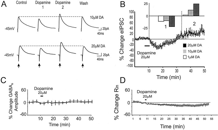
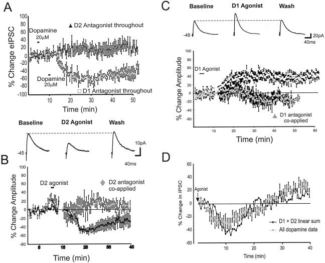
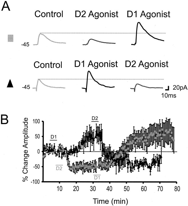
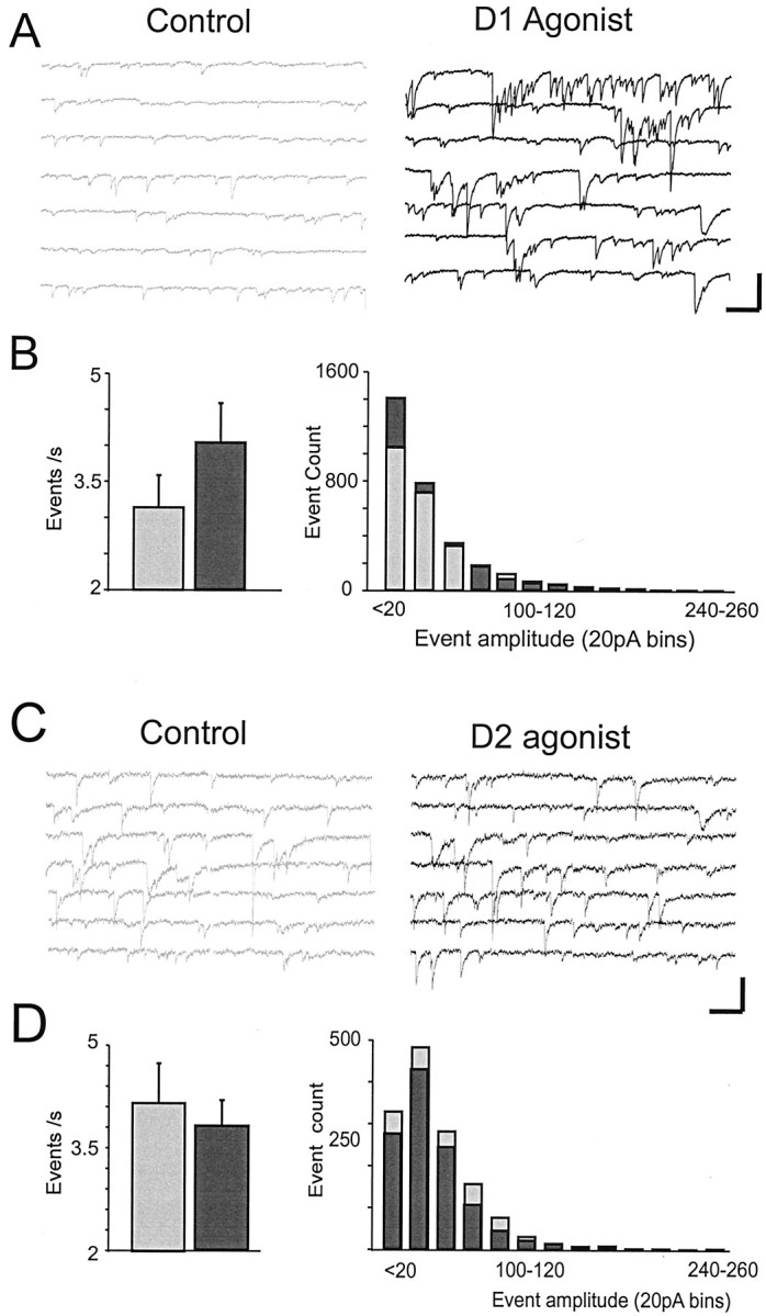
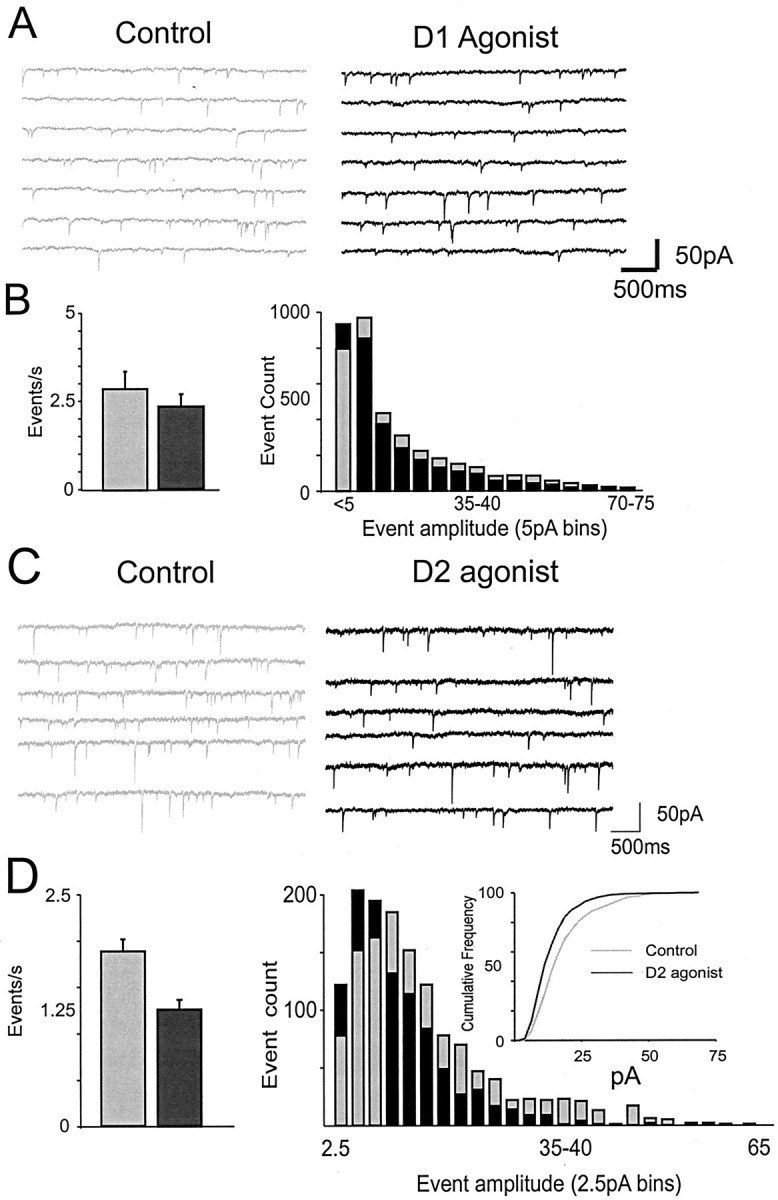
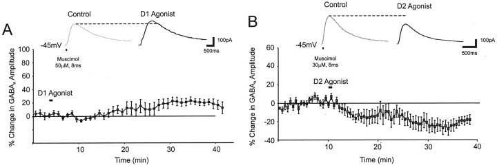
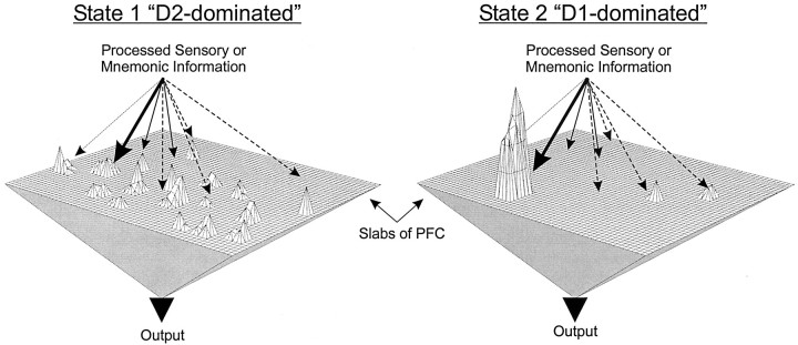
Dopamine regulates the activity of neural networks in the prefrontal cortex that process working memory information, but its precise biophysical actions are poorly understood. The present study characterized the effects of dopamine on GABAergic inputs to prefrontal pyramidal neurons using whole-cell patch-clamp recordings in vitro. In most pyramidal cells, dopamine had a temporally biphasic effect on evoked IPSCs, producing an initial abrupt decrease in amplitude followed by a delayed increase in IPSC amplitude. Using receptor subtype-specific agonists and antagonists, we found that the initial abrupt reduction was D2 receptor-mediated, whereas the late, slower developing enhancement was D1 receptor-mediated. Linearly combining the effects of the two agonists could reproduce the biphasic dopamine effect. Because D1 agonists enhanced spontaneous (sIPSCs) but did not affect miniature (mIPSCs) IPSCs, it appears that D1 agonists caused larger evoked IPSCs by increasing the intrinsic excitability of interneurons and their axons. In contrast, D2 agonists had no effects on sIPSCs but did produce a significant reduction in mIPSCs, suggestive of a decrease in GABA release probability. In addition, D2 agonists reduced the postsynaptic response to a GABA(A) agonist. D1 and D2 receptors therefore regulated GABAergic activity in opposite manners and through different mechanisms in prefrontal cortex (PFC) pyramidal cells. This bidirectional modulation could have important implications for the computational properties of active PFC networks.


Figures

Fig. 1.
Fig. 1.
Dopamine has temporally biphasic effects on eIPSCs. A, Representative traces from neurons voltage-clamped at −45 mV using QX-314-filled electrodes in the presence of APV and CNQX. Application of 10 (top) or 20 (bottom) μm dopamine initially depressed eIPSC amplitude (Dopamine 1) but subsequently increased amplitude (Dopamine 2) before recovery.B, Mean and SEM group data showing the change in eIPSC amplitude over time. Dopamine (20 μm) produced an initial reduction (1) before an enhancement (2) in eIPSC amplitude. Inset, The effects were dose dependent. A significant (p < 0.05) biphasic change in eIPSC amplitude was observed at 10 and 20 μm dopamine but not at 1 μm. C, Dopamine (20 μm) had no effect on the response to focal puff application of the GABAB agonist baclofen (1 mm in ACSF;n = 7). D, Dopamine (20 μm) produced a long-lasting decrease but no rebound increase in Rin, as assessed with intracellular voltage steps of −10 mV.
Fig. 2.
Fig. 2.
Dopamine agonist effects on eIPSC amplitude.A, Counterclockwise from top left, Application of dopamine (20 μm) in the presence of a D1 antagonist (10 μm SCH-23390) produced a pure depression of eIPSC amplitude (gray triangles;n = 3), whereas application of dopamine in the presence of a D2 antagonist (10 μm sulpiride or raclopride; n = 12) produced only a slight increase in eIPSC amplitude. B, Top, Representative traces showing that application of a D2 agonist (quinpirole 10 μm; n = 8) produced a pure decrease in eIPSC amplitude. Bottom, Mean and SEM group data showing that the D2 agonist reduced the average eIPSC amplitude (black triangles) and that this effect was blocked by coapplication of a D2 antagonist (gray triangles). C, Top, Representative traces showing that application of a D1 agonist (10 μm SKF-81297; n = 12) produced a pure increase in eIPSC amplitude. Bottom, Mean and SEM group data showing that the D1 agonist increased the average eIPSC amplitude (black squares) and that this effect was blocked by coapplication of a D1 antagonist (gray triangles). D, The dopamine effect on eIPSCs (gray triangles; n = 20) is similar to the linear sum (black circles) of the effect of the D1 agonist and D2 agonist (graphs shown in B andC) aligned to the time of drug offset.
Fig. 3.
Fig. 3.
eIPSC amplitude can be upregulated and downregulated by D1 and D2 agonists. A,Top, Representative traces showing that a D2 agonist produced a reduction in eIPSC amplitude that was reversed by a D1 agonist into an increase. Bottom, Representative traces showing that a D1 agonist produced an increase in eIPSC amplitude that was reversed by a D2 agonist into a decrease. B, Mean and SEM group data showing that the D1 agonist produced an increase in eIPSC amplitude that was changed to a decrease when followed 20 min later by a D2 agonist (black triangles;n = 7). Relative to the time scale on the graph, the D1 agonist was applied at 3–8 min, whereas the D2 agonist was applied at 27–30 min. The gray squares show the effects of the opposite experimental protocol whereby a D2 agonist was applied before a D1 agonist (n = 4). Relative to the time scale on the graph, the D2 agonist was applied at 8–15 min, whereas the D1 agonist was applied at 30–35 min. The difference in application times for the two agonists in the two experiments was to control for the temporal differences in the effect of each drug (see Fig.2B,C).
Fig. 4.
Fig. 4.
Dopamine agonist modulation of sIPSCs.A, Representative traces showing that the D1 agonist (black, right) increased the frequency of sIPSCs relative to the control condition (gray,left). Responses were recorded using CsCl- and QX-314-filled electrodes in the presence of APV and CNQX. Calibration: 100 pA, 600 msec. B, Left, Histogram of sIPSC frequency generated from all cells sampled for a 5 min period before D1 agonist application and for a 5 min period 10 min after D1 agonist application. D1 agonists increased sIPSC frequency.Right, Histogram from all cells showing the number of events in 20 pA bins for the control condition (gray bars) versus the D1 agonist condition (black bars). C, Representative traces showing sIPSCs in the control condition (gray,left) and after application of the D2 agonist (black, right). D,Left, Histogram of sIPSC frequency generated from all cells sampled for a 5 min period before D2 agonist application and for a 5 min period immediately after D2 agonist application.Right, Histogram from all cells showing the number of events in 20 pA bins for the control condition (gray bars) versus the D2 agonist condition (black bars). D2 agonists had no significant effects on sIPSCs.
Fig. 5.
Fig. 5.
Effects of D1 and D2 agonists on mIPSCs.A, Representative traces showing mIPSCs in the control condition (gray, left) and after application of a D1 agonist (black,right). Responses were recorded using CsCl-filled electrodes in the presence of APV and CNQX and TTX. B,Left, Histogram of mIPSC frequency generated from all cells sampled for a 5 min period before D1 agonist application and for a 5 min period 10 min after D1 agonist application.Right, Histogram generated from all cells showing the number of events in 5 pA bins for the control condition (gray bars) versus the D1 condition (black bars). D1 agonists had no effects on mIPSCs. C, Representative traces showing that the D2 agonist (black, right) decreased the frequency of mIPSCs relative to the control condition (gray,left). D, Left, Histogram of mIPSC frequency generated from all cells sampled for a 5 min period before D2 agonist application and for a 5 min period immediately after D2 agonist application. Right, Histogram generated from all cells showing the number of events in 2.5 pA bins for the control condition (gray bars) versus the D2 condition (black bars). Inset, Same data replotted as a cumulative frequency plot to emphasize the leftward shift induced by a D2 agonist. D2 agonists decreased mIPSC frequency and amplitude.
Fig. 6.
Fig. 6.
The effects of D1 and D2 agonists on the postsynaptic GABAA current. A,Top, Representative traces showing that the response to puff application of the GABAA agonist muscimol (20–50 μm in ACSF containing APV and CNQX) on the perisomatic region was slightly enhanced by the D1 agonist. Bottom, Group data (mean and SEM) showing a small delayed increase in the postsynaptic GABAA response by a D1 agonist (n = 8). B, Top, Representative traces showing that the response to puff application of the GABAA agonist muscimol was reduced by the D2 agonist.Bottom, Group data (mean and SEM) showing an abrupt decrease in the postsynaptic GABAA response induced by a D2 agonist (n = 10).
Fig. 7.
Fig. 7.
Theoretical implications of a bidirectional change in inhibition in the PFC. Left, In state 1, the D2 modulation predominates, and there is a net reduction in inhibition. As a result, multiple inputs impinging on the PFC have access to the working memory buffers, allowing multiple representations (i.e., sustained activity driven by recurrent excitation that encodes working memory information) to be held in PFC networks nearly simultaneously.Right, In state 2, the D1 modulation predominates, and there is a net increase in inhibition. As a result, inputs have difficulty accessing PFC networks. However, particularly strong inputs, which can overcome the effects of heightened inhibition, benefit from the simultaneous D1-mediated increases in long-lasting inward currents (i.e., persistent Na+ and NMDA currents), which produce very active and stable network representations, even after the offset of the initiating stimulus (Yang and Seamans, 1996; Durstewitz et al., 2000; Seamans et al., 2001). In this way, dopamine may first allow an exploration of the input space (state 1), entertaining multiple network representations nearly simultaneously. Subsequent transition into state 2 shuts off the influence of weak inputs on PFC networks and strongly stabilizes one or a limited set of representations, which would then have complete control of PFC output.

Similar articles

Cited by

References

    1. Abekawa T, Ohmori T, Ito K, Koyama T. D1 dopamine receptor activation reduces extracellular glutamate and GABA concentrations in the medial prefrontal cortex. Brain Res. 2000;867:250–254. - PubMed
    1. Benes FM, Vincent SL, Molloy R. Dopamine-immunoreactive axon varicosities form nonrandom contacts with GABA-immunoreactive neurons of rat medial prefrontal cortex. Synapse. 1993;15:285–295. - PubMed
    1. Benes FM, Vincent SL, Molloy R, Khan Y. Increased interaction of dopamine-immunoreactive varicosities with GABA neurons of rat medial prefrontal cortex occurs during the postweanling period. Synapse. 1996;23:237–245. - PubMed
    1. Bourdelais AJ, Deutch AY. The effects of haloperidol and clozapine on extracellular GABA levels in the prefrontal cortex of the rat: an in vivo microdialysis study. Cereb Cortex. 1994;4:69–77. - PubMed
    1. Delgado A, Sierra A, Querejeta E, Valdiosera RF, Aceves J. Inhibitory control of the GABAergic transmission in the rat neostriatum by D2 dopamine receptors. Neuroscience. 2000;95:1043–1048. - PubMed

Publication types

MeSH terms