. 2001 May 15;21(10):3674-87.
doi: 10.1523/JNEUROSCI.21-10-03674.2001.

Susceptibility to kindling and neuronal connections of the anterior claustrum

Affiliations

Susceptibility to kindling and neuronal connections of the anterior claustrum

X Zhang et al. J Neurosci. .

Abstract

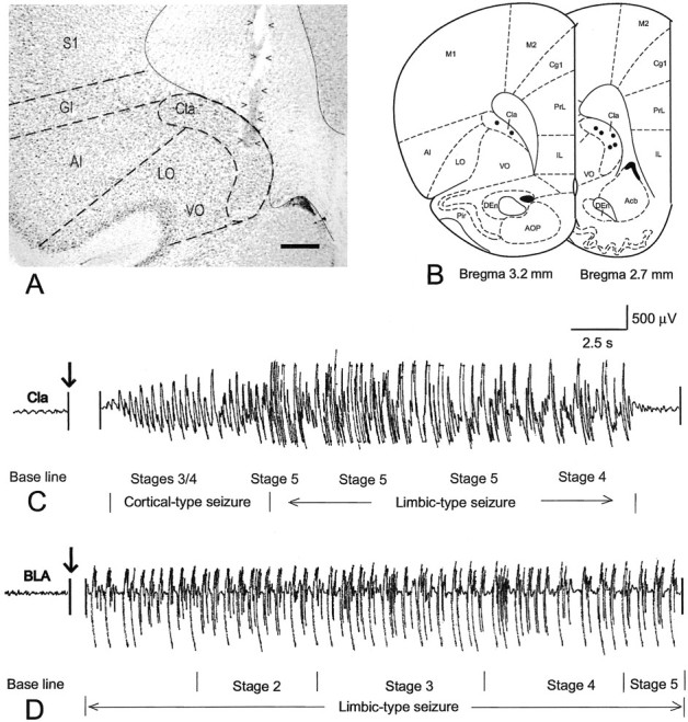
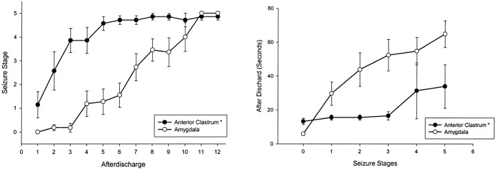
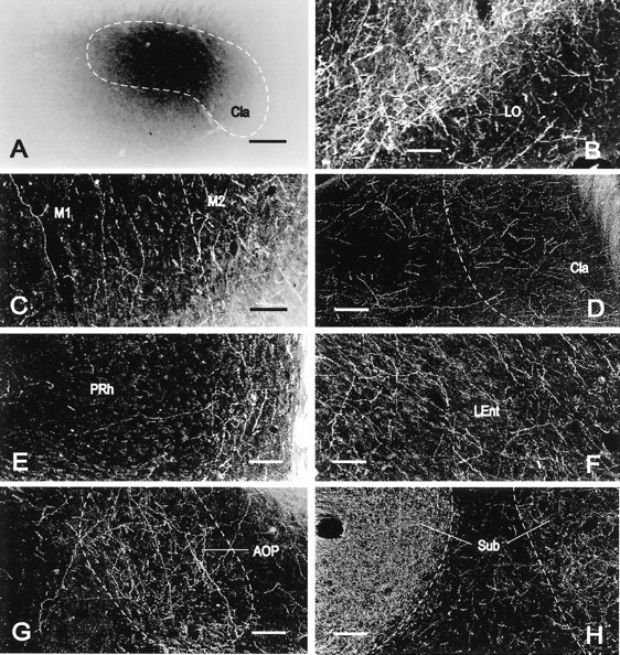
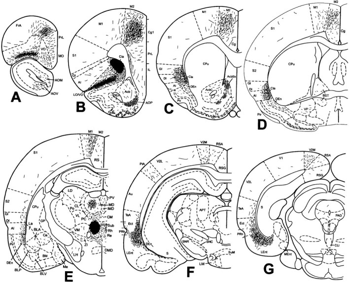
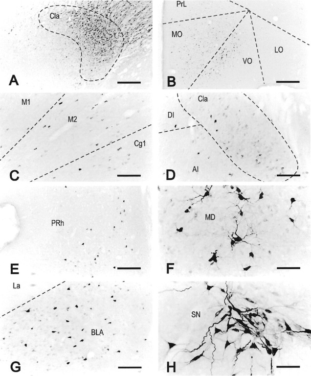
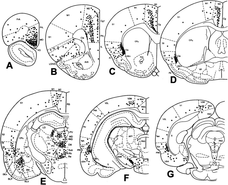
The claustrum has been implicated in the kindling of generalized seizures from limbic sites. We examined the susceptibility of the anterior claustrum itself to kindling and correlated this with an anatomical investigation of its afferent and efferent connections. Electrical stimulation of the anterior claustrum resulted in a pattern of rapid kindling with two distinct phases. Early kindling involved extremely rapid progression to bilaterally generalized seizures of short duration. With repeated daily kindling stimulations, early-phase generalized seizures abruptly became more elaborate and prolonged, resembling limbic-type seizures as triggered from the amygdala. We suggest that the rapid rate of kindling from the anterior claustrum is an indication that the claustrum is functionally close to the mechanisms of seizure generalization. In support of our hypothesis, we found significant afferent, efferent, and often reciprocal connections between the anterior claustrum and areas that have been implicated in the generation of generalized seizures, including frontal and motor cortex, limbic cortex, amygdala, and endopiriform nucleus. Additional connections were found with various other structures, including olfactory areas, nucleus accumbens, midline thalamus, and brainstem nuclei including the substantia nigra and the dorsal raphe nucleus. The anatomical connections of the anterior claustrum are consistent with its very high susceptibility to kindling and support the view that the claustrum is part of a forebrain network of structures participating in the generalization of seizures.


Figures

Fig. 1.
Fig. 1.
Kindling site and EEG pattern. A, Bright-field microphotograph showing the location of an implanted electrode. Arrowheads indicate the track of the electrode, with the electrode tip located within the anterior claustrum. B, Schematic representation of the location of electrode tips (black dots) in the anterior claustrum in the seven rats that received kindling stimulation. C, A typical EEG profile associated with a seizure kindled from the anterior claustrum. After electrical stimulation the rat almost immediately exhibited stages 3/4 seizures, several episodes of stage 5 seizures, and one episode of stage 4 seizures in sequence, which were accompanied by a relatively short period of cortical-type EEG seizure and a relatively long-lasting limbic-type seizure. D, A typical EEG profile of a seizure kindled from the basolateral amygdaloid nucleus. After stimulation the rat showed a limbic-type EEG seizure lasting for 2 min, whereas no obvious behavioral seizure appeared until 10 sec after stimulation, at which time the rat began to sequentially show stages 2, 3, 4, and 5 seizures. Acb, Accumbens nucleus; AI, agranular insular cortex;AOP, anterior olfactory nucleus, posterior part;Cg1, cingulate cortex, area 1; Cla, claustrum; DEn, dorsal endopiriform nucleus;GI, granular insular cortex; IL, infralimbic cortex; LO, lateral orbital cortex;M1, primary motor cortex; M2, secondary motor cortex; Pir, piriform cortex; PrL, prelimbic cortex; S1, primary somatosensory cortex;VO, ventral orbital cortex. Also see Results for a more complete account of the contralateral afferent connections. Scale bar, 520 μm.
Fig. 2.
Fig. 2.
Kindling profiles in the anterior claustrum and basolateral amygdaloid nucleus. A, Behavioral seizure stages plotted against the corresponding afterdischarges.B, Duration of afterdischarges plotted against motor seizures.
Fig. 3.
Fig. 3.
Localization of PHA-L immunoreactivity in the rat brain. Bright-field (A) and dark-field (BH) microphotographs of coronal sections showing an ejection site and the distribution of PHA-L-immunoreactive axons in representative brain regions in rat 99217. A, PHA-L occupied the major part of the anterior claustrum with minimal spread outside the morphological limits of the anterior claustrum. B, Layer II of the lateral orbital cortex was occupied with a high density of labeled fibers, whereas layer I exhibited low to moderate density of labeled axons.C, In contrast with the primary motor cortex (on theleft side of the image), with a low concentration of labeled fibers oriented perpendicularly to the cortical surface, many axons with a moderate density of varicosities were distributed with no specific direction in the secondary motor cortex (on the right side of the image). D, The middle part of the claustrum exhibited many fine, labeled axons, some of which ran laterally into the adjacent insular cortex toward the cortical surface.E, Although many fibers oriented parallel to the cortical surface in deep layers of the perirhinal cortex (on theright side of the image), some axons were oriented perpendicularly to the cortical surface in the superficial layers of the perirhinal cortex (on the left side of the image).F, Numerous axons with many varicosities ran dorsoventrally in deep layers of the posterior part of the lateral entorhinal cortex. G, Many labeled fibers were concentrated within the morphological limits of the posterior part of the anterior olfactory nucleus. H, The submedius thalamic nucleus ipsilateral to the ejection site (on the left side of the image) was fully occupied by an especially high density of numerous terminal-like axons, whereas the contralateral submedius thalamic nucleus (on the right side of the image) contained a much lower concentration of terminal-like axons.LEnt, Lateral entorhinal cortex; PRh, perirhinal cortex; Sub, submedius thalamic nucleus. All other abbreviations are as indicated in Figure 1. Scale bars:A, 290 μm; BH, 110 μm.
Fig. 4.
Fig. 4.
Projections of the anterior claustrum in rat 99217. Schematic representation of the density and localization of PHA-L-labeled fibers determined by immunohistochemistry was plotted from seven representative coronal sections corresponding to bregma 4.70, 2.70, 1.20, −0.26, −2.56, −4.80, and −6.80 mm (AG, respectively). Theirregular dark area in B indicates the ejection site, which is illustrated in Figure 3A. The full extent of labeled axons contralateral to the injection is not shown (see Results for more complete account of the contralateral projection). This schematic was generated on an IBM-compatible computer using Paxinos and Watson (1998). AcbC, Accumbens nucleus, core; AcbSh, accumbens nucleus, shell;AOM, anterior olfactory nucleus, medial part;AOV, anterior olfactory nucleus, ventral part;APT, anterior pretectal nucleus; Au, secondary auditory cortex; BLA, basolateral amygdaloid nucleus, anterior part; BLP, basolateral amygdaloid nucleus, posterior part; BLV, basolateral amygdaloid nucleus, ventral part; BM, basomedial amygdaloid nucleus; BST, bed nucleus of the stria terminalis;Ce, central amygdaloid nucleus; Cg, cingulate cortex; CL, centrolateral thalamic nucleus;Cli, caudal linear nucleus of the raphe;CM, central medial thalamic nucleus; CPu, caudate putamen; DI, dysgranular insular cortex;DMD, dorsomedial hypothalamic nucleus, dorsal part;Ect, ectorhinal cortex; FrA, frontal association cortex; GP, globus pallidus;IMD, intermediodorsal thalamic nucleus;La, lateral amygdaloid nucleus; LD, laterodorsal thalamic nucleus; LH, lateral hypothalamic area; LM, lateral mammillary nucleus; MD, mediodorsal thalamic nucleus; Me, medial amygdaloid nucleus; Ment, medial entorhinal cortex;MO, medial orbital cortex; PAG, periaqueductal gray; PC, paracentral thalamic nucleus;Po, posterior thalamic nuclear group;PtA, parietal association cortex; PV, paraventricular thalamic nucleus; Re, reuniens thalamic nucleus; Rh, rhomboid thalamic nucleus;RS, retrosplenial cortex; RSA, retrosplenial agranular cortex; RSG, retrosplenial granular cortex; S, subiculum; S2, secondary somatosensory cortex; SNC, substantia nigra, pars compacta; SNR, substantia nigra, pars reticulata;SuM, supramammillary nucleus; TeA, temporal association cortex; Tu, olfactory tubercle;V1, primary visual cortex; V2L, secondary visual cortex, lateral part; V2M, secondary visual cortex, medial part; VL, ventrolateral thalamic nucleus;VM, ventromedial thalamic nucleus; VP, ventral pallidum; ZI, zona incerta. All other abbreviations are as indicated in previous figures.
Fig. 5.
Fig. 5.
Localization of FG immunoreactivity in rat 99143. Representative bright-field microphotographs of coronal sections showing an ejection site that occupied the major part of the anterior claustrum (A) and the distribution of FG-immunoreactive neurons in the orbital cortex (B), motor and cingulate cortices (C), middle part of the claustrum (D), perirhinal cortex (E), central part of the mediodorsal thalamic nucleus (F), basolateral amygdaloid nucleus (G), and substantia nigra (H). The orientation of all images is such that the left side of the image is to the lateral side of the brain, right side to medial, and top side to dorsal. All other abbreviations are as indicated in previous figures. SN, Substantia nigra. Scale bars:A, B, 435 μm;CE, G, 174 μm;F, H, 87 μm.
Fig. 6.
Fig. 6.
Afferent connections of the anterior claustrum in rat 99143. The distribution of FG-immunoreactive cells was plotted onto a series of standard drawings of the rat brain (Paxinos and Watson, 1998) at the same levels as in Figure 4. The dark gray area in B indicates the ejection site, which is illustrated in Figure 5A. See Results for a more complete account of the contralateral afferent connections. For abbreviations, see previous figures.

Similar articles

Cited by

References

    1. Bayer SA, Altman J. Development of the endopiriform nucleus and the claustrum in the rat brain. Neuroscience. 1991;45:391–412. - PubMed
    1. Bayer SA, Altman J, Russo RJ, Zhang X. Timetables of neurogenesis in the human brain based on experimentally determined patterns in the rat. Neurotoxicology. 1993;14:83–144. - PubMed
    1. Beck CHM, Fibiger HC. Conditioned fear-induced changes in behavior and in the expression of the immediate early gene c-fos: with and without diazepam pretreatment. J Neurosci. 1995;15:709–720. - PMC - PubMed
    1. Beckstead RM. An autoradiographic examination of corticocortical and subcortical projections of the mediodorsal-projection (prefrontal) cortex in the rat. J Comp Neurol. 1979;184:43–61. - PubMed
    1. Behan M, Haberly LB. Intrinsic and efferent connections of the endopiriform nucleus in rat. J Comp Neurol. 1999;408:532–548. - PubMed

Publication types

MeSH terms

Substances