. 2001 May 1;20(9):2191-201.
doi: 10.1093/emboj/20.9.2191.

PACS-1 binding to adaptors is required for acidic cluster motif-mediated protein traffic

Affiliations

PACS-1 binding to adaptors is required for acidic cluster motif-mediated protein traffic

C M Crump et al. EMBO J. .

Abstract

PACS-1 is a cytosolic protein involved in controlling the correct subcellular localization of integral membrane proteins that contain acidic cluster sorting motifs, such as furin and human immunodeficiency virus type 1 (HIV-1) NEF: We have now investigated the interaction of PACS-1 with heterotetrameric adaptor complexes. PACS-1 associates with both AP-1 and AP-3, but not AP-2, and forms a ternary complex between furin and AP-1. A short sequence within PACS-1 that is essential for binding to AP-1 has been identified. Mutation of this motif yielded a dominant-negative PACS-1 molecule that can still bind to acidic cluster motifs on cargo proteins but not to adaptor complexes. Expression of dominant-negative PACS-1 causes a mislocalization of both furin and mannose 6-phosphate receptor from the trans-Golgi network, but has no effect on the localization of proteins that do not contain acidic cluster sorting motifs. Furthermore, expression of dominant-negative PACS-1 inhibits the ability of HIV-1 Nef to downregulate MHC-I. These studies demonstrate the requirement for PACS-1 interactions with adaptor proteins in multiple processes, including secretory granule biogenesis and HIV-1 pathogenesis.


Figures

None
Fig. 1. PACS-1 associates with AP-1 and AP-3 adaptor complexes. (A) BSC-40 cells were infected with wild-type or recombinant vaccinia viruses expressing PACS-1HA, and proteins were immunoprecipitated from cytosol with anti-HA antibodies. Immunoprecipitated samples and cell lysates were analyzed by western blotting using antisera specific to AP-1 (anti-γ-adaptin; row 1), AP-2 (anti-α-adaptin; row 2), AP-3 (anti-δ-adaptin; row 3) and the HA tag (HA-11; row 4). (B) A7 cells were fixed and co-stained with antibodies against PACS-1 and AP-1 (anti-γ-adaptin; a–c) or AP-3 (anti-AP-3; d–f). The co-localization of PACS-1 (green) with AP-1 or AP-3 (red) is shown in the merged images (c and f, respectively; examples of co-localization are indicated by arrows).
None
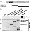
Fig. 2. AP-1 interacts with the FBR domain of PACS-1. (A) Schematic representation of PACS-1 and GST–PACS-1 fusion proteins (ARR, atrophin-1-related region; FBR, furin-binding region; MR, middle region; CTR, C-terminal region). Predicted coiled-coil domains are shown (zigzags). (B) GST–PACS-1 fusion proteins and GST alone were incubated with BSC-40 cytosol, isolated with glutathione resin and analyzed by western blotting using anti-γ-adaptin (upper panel). The load of the different GST proteins used is shown (lower panel).
None
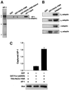
Fig. 3. PACS-1 interacts directly with purified AP-1, in vitro translated µ1 and σ1, and forms a ternary complex between the furin cytosolic domain, PACS-1 and AP-1. (A) SDS–PAGE and Coomassie Blue staining of purified AP-1 is shown (panel 1). GST–PACS-1FBR, GST–PACS-1FBR-Admut and GST alone were incubated with purified AP-1, isolated with glutathione resin and analyzed by western blotting using anti-γ-adaptin (panel 2). (B) GST–PACS-1FBR and GST alone were incubated with 35S-labeled, in vitro translated µ1-, σ1-, β1- and γ-adaptins, isolated with glutathione resin, separated by SDS–PAGE and analyzed by autoradiography. (C) GST–Fur-cd(DDD) (phosphoryl ation mimic mutant) was incubated with purified AP-1 in the presence or absence of Trx-PACS-1FBR, isolated with glutathione resin and analyzed by western blotting using anti-γ-adaptin. Chemiluminescent signals were quantified using the NIH gel analysis software and are expressed as arbitrary units normalized to non-specific GST signal. A representative blot is shown (lower panel; the background level of AP-1’s interaction with GST alone is due to the low stringency conditions required to maintain ternary complex formation in these assays).
None
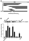
Fig. 4. Sequence determinants required for PACS-1 interaction with AP-1. (A) Schematic representation of the FBR region of PACS-1 (residues 117–266) and the various mutants generated (Δ1–Δ8 and Admut). (B) GST fusion proteins of each of these constructs were incubated with lysate, isolated with glutathione resin and analyzed by western blotting using anti-γ-adaptin. Interaction between each GST fusion protein and AP-1 was quantified using the NIH gel analysis software and is expressed relative to AP-1’s interaction with GST–PACS-1FBR.
None
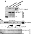
Fig. 5. Cargo binding and peptide competition. (A) GST–PACS-1FBR, GST–PACS-1FBR-Admut and GST alone were incubated with CK2-phosphorylated Trx-furin-cd, isolated with glutathione resin and analyzed by western blotting using anti-tetra-His. The load of the GST proteins used in these binding assays is shown (lower panel). (B) GST–PACS-1FBR and GST were incubated with lysate in the presence of 50 or 500 µM control peptide (Con-pep261–274) or competing peptide (ABP-pep166–179), isolated with glutathione resin and analyzed by western blotting using anti-γ-adaptin. The load of the GST proteins used in these binding assays is shown (lower panel).
None
Fig. 6. PACS-1Admut can not co-immunoprecipitate AP-1 or AP-3. BSC-40 cells were infected with wild-type or recombinant vaccinia viruses expressing PACS-1 or PACS-1Admut, and proteins were immunoprecipitated with anti-HA. Samples were analyzed by western blotting using antisera specific to AP-1 (anti-γ-adaptin; row 1), AP-3 (anti-δ-adaptin; row 2) and the HA-tag (HA-11; row 3).
None
Fig. 7. PACS-1 or PACS-1Admut expression does not affect endocytosis. (A) Cells were infected with wild-type or recombinant vaccinia viruses expressing PACS-1 or PACS-1Admut, or were mock infected. The rate of 125I-labeled transferrin internalization was monitored. Each time point was performed in quadruplicate and corrected for non-specific uptake. The following linear equations for each data set were determined, with the gradient functions representing the rate of transferrin uptake: PACS-1, y = 0.150x + 0.473; Admut, y = 0.162x + 0.254; wild-type, y = 0.155x + 0.341; mock infected, y = 0.158x + 0.400. (B) Cells were infected with wild-type or recombinant vaccinia viruses expressing PACS-1 or PACS-1Admut, or were mock infected. Cells were incubated with rhodamine-labeled transferrin, fixed and analyzed by fluorescence microscopy.
None
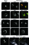
Fig. 8. Expression of dominant-negative PACS-1 disrupts the subcellular localization of furin and CI-MPR. A7 or AtT-20 cells were infected with recombinant vaccinia viruses expressing PACS-1 or PACS-1Admut alone (DL) or together with vaccinia viruses expressing FLAG-tagged furin (AC and MO). Cells were fixed, permeabilized and double labeled with anti-FLAG tag (M1; A–C and M–O) or anti-CI-MPR (D–F) and anti-TGN46 (C and F) or anti-ACTH (O). Further cells were labeled with anti-mannosidase II (G and J), anti-lamp-1 (H and K) or anti-γ-adaptin (I and L). Cells were analyzed by fluorescence microscopy, and merged images of green and red signals are shown (C, F and O inset).
None
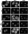
Fig. 9. Expression of dominant-negative PACS-1 disrupts the ability of HIV-1 Nef to downregulate MHC-I. HeLa-CD4 cells were infected with vaccinia virus alone (AC), viruses expressing Nef (DF) or NefEEEE65AAAA (GI), or co-infected with viruses expressing Nef and PACS-1 (JL) or Nef and PACS-1Admut (MO). Cells were fixed, permeabilized and labeled with anti-CD4 (column 1) or double labeled with anti-MHC-I (column 2) and anti-TGN46 (column 3) followed by fluorescently labeled secondary antisera. The expression of PACS-1 alone had no effect on MHC-I or CD4 localization (data not shown).

Similar articles

Cited by

References

    1. Alconada A., Bauer,U., Sodeik,B. and Hoflack,B. (1999) Intracellular traffic of herpes simplex virus glycoprotein gE: characterization of the sorting signals required for its trans-Golgi network localization. J. Virol., 73, 377–387. - PMC - PubMed
    1. Anderson E.D., Thomas,L., Hayflick,J.S. and Thomas,G. (1993) Inhibition of HIV-1 gp160-dependent membrane fusion by a furin-directed α1-antitrypsin variant. J. Biol. Chem., 268, 24887–24891. - PubMed
    1. Austin C., Hinners,I. and Tooze,S.A. (2000) Direct and GTP-dependent interaction of ADP-ribosylation factor 1 with clathrin adaptor protein AP-1 on immature secretory granules. J. Biol. Chem., 275, 21862–21869. - PubMed
    1. Chen H.J., Yuan,J. and Lobel,P. (1997) Systematic mutational analysis of the cation-independent mannose 6-phosphate/insulin-like growth factor II receptor cytoplasmic domain. An acidic cluster containing a key aspartate is important for function in lysosomal enzyme sorting. J. Biol. Chem., 272, 7003–7012. - PubMed
    1. Dell’Angelica E.C., Mullins,C. and Bonifacino,J.S. (1999) AP-4, a novel protein complex related to clathrin adaptors. J. Biol. Chem., 274, 7278–7285. - PubMed

Publication types

MeSH terms

Substances