. 2001 May 1;20(9):2214-23.
doi: 10.1093/emboj/20.9.2214.

Patched1 interacts with cyclin B1 to regulate cell cycle progression

Affiliations

Patched1 interacts with cyclin B1 to regulate cell cycle progression

E A Barnes et al. EMBO J. .

Abstract

The initiation of mitosis requires the activation of M-phase promoting factor (MPF). MPF activation and its subcellular localization are dependent on the phosphorylation state of its components, cdc2 and cyclin B1. In a two-hybrid screen using a bait protein to mimic phosphorylated cyclin B1, we identified a novel interaction between cyclin B1 and patched1 (ptc1), a tumor suppressor associated with basal cell carcinoma (BCC). Ptc1 interacted specifically with constitutively phosphorylated cyclin B1 derivatives and was able to alter their normal subcellular localization. Furthermore, addition of the ptc1 ligand, sonic hedgehog (shh), disrupts this interaction and allows cyclin B1 to localize to the nucleus. Expression of ptc1 in 293T cells was inhibitory to cell proliferation; this inhibition could be relieved by coexpression of a cyclin B1 derivative that constitutively localizes to the nucleus and that could not interact with ptc1 due to phosphorylation-site mutations to ALA: In addition, we demonstrate that endogenous ptc1 and endogenous cyclin B1 interact in vivo. The findings reported here demonstrate that ptc1 participates in determining the subcellular localization of cyclin B1 and suggest a link between the tumor suppressor activity of ptc1 and the regulation of cell division. Thus, we propose that ptc1 participates in a G(2)/M checkpoint by regulating the localization of MPF.


Figures
None
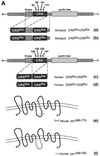
Fig. 1. (A) Schematic representation of cyclin B1 and patched1 constructs used in the yeast two-hybrid screen. (a–d) Cyclin B1 constructs containing a tandem repeat of the CRS domain. Ser residues (S) of the CRS domain were mutated to Ala or Glu to mimic the unphosphorylated or phosphorylated state of cyclin B1, respectively. (a and b) Constructs representing the Xenopus laevis CRS–CRS mutants with the destruction box (D-box) domain of cyclin B1 included. (c and d) Constructs representing the human CRS–CRS mutants. (e) Mouse ptc1 clone isolated from the two-hybrid screen. (f) Human ptc1 construct synthesized for further testing in the yeast two-hybrid system. (B) Schematic representation of cyclin B1 constructs used in mammalian cell studies. (a and b) Constructs representing the human CRS mutants. (c and d) Human CRS mutants with an appended NLS of Xenopus nucleoplasmin at the N-terminus. (e and f) Full-length Xenopus cyclin B1 derivatives with an appended NLS are represented.
None
Fig. 2. Patched1 intracellular loop binds endogenous cyclin B1. (A) Mammalian GST fusion genes encoding either GST alone (GST) or GST fused to the intracellular loop of ptc1 (GST–ptc1599–750) were transfected into 293T cells and immunoprecipitated with cyclin B1 antibody. Samples were analyzed by 10% SDS–PAGE and immunoblotted with mAb GST (upper panel) followed by mAb cyclin B1 (lower panel). (B) Expression of GST fusion proteins in lysates.
None
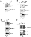
Fig. 3. Patched1 interacts with cyclin B1. (A) Endogenous ptc1 and endogenous cyclin B1 interact in 293T cells. Lysates were immuno precipitated as indicated. Upper panel, immunoblot of ptc1 shows ptc1 in the lysate and immunoprecipitate. Lane 3 indicates the use of ptc1 blocking peptide to discriminate antibody specificity. Lower panel, immunoblot of cyclin B1 showing cyclin B1 in lysate and ptc1 immunoprecipitate, but not the peptide blocked lane. (B) Patched1 binds endogenous cyclin B1. 293T cells were transfected with Myc–ptc1, immunoprecipitated with cyclin B1 antibody and immuno blotted as indicated. Lane 3 indicates the use of cyclin B1 blocking peptide to discriminate antibody specificity. (C) Patched1, cyclin B1 and cdc2 form a complex. 293T cells were transfected with Myc–ptc1, immunoprecipitated with cdc2 antibody and immunoblotted as indicated. (D) Active MPF complex associates with patched1. 293T cells were transfected with Myc–ptc1 as indicated. Upper panel, lysates were immunoprecipitated and assayed for histone H1 kinase activity. Middle panel, anti-Myc immunoblot displaying expression of Myc–ptc1. Lower panel, anti-cdc2 immunoblot displaying the presence of cdc2 in lane 1 (positive control) and lane 3 (ptc1-transfected cells). The lower band represents the IgG band from mouse Myc antiserum as detected by mouse cdc2 antiserum.
None
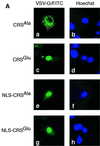
Fig. 4. Patched1 colocalizes with the CRSGlu domain of cyclin B1 at the cell membrane. (A) Localization of human CRS and NLS–CRS constructs in COS-1 cells detected by immunofluorescence. (a) CRSAla construct expressed in cytoplasm. (c) CRSGlu construct expressed in nucleus. (e and g) NLS–CRSAla and NLS–CRSGlu constructs expressed in nucleus. (b, d, f and h) Nuclei detected with Hoechst dye. (B) Localization of Myc-tagged ptc1 in COS-1 cells detected by immunofluorescence. (a) Mock-transfected cell. (c) Myc–ptc1 expressed at membrane. (b and d) Nuclei detected with Hoechst dye. (C) Altered localization of NLS–CRSGlu due to cotransfection of ptc1. (a) NLS–CRSAla remains nuclear in the presence of ptc1 expression (b). In contrast (d), NLS–CRSGlu associates with the cell membrane due to ptc1 expression (e). (f) NLS–CRSGlu and ptc1 colocalization. (g, h and i) Sonic hedgehog restores nuclear accumulation of NLS–CRSGlu. (g) NLS–CRSGlu retains nuclear localization in the presence of ptc1 (h) with shh-N exposure. (c and i) Nuclei detected with Hoechst dye.
None
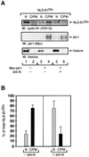
Fig. 5. Cyclin B1 localization mediated by patched1 expression and shh exposure. (A) Upper panel, expression of NLS–B1Glu protein in cellular fractions. Middle panel, cytoplasmic/plasma membrane (C/PM) expression of ptc1 in cotransfected cells. Lower panel, nuclear (N) expression of endogenous histone proteins. (B) Quantitation of data presented in lanes 3–6 of (A) representing the average of three independent cellular fractionation experiments. The standard deviation is shown.
None
Fig. 6. Anti-proliferative function of the cyclin B1–ptc1 complex. (A) Growth curve of 293T cells transfected with the indicated constructs. There is a decrease in cell proliferation with cotransfection of ptc1 and NLS–B1Glu. Error bars represent the standard error of the mean. (B) Upper panel, expression of Myc-tagged ptc1 in cell lysates obtained from the last day of the growth curve. Lower panel, expression of VSV-G-tagged cyclin B1 derivatives in cell lysates. (C) Decrease in mitotic index in cells cotransfected with ptc1 and NLS–B1Glu. The percentage of cells showing a mitotic phenotype from three independent transfections with the standard deviation is shown. (D) Increase in cell proliferation with cellular exposure to shh-N. The average value of two experiments is shown.
None
Fig. 7. Model of patched1 regulation of MPF. G1 phase, cdc2 is present in abundance throughout the cell cycle. S phase, cyclin B1 is synthesized and begins to accumulate in the cytoplasm by late S phase. Cyclin B1 binds to cdc2 via a region termed the cyclin box. G2 phase, before forming an active MPF complex, cyclin B1–cdc2 enters and rapidly exits the nucleus due to a NES located within the CRS domain. G2/M transition, cyclin B1 becomes phosphorylated, which creates a fully active MPF complex (MPF*). ptc1 binds to phosphorylated cyclin B1 and sequesters an active MPF complex in pre-mitotic cells which inhibits the translocation of MPF to the nucleus. Cellular exposure to shh promotes the degradation of ptc1, which in turn facilitates the release of cyclin B1. MPF is now available for nuclear import.

Similar articles

Cited by

References

    1. Borgne A. and Meijer,L. (1996) Sequential dephosphorylation of p34cdc2 on Thr-14 and Tyr-15 at the prophase/metaphase transition. J. Biol. Chem., 271, 27847–27854. - PubMed
    1. Borgne A., Ostvold,A.C., Flament,S. and Meijer,L. (1999) Intra-M phase-promoting factor phosphorylation of cyclin B at the prophase/metaphase transition. J. Biol. Chem., 274, 11977–11986. - PubMed
    1. Carpenter D., Stone,D.M., Brush,J., Ryan,A., Armanini,M., Frantz,G., Rosenthal,A. and de Sauvage,F.J. (1998) Characterization of two patched receptors for the vertebrate hedgehog protein family. Proc. Natl Acad. Sci. USA, 95, 13630–13634. - PMC - PubMed
    1. Chen Y. and Struhl,G. (1996) Dual roles for patched in sequestering and transducing hedgehog. Cell, 87, 553–563. - PubMed
    1. Chidambaram A., Goldstein,A.M., Gailani,M.R., Gerrard,B., Bale,S.J., DiGiovanna,J.J., Bale,A.E. and Dean,M. (1996) Mutations in the human homologue of the drosophila/patched gene in caucasian and African-American nevoid basal cell carcinoma syndrome patients. Cancer Res., 56, 4599–4601. - PubMed

Publication types

MeSH terms