. 2001 May 1;20(9):2236-45.
doi: 10.1093/emboj/20.9.2236.

Association and spreading of the Drosophila dosage compensation complex from a discrete roX1 chromatin entry site

Affiliations

Association and spreading of the Drosophila dosage compensation complex from a discrete roX1 chromatin entry site

Y Kageyama et al. EMBO J. .

Abstract

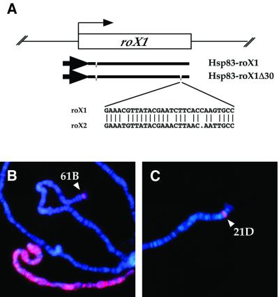
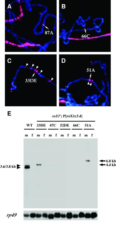
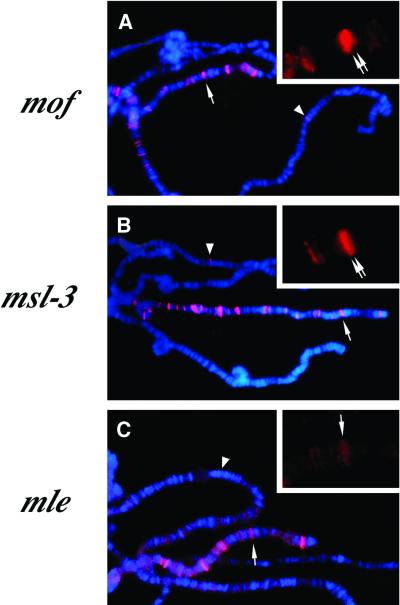
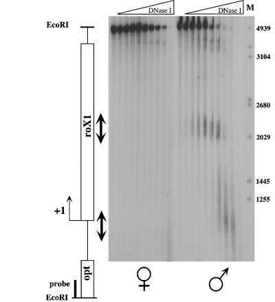
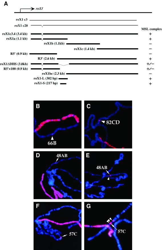
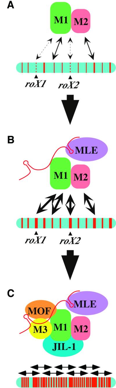
In Drosophila, dosage compensation is controlled by the male-specific lethal (MSL) complex consisting of MSL proteins and roX RNAs. The MSL complex is specifically localized on the male X chromosome to increase its expression approximately 2-fold. We recently proposed a model for the targeted assembly of the MSL complex, in which initial binding occurs at approximately 35 dispersed chromatin entry sites, followed by spreading in cis into flanking regions. Here, we analyze one of the chromatin entry sites, the roX1 gene, to determine which sequences are sufficient to recruit the MSL complex. We found association and spreading of the MSL complex from roX1 transgenes in the absence of detectable roX1 RNA synthesis from the transgene. We mapped the recruitment activity to a 217 bp roX1 fragment that shows male-specific DNase hypersensitivity and can be preferentially cross-linked in vivo to the MSL complex. When inserted on autosomes, this small roX1 segment is sufficient to produce an ectopic chromatin entry site that can nucleate binding and spreading of the MSL complex hundreds of kilobases into neighboring regions.


Figures

None
Fig. 1. A cDNA fragment of roX1 recruits the MSL complex. (A) Diagram of the 3.4 kb roX1c20 cDNA driven by the Hsp83 promoter and a deletion derivative lacking a 30 bp segment that shares 25/30 identity with roX2. An intron near the 5′ end is indicated in each construct. The sequence comparison of roX1 and roX2 in the 30 bp region deleted in roX1Δ30 is shown below. (B) Polytene chromosomes from a [Hsp83–roX1]61B male. (C) Polytene chromosomes from a [Hsp83–roX1Δ30]21D male. Chromosome squashes were immuno stained with rabbit anti-MSL1 (red) and counterstained with 4′,6- diamine-2-phenylindole (DAPI) (blue). Both nuclei show a strong site of MSL localization at the insertion site of the respective transgenes (arrowheads). A heavily stained X chromosome is also seen in (B).
None
Fig. 2. Localization of MSL complexes to roX1c3.4 transgenes. MSL1 localization in male nuclei, detected by immunostaining with anti-MSL1 antibodies (red). (A) Expressed antisense construct [Hsp83–roX1AS]87A, (B) [roX1c3.4]66C, from which transcription has not been detected, (C) [roX1c3.4]33DE, (D) [roX1c3.4]51A. The [roX1c3.4] transgenes show MSL1 binding at the insertion sites of the transgenes (B–D, arrows), as well as in neighboring regions (C and D, arrowheads). (E) Northern analysis shows fusion RNA expression of roX1 from flanking sequences in the [roX1c3.4]33DE and [roX1c3.4]51A lines, in which relatively frequent spreading was also observed. All transgenic lines carry a mutant allele (roX1ex6) at their endogenous roX1 locus. The [roX1c3.4]66C, 52DE and 47C lines do not express detectable roX1 RNA. The wild-type strain (y w; WT) expresses endogenous roX1 RNA.
None
Fig. 3. MSL complex binding at roX1 transgenes requires MLE. Females carrying [Hsp83–MSL2] and [roX1c3.4]51A in addition to being homozygous mutant for mof (A), msl-3 (B) or mle (C). Polytene chromosomes were stained with anti-MSL1 antibodies (red) and DAPI (blue). In each of these mutants the MSL complex associates with the X chromosome only at chromatin entry sites. Arrowheads and arrows represent the roX1 transgene and the endogenous roX1 locus at 3F, respectively. Top right inset in each panel shows an enlarged image of anti-MSL1 immunostaining of the endogenous roX1 locus.
None
Fig. 4. DNase I hypersensitivity within the roX1 locus. DNase I cleavages were mapped within an EcoRI fragment containing the roX1 gene, schematized to the left (open box; +1 indicates the approximate 5′ end of the gene). Nuclei from female (left) or male (right) adult flies were treated with increasing concentrations of DNase I. DNA was isolated and digested to completion with EcoRI. DNase I cleavages were revealed by Southern blotting using a probe adjacent to an EcoRI site within the opt gene. Arrows highlight male-specific DNase I hypersensitive sites in roX1 chromatin. M, DNA size markers (bp).
None
Fig. 5. Deletion analysis used to map MSL complex-recruiting activity within roX1 transgenes. (A) Diagram of the roX1 fragments tested in transgenic assays. (B–G) Male polytene chromosomes from each transgenic strain were immunostained with anti-MSL1 (red) and counterstained by DAPI (blue). The integration site of each transgene is represented by an arrowhead. Two constructs that share a 300 bp overlap and show strong anti-MSL staining were [roX1a]66B (B) and [roX1.R3′]82CD (C). A 217 bp fragment [roX1-S]48AB (D and E) and a 9× tandem repeat of roX1-S [roX1-SM]57C, F and G) also showed MSL1 localization to the transgene insertion site. Spreading from these small roX1 derivatives is seen in (E) and (G) (arrowheads).
None
Fig. 6. Mapping of in vivo interactions of MSL proteins within the roX1 gene by chromatin immunoprecipitation. (A) Restriction map of roX1 cDNA. (B) Raw data of a representative experiment. Left panel: SYBR gold stained agarose gel containing various fragments derived from the roX1 cDNA. The cDNA was excised from pBluescript with XhoI and XbaI and gel purified (lane 8). This fragment was further digested with XmnI (lane 1), RsaI (lane 2), Esp3I–ScaI (lane 3), Esp3I–BstEII (lane 4), BanII–NsiI (lane 5), HhaI–HindIII (lane 6), EcoRI–HindIII (lane 7). Center panel: Southern blot probed with amplified DNA isolated by immunoprecipitation of SL-2 chromatin with anti-MSL2 antibody. Right panel: Southern blot using a DNA probe isolated from a mock immunoprecipitation of SL-2 chromatin (lacking a specific antibody). (C) Summary of one representative experiment. Each column shows the normalized hybridization profile obtained for the chromatin immunoprecipitation of the protein indicated above the column. Each row shows the data derived from one roX1 digest. Row 1, XmnI (see lane 1, B); row 2, RsaI (see lane 2, B); row 3, HhaI–HindIII (see lane 6, B). The y-axes of the diagrams show 1/100th of the normalized PhosphorImager counts. Note the scale changes between columns. DHS, position of the DNase I hypersensitive site as mapped in Figure 4. Asterisks indicate two fragments that could not be separated on the gel. The value indicated assumes that both fragments contribute the same signal, which is not necessarily the case.
None
Fig. 7. Model for MSL complex assembly and spreading on the X chromosome. (A) MSL1 and MSL2 show a weak interaction with most chromatin entry sites in the absence of MLE, but roX1 and roX2 are particularly poor targets under these conditions. (B) When MLE is present, roX2 RNA can be incorporated into complexes, potentially altering the conformation to allow strong binding to chromatin entry sites. (C) When complete complexes are present, containing at least five MSL proteins and one or more roX RNAs, binding and spreading occur in a banded pattern along the length of the X chromosome.

Similar articles

Cited by

References

    1. Akhtar A. and Becker,P.B. (2000) Activation of transcription through histone H4 acetylation by MOF, an acetyltransferase essential for dosage compensation in Drosophila. Mol. Cell, 5, 367–375. - PubMed
    1. Akhtar A., Zink,D. and Becker,P.B. (2000) Chromodomains are protein–RNA interaction modules. Nature, 407, 405–409. - PubMed
    1. Amrein H. and Axel,R. (1997) Genes expressed in neurons of adult male Drosophila. Cell, 88, 459–469. - PubMed
    1. Bone J.R., Lavender,J., Richman,R., Palmer,M.J., Turner,B.M. and Kuroda,M.I. (1994) Acetylated histone H4 on the male X chromosome is associated with dosage compensation in Drosophila. Genes Dev., 8, 96–104. - PubMed
    1. Church G.M. and Gilbert,W. (1984) Genomic sequencing. Proc. Natl Acad. Sci. USA, 81, 1991–1995. - PMC - PubMed

Publication types

MeSH terms

LinkOut - more resources