. 2001 May 1;20(9):2254-72.
doi: 10.1093/emboj/20.9.2254.

TGF-beta-induced repression of CBFA1 by Smad3 decreases cbfa1 and osteocalcin expression and inhibits osteoblast differentiation

Affiliations

TGF-beta-induced repression of CBFA1 by Smad3 decreases cbfa1 and osteocalcin expression and inhibits osteoblast differentiation

T Alliston et al. EMBO J. .

Abstract

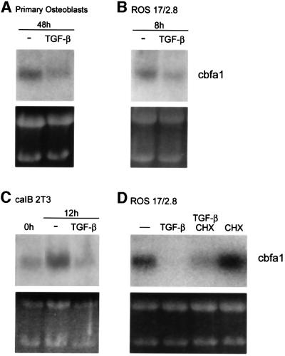
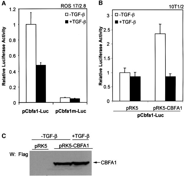
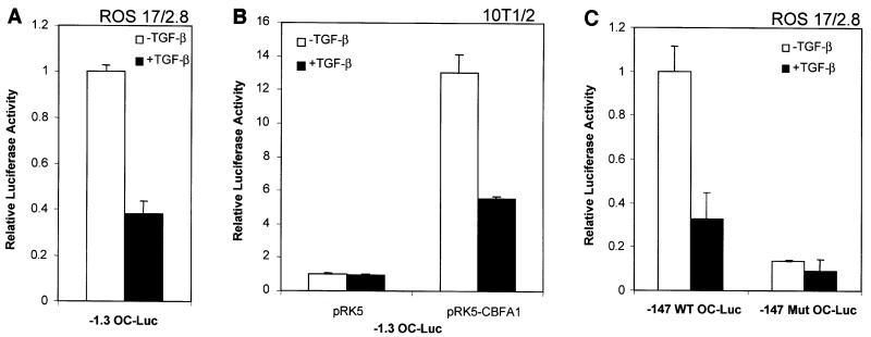
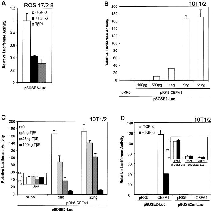
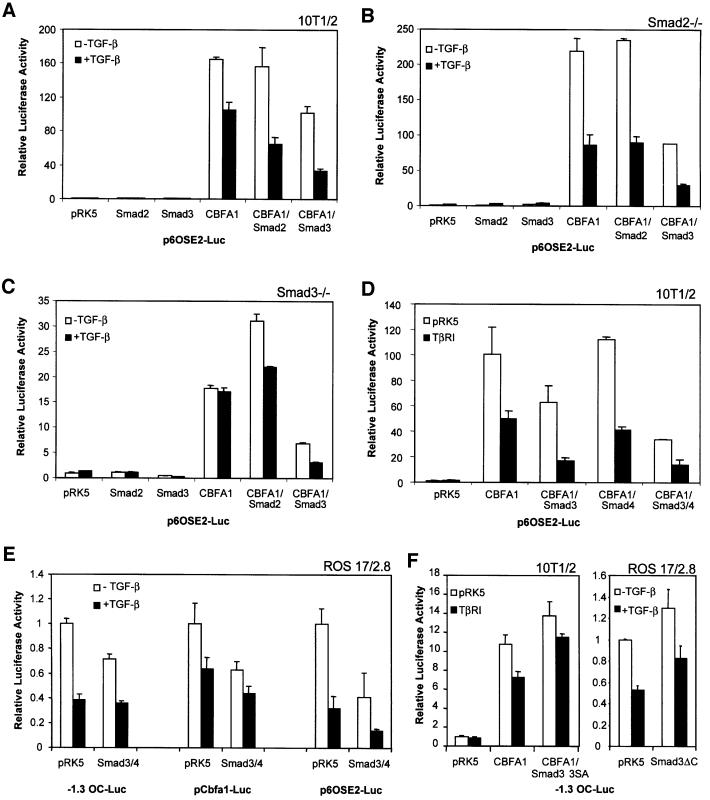
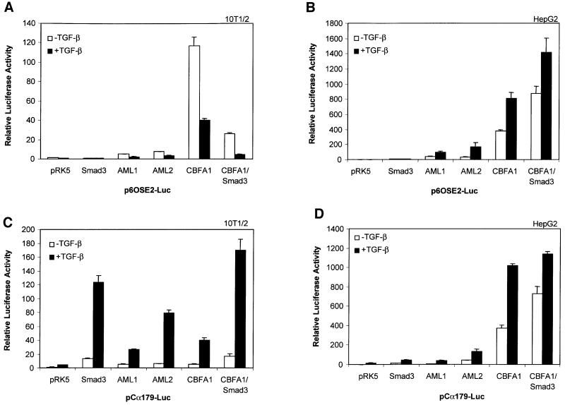
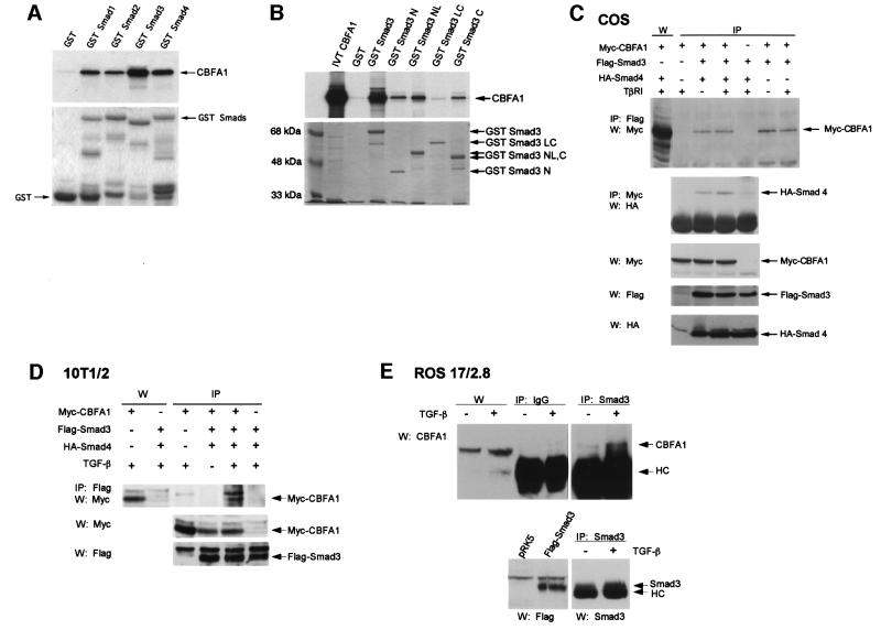
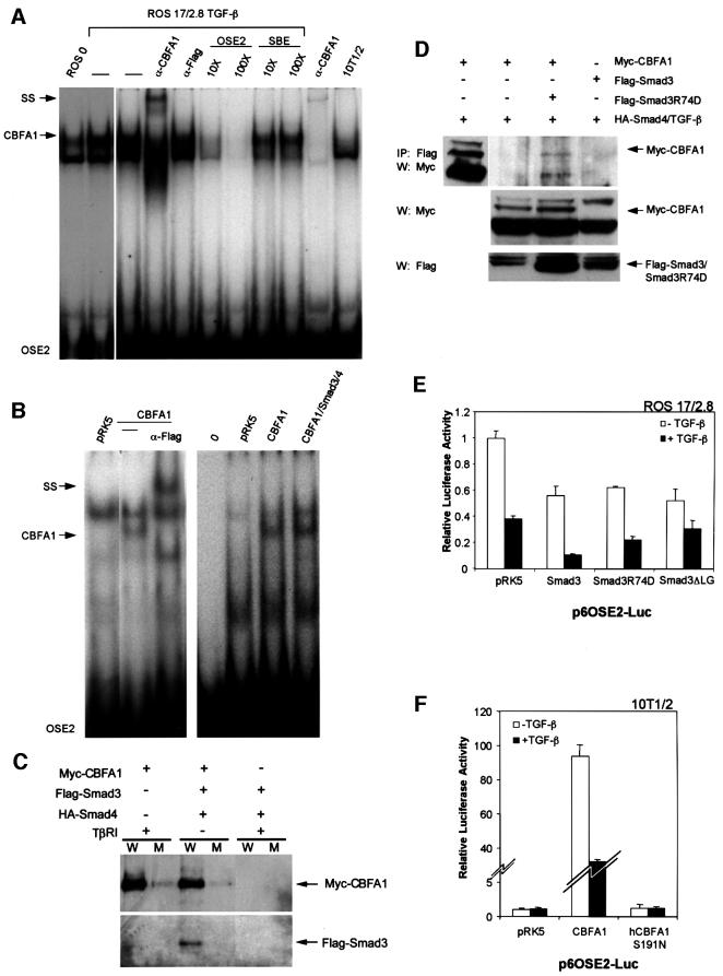
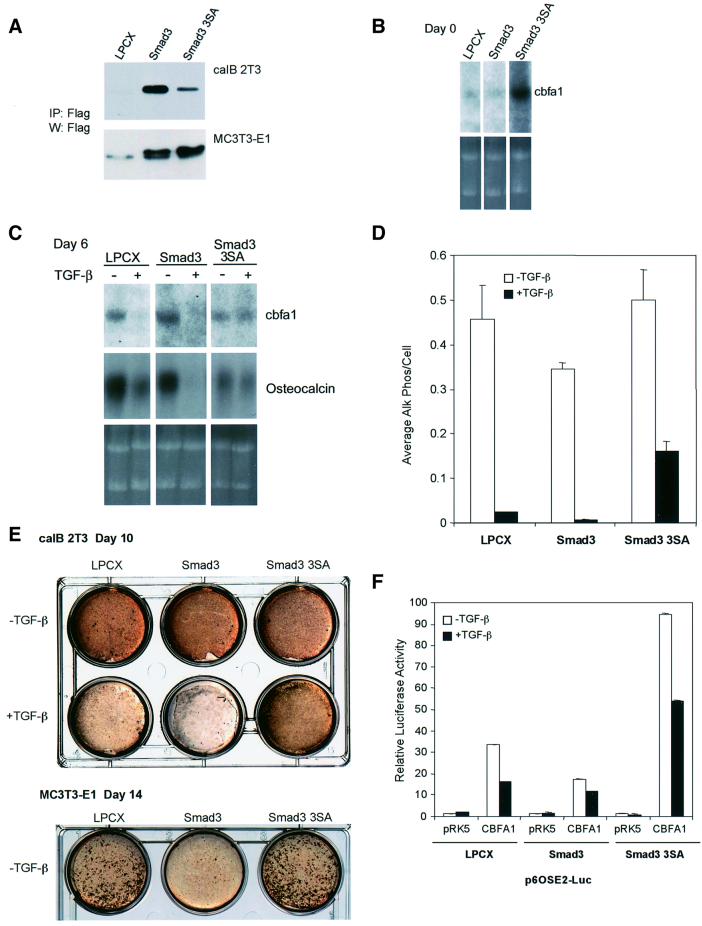
Transforming growth factor-beta (TGF-beta), a secreted factor present at high levels in bone, inhibits osteoblast differentiation in culture; yet, the mechanism of this inhibition remains unclear. We studied the effects of TGF-beta and its effectors, the Smads, on the expression and function of the osteoblast transcription factor CBFA1. TGF-beta inhibited the expression of the cbfa1 and osteocalcin genes, whose expression is controlled by CBFA1 in osteoblast-like cell lines. This inhibition was mediated by Smad3, which interacts physically with CBFA1 and represses its transcriptional activity at the CBFA1-binding OSE2 promoter sequence. The repression of CBFA1 function by Smad3 contrasts with previous observations that Smads function as transcription activators. This repression occurred in mesenchymal but not epithelial cells, and depended on the promoter sequence. Smad3-mediated repression of CBFA1 provides a central regulatory mechanism for the inhibition of osteoblast differentiation by TGF-beta, since it inhibits both cbfa1 transcription and transcriptional activation of osteoblast differentiation genes by CBFA1. Altering Smad3 signaling influenced osteoblast differentiation in the presence or absence of TGF-beta, implicating Smad3/TGF-beta-mediated repression in autocrine regulation of osteoblast differentiation.


Figures

None
Fig. 1. TGF-β inhibits the expression of cbfa1 mRNA independently of new protein synthesis. The cbfa1 mRNA, visualized following northern blot hybridization and autoradiography, is shown. (A) Primary mouse calvarial osteoblasts cultured for 48 h in differentiation medium in the presence of TGF-β (5 ng/ml) express reduced levels of cbfa1 mRNA. (B) Treatment with TGF-β for 8 h decreases the level of cbfa1 mRNA in ROS 17/2.8 cells. (C) TGF-β treatment of caIB 2T3 cells during the 12 h period in differentiation medium represses cbfa1 mRNA expression. (D) ROS 17/2.8 cells were treated with TGF-β for 8 h as in (B) in the absence or presence of cycloheximide (CHX) (10 µg/ml) or with cycloheximide alone. Ethidium bromide-stained gels are included to show similar loading of RNA.
None
Fig. 2. TGF-β inhibits transcription from the cbfa1 promoter. ROS 17/2.8 (A) or 10T1/2 (B) cells were transfected with the pCbfa1-Luc or pCbfa1m-Luc reporter plasmids and expression plasmids, as indicated. 10T1/2 cells (B) were cotransfected with the CBFA1 expression plasmid (pRK5-CBFA1) or the control empty plasmid (pRK5). Cells were cultured in the presence or absence of TGF-β (1 ng/ml) 16 h after transfection. Forty-eight hours after transfection, cells were harvested and reporter activities measured. Values normalized for transfection efficiency are shown as fold induction relative to basal promoter activity as described in Materials and methods. (C) Whole 10T1/2 cell lysates were prepared from a parallel transfection experiment for visualization of CBFA1 protein levels by western analysis using Flag antibody.
None
Fig. 3. Inhibition of transcription from the osteocalcin promoter by TGF-β requires CBFA1. ROS 17/2.8 (A and C) and 10T1/2 (B) cells were transfected with the designated osteocalcin promoter/reporter plasmids followed by treatment with or without TGF-β (1 ng/ml). Luciferase expression was scored as in Figure 2. 10T1/2 cells (B) were cotransfected with the CBFA1 expression plasmid (pRK5-CBFA1) or the control empty plasmid (pRK5).
None
Fig. 4. TGF-β inhibits transcription from the OSE2 sequence by CBFA1. (A) Transiently transfected ROS 17/2.8 cells were treated with TGF-β (1 ng/ml; black bar) or cotransfected with an expression plasmid for a constitutively activated form of the TGF-β type I receptor (TβRI) (gray bar). (B) 10T1/2 cells were transfected with different quantities of pRK5-CBFA1. (C) 10T1/2 cells were transfected with 5 or 25 ng of pRK5-CBFA1, or the empty vector pRK5, in the presence of increasing quantities of TβRI expression plasmid. The inset shows the lack of effect of increased TβRI expression on basal promoter activity in 10T1/2 cells (note difference in scale). (D) A reporter plasmid containing six copies of a mutant OSE2 site, p6OSE2m-Luc, has reduced basal and CBFA1-inducible transactivation (see inset), when compared with p6OSE2-Luc. Luciferase expression was scored as in Figure 2.
None
Fig. 5. Smad3 inhibits CBFA1 function and mediates the inhibitory effect of TGF-β (1 ng/ml). Cells were transiently transfected with expression plasmids and luciferase reporter plasmids, as shown. Luciferase expression from the reporter plasmids was scored as in Figure 2. (A, D and F) 10T1/2 cells, (B) Smad2–/– mouse embryonic fibroblasts, (C) Smad3–/– mouse embryonic fibroblasts, (E and F) ROS 17/2.8 cells.
None
Fig. 6. Effects of Smad3 and TGF-β on the function of CBFA1 and runt family transcription factors depend on the cell type and promoter. 10T1/2 cells (A and C) or HepG2 cells (B and D) were transiently transfected with the indicated expression plasmids and the luciferase reporter plasmids p6OSE2-Luc (A and B) or pCα179-Luc (C and D). Transactivation from these promoters was measured in the absence or presence of TGF-β (1 ng/ml). Luciferase expression from the reporter plasmids was scored as in Figure 2.
None
Fig. 7. CBFA1 interacts with Smad3. (A and B) Interaction of 35S-labeled, in vitro translated CBFA1 with GST–Smad fusion proteins, as shown. Interacting 35S-labeled CBFA1 (arrow) is visualized following gel electrophoresis and autoradiography. Five percent of the input reaction volume of 35S-labeled CBFA1 (IVT CBFA1) was loaded as control in lane 1 of (B). Below the autoradiograms are photographs of Coomassie Blue-stained gels to show the integrity and equal loading of the fusion proteins. (C) Interaction of Smad3 and Smad4 with CBFA1 in vivo, as assessed using co-immunoprecipitations. Cell lysates from transfected COS-1 cells expressing the indicated tagged proteins were used for immunoprecipitations (IP), followed by western blotting (W). The top panel shows the interaction of Myc-CBFA1 with Flag-Smad3, as assessed by immunoprecipitations with anti-Flag antibody-coupled Sepharose beads, followed by western blotting using anti-Myc antibody. The first lane visualizes Myc-tagged CBFA1 by western blotting without prior immunoprecipitation. The second panel shows interaction of HA-Smad4 with Myc-CBFA1, as assessed by immunoprecipitations with anti-Myc antibody, followed by western blotting using anti-HA antibody. The three lower panels show the relative expression levels of the tagged proteins in each lysate, as assessed by western blotting. (D) Interaction of Flag-Smad3 and Myc-CBFA1 in transfected 10T1/2 cells in the presence or absence of TGF-β (5 ng/ml), as assessed in (C). The first two lanes show the expression of Myc-tagged CBFA1, visualized by western blotting without prior immunoprecipitation. The two lower panels visualize the expression of Myc-CBFA1 and Flag-Smad3 by western analysis. (E) Interaction of endogenous CBFA1 and endogenous Smad3 in ROS 17/2.8 cells. This interaction was detectable by immunoprecipitation using a Smad3 antibody, followed by anti-CBFA1 western blotting, in TGF-β-treated (5 ng/ml), but not in untreated ROS 17/2.8 cell lysates. No CBFA1 was detected when rabbit anti-mouse IgG was used instead of anti-Smad3 antibody. The IgG heavy chain band is marked HC. CBFA1 levels in the lysates were detected by western analysis (left two lanes), whereas Smad3 levels were visualized by Smad3 immunoprecipitation followed by western analysis with a Smad3 antibody (lower panel, right two lanes), using Flag-Smad3 expressed in transfected 10T1/2 cells as reference (lower panel, left two lanes).
None
Fig. 8. Binding of CBFA1 and Smad3 to the OSE2 DNA sequence. (A) Electrophoretic mobility shift assays using untreated (lane 1) or TGF-β-treated (5 ng/ml; lane 2) ROS 17/2.8 cell extracts allowed visualization of endogenous CBFA1 binding (arrow) to a 32P-labeled OSE2 oligonucleotide. The identity of the CBFA1-containing complex was confirmed by its absence in 10T1/2 cells, which lack CBFA1 expression (lane 10), and its partial disappearance and supershift (SS) following incubation with an anti-CBFA1 antibody (α-CBFA1, lane 4), but not with an unrelated antibody (α-Flag, lane 5). Unlabeled OSE2 competitor oligonucleotide, at 10- to 100-fold molar excess, competed (lanes 6 and 7), whereas an unrelated competitor oligonucleotide did not compete (lanes 8 and 9) for the radiolabeled OSE2–CBFA1 complex. (B) Electrophoretic mobility shift assays show CBFA1 binding to an OSE2 oligonucleotide, both in the absence or presence of Smad3 and Smad4. COS-1 cells were transfected with expression plasmids for Flag-CBFA1, with or without Smad3/4 (as shown) or the pRK5 control plasmid. The radiolabeled OSE2 oligonucleotide was incubated alone (0) or in the presence of cell lysates. Expression of Flag-CBFA1 resulted in the formation of a distinct band (lanes 2 and 6), which could be supershifted (SS) using an anti-Flag antibody (lane 3). This OSE2–CBFA1 complex was also apparent in the presence of overexpressed Smad3/4 (lane 7). (C) Binding of CBFA1 to an OSE2 oligonucleotide, in the absence or presence of equimolar levels of Smad3 and Smad4. Biotinylated wild-type (W) or mutant (M) OSE2 oligonucleotides were incubated with the same lysates as in Figure 7C, and the interactions of Myc-CBFA1 and Flag-Smad3 with the oligonucleotides were assessed by western blotting. CBFA1 binds to the wild-type oligonucleotide, both in the presence or absence of Smad3/4, but not to the mutant oligonucleotide. Furthermore, Smad3 interacts with the OSE2 nucleotide only in the presence of CBFA1. (D) The DNA binding-defective Smad3R74D interacts with CBFA1 in transfected 10T1/2 cells. Immunoprecipitation assays, performed as in Figure 7C and D, revealed coprecipitation of Myc-CBFA1 (arrow) with Flag-Smad3R74D (top panel). Western analysis without prior immunoprecipitation visualized the migration of Myc-CBFA1 (top panel, left lane), and the expression levels of CBFA1 and Smad3 or Smad3 R74D (lower panels). (E) DNA binding of Smad3 is not essential for TGF-β/Smad3-mediated repression of transcription from the OSE2 sequence by CBFA1. ROS 17/2.8 cells were transfected with the p6OSE2-Luc reporter and wild-type or mutant Smad3 expression plasmids. Luciferase expression in the absence or presence of TGF-β(1 ng/ml) was scored as in Figure 2. (F) DNA binding of CBFA1 is essential for transcriptional activity and Smad3-mediated repression. 10T1/2 cells were transfected with the p6OSE2-Luc reporter and an expression plasmid for wild-type CBFA1 or the DNA binding-defective mutant hCBFA1 S191N. Luciferase expression in the absence or presence of TGF-β (1 ng/ml) was scored as in Figure 2.
None
Fig. 9. Stable expression of wild-type or dominant-negative Smad3 in caIB 2T3 cells and MC3T3-E1 cells alters osteoblast differentiation. (A) Immunoprecipitation followed by western blot analysis demonstrates expression of Flag-tagged Smad3 or Smad3-3SA in cell lysates of stably infected caIB 2T3 and MC3T3-E1 cells. LPCX is the control empty vector. (B) Alterations in Smad3 signaling affect endogenous cbfa1 mRNA expression in caIB 2T3 cells at day 0, i.e. prior to exposure to differentiation conditions, in LPCX control cells, or cells stably expressing Smad3 or Smad3-3SA. The top panel shows cbfa1 northern hybridizations, while the lower panel shows the ethidium bromide-stained gel. (C) Alterations in Smad3 signaling affect endogenous cbfa1 and osteocalcin mRNA expression in differentiating caIB 2T3 cells at day 6 in differentiation conditions, in the absence or presence of TGF-β. The top panels show hybridizations for cbfa1 or osteocalcin mRNA using RNA from LPCX control cells or cells stably expressing Smad3 or Smad3-3SA, while the lower panel shows the ethidium bromide-stained gel. (D) Alterations in Smad3 signaling affect alkaline phosphatase activity in caIB 2T3 cells and the extent of inhibition by TGF-β (5 ng/ml). Cells were incubated in differentiation medium for 6 days in the presence or absence of TGF-β (5 ng/ml). Cell lysates were then assayed for alkaline phosphatase activity. Values are expressed per cell. (E) Alterations in Smad3 signaling affect matrix mineralization in caIB 2T3 and MC3T3-E1 cells and the extent of inhibition by TGF-β (5 ng/ml) in caIB 2T3 cells. Confluent cells were incubated for 10 (caIB 2T3 cells) or 14 days (MC3T3-E1 cells) in differentiation medium, in the presence or absence of TGF-β, and then stained for mineralization (brown) using the von Kossa method. (F) Alterations in Smad3 signaling affect the transactivation of the p6OSE2-reporter by CBFA1. Stably infected LPCX control or stable caIB 2T3 cells expressing Smad3 or Smad3-3SA were transiently transfected with the p6OSE2-Luc reporter plasmid, with or without an expression plasmid for CBFA1 (pRK5-CBFA1). Luciferase expression in the absence or presence of TGF-β (1 ng/ml) was scored as in Figure 2.

Similar articles

Cited by

References

    1. Akiyoshi S., Inoue,H., Hanai,J., Kusanagi,K., Nemoto,N., Miyazono,K. and Kawabata,M. (1999) c-Ski acts as a transcriptional co-repressor in transforming growth factor-β signaling through interaction with Smads. J. Biol. Chem., 274, 35269–35277. - PubMed
    1. Alliston T.N. and Derynck,R. (2000) Transforming growth factor-β in skeletal development and maintenance. In Canalis,E. (ed.), Skeletal Growth Factors. Lippincott Williams & Wilkins, Philadelphia, PA, pp. 233–249.
    1. Alliston T.N., Maiyar,A.C., Buse,P., Firestone,G.L. and Richards,J.S. (1997) Follicle stimulating hormone-regulated expression of serum/glucocorticoid-inducible kinase in rat ovarian granulosa cells: a functional role for the Sp1 family in promoter activity. Mol. Endocrinol., 11, 1934–1949. - PubMed
    1. Aronson B.D., Fisher,A.L., Blechman,K., Caudy,M. and Gergen,J.P. (1997) Groucho-dependent and -independent repression activities of Runt domain proteins. Mol. Cell. Biol., 17, 5581–5587. - PMC - PubMed
    1. Ausubel F.M., Brent,R., Kingston,R.E., Moore,D.D., Seidman,J.G., Smith,J.A. and Struhl,K. (1994) Current Protocols in Molecular Biology. John Wiley & Sons, New York, NY.

Publication types

MeSH terms