. 2001 May 1;20(9):2293-303.
doi: 10.1093/emboj/20.9.2293.

RNA binding in an Sm core domain: X-ray structure and functional analysis of an archaeal Sm protein complex

Affiliations

RNA binding in an Sm core domain: X-ray structure and functional analysis of an archaeal Sm protein complex

I Törö et al. EMBO J. .

Abstract

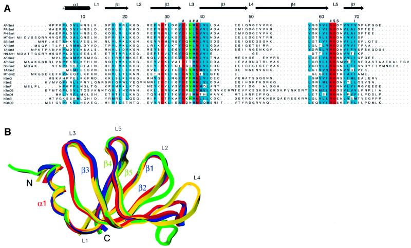
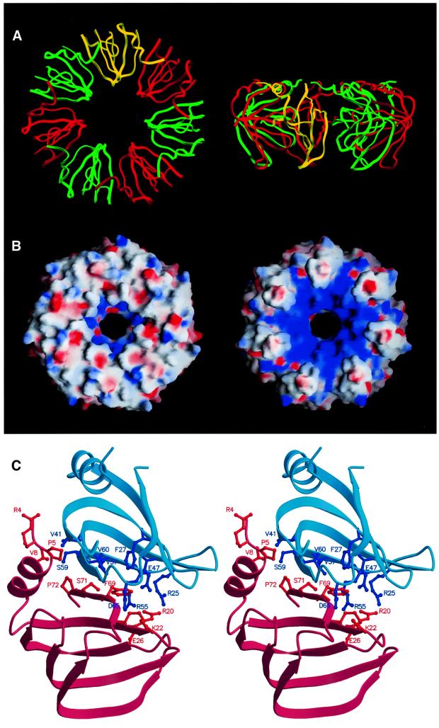
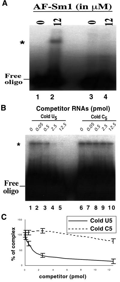
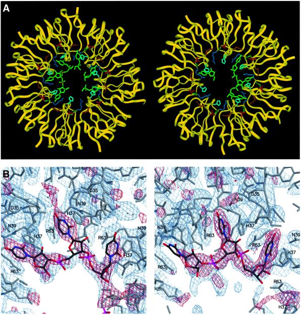
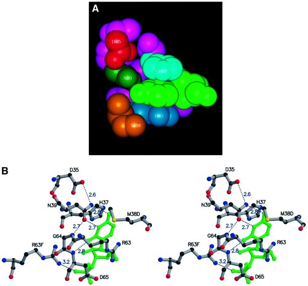
Eukaryotic Sm and Sm-like proteins associate with RNA to form the core domain of ribonucleoprotein particles involved in pre-mRNA splicing and other processes. Recently, putative Sm proteins of unknown function have been identified in Archaea. We show by immunoprecipitation experiments that the two Sm proteins present in Archaeoglobus fulgidus (AF-Sm1 and AF-Sm2) associate with RNase P RNA in vivo, suggesting a role in tRNA processing. The AF-Sm1 protein also interacts specifically with oligouridylate in vitro. We have solved the crystal structures of this protein and a complex with RNA. AF-Sm1 forms a seven-membered ring, with the RNA interacting inside the central cavity on one face of the doughnut-shaped complex. The bases are bound via stacking and specific hydrogen bonding contacts in pockets lined by residues highly conserved in archaeal and eukaryotic Sm proteins, while the phosphates remain solvent accessible. A comparison with the structures of human Sm protein dimers reveals closely related monomer folds and intersubunit contacts, indicating that the architecture of the Sm core domain and RNA binding have been conserved during evolution.


Figures

None
Fig. 1. The Sm fold is conserved between Archaea and eukaryotes. (A) Sequence alignment of archaeal Sm proteins with the human canonical Sm (hSm) proteins G, E, F, D2, D1, B and D3. Shown on the top is the secondary structure assignment in AF-Sm1 as determined by X-ray analysis. Loop regions are labelled L1–L5, β-strands β1–β5; α1 denotes the N-terminal α-helix. β-strands 1, 2 and 3 constitute the first, and strands 4 and 5 the second half of the bipartite Sm domain. Residues fully (N39) or almost fully conserved throughout the Sm and Lsm protein family are marked in red, with highly conserved residues in blue. Residues forming the uracil-binding pocket are labelled ‘#’ or ‘$’ (if they interact through their main chain amide groups); His37, in stacking contact with the base, and the corresponding tyrosine or phenylalanine residues in human Sm proteins, are shown in green. The names of the archaeal proteins indicate the species (AF, Archaeoglobus fulgidus; PA, Pyrococcus abyssi; PH, Pyrococcus horikoshi; MT, Methanobacterium thermoautotrophicum; SS, Sulfolobus solfataricus; AP, Aeropyrum pernix; TA, Thermoplasma acidophilum; HN, Halobacterium sp. NRC-1) and the subfamily (Sm1- or Sm2-type). (B) Superposition of the Cα traces of the archaeal PA-Sm1 (our unpub lished results), AF-Sm1 and AF-Sm2 proteins with human D3 (Kambach et al., 1999a) shown in red, blue, green and yellow, respectively. Differences are restricted to the N- and C-termini as well as the loop regions, in particular loop 4.
None
Fig. 2. Structure of the AF-Sm1 heptamer. (A) Ribbon representation of the AF-Sm1 heptamer (top and side view). For clarity, the monomers are drawn alternately in red and green, and one monomer is depicted in yellow. (B) Electrostatic surface charge potential showing the two faces of the seven-membered ring. Shown on the left is the side binding the RNA and containing the N-terminal helix (corresponding to the top view shown in A). It is relatively flat, while the other side exhibits pronounced positively charged grooves emanating from the centre (as indicated by the blue colour). The figure was produced with GRASP (Nicholls et al., 1991). (C) Dimer contacts in the AF-Sm1 heptamer. The molecules are shown as ribbons of different colours for the pair of interacting molecules. Side chains involved in contacts (<4.0 Å), mainly located on β-strands 4 and 5 as well as the N-terminal helix, are represented in ball-and-stick mode. The figure was produced with MOLSCRIPT (Kraulis, 1991).
None
Fig. 3. In vitro binding of oligo(U) to AF-Sm1. (A) Direct binding assay by gel shift. Radiolabelled RNA was incubated with or without AF-Sm1 and complexes were resolved following native gel electro phoresis. U5, but not C5, produces a bandshift. A 75 fmol concentration of 32P-labelled oligouridine (lanes 1 and 2) or oligocytidine (lanes 3 and 4) was incubated alone (lanes 1 and 3) or with 1 µg of purified AF-Sm1 (lanes 2 and 4). (B) Competition experiments demonstrate the specificity of the interaction. Various concentrations of cold oligonucleotides (indicated above each lane) were incubated with the 32P-labelled oligouridine (U5) probe and 12 µM AF-Sm1. Complex formation was assayed following native gel electrophoresis. The data demonstrate successful competition by U5 (lanes 1–5) and not by C5 (lanes 6–10). This reveals specific binding of AF-Sm1 to U5. (C) Quantification of competition experiments. Cold U5 displaces AF-Sm1 from 32P-labelled U5 (solid line, n = 2). In comparison, cold C5 does not displace it except when it is present in great excess (dashed line, n = 2). Error bars are derived from two independent experiments. The slower migrating band (marked by *) corresponds to the protein–RNA complex while the faster migrating band corresponds to the free probe.
None
Fig. 4. The AF-Sm1–U5 complex. (A) The two AF-Sm1 heptamers in the asymmetric unit are shown in a ribbon plot representation (yellow), and the bound oligonucleotides and residues forming the uracil-binding pocket in stick mode, with the oligo(U) coloured green, Asp35 red, His37 light blue, Met38 gold, Asn39 dark green and Arg63 blue. Continuous electron density is present for a trinucleotide as well as for three isolated uridines in the first ring (shown on the left), and for a trinucleotide and a single uridine in the second ring. (B) Portions of the 2FoFc (blue) and FoFc (red) omit maps not including the RNA, contoured at 1σ and 2.6σ, respectively (ring 1 on the left). Atom-type colouring is used for the oligo(U), while the protein is shown in grey. Residues interacting with the bases are labelled.
None
Fig. 5. The base-binding pocket. van der Waals (A) and ball-and-stick stereo (B) representation of the residues forming the uracil-binding pocket. The colour code in (A) is as in Figure 4A. The uracil is sandwiched between His37 and Arg63 and forms specific hydrogen bonds with Asn39 and the backbone NH group of Asp65 (distances in Å are indicated). Note that M38D and R63F belong to neighbouring protein subunits.
None
Fig. 6. Sm proteins from A.fulgidus are expressed in vivo and co-precipitate. (A) Antibodies directed against AF-Sm2 do not cross-react with AF-Sm1. A 0.5 µg aliquot of recombinant AF-Sm1 (Input, lane 1) was incubated with protein A-coated agarose beads that had been previously coupled to antibodies directed against AF-Sm1 (α-AF-Sm1, lanes 4 and 5), AF-Sm2 (α-AF-Sm2, lanes 6 and 7) or, as control, no antibodies (lanes 2 and 3). The AF-Sm1 protein present in the pellet (P, lanes 3, 5 and 7) and supernatant (S, lanes 2, 4 and 6) fractions of each immunoprecipitation reaction was detected by western blotting following gel electrophoresis using α-AF-Sm1 antibodies. Smearing of the AF-Sm1 protein results from incomplete denaturation in standard gel conditions (data not shown). No cross-reaction of α-AF-Sm2 antibodies with AF-Sm1 was observed. (B) Antibodies directed against AF-Sm1 do not cross-react with AF-Sm2. The procedure described in (A) was repeated using 0.5 µg of AF-Sm2 and detection with α-AF-Sm2 antibodies. Smearing of the AF-Sm2 protein results from incomplete denaturation in standard gel conditions (data not shown). No cross-reaction of α-AF-Sm1 antibodies with AF-Sm2 was observed. (C) Total lysate from A.fulgidus cells was prepared as indicated in Materials and methods and used in immunoprecipitation reactions (IP) with α-AF-Sm1 (lane 3), α-AF-Sm2 (lane 5) and the corresponding pre-immune serum (PI α-AF-Sm1 and PI α-AF-Sm2, lanes 2 and 4, respectively). An aliquot of the total lysate before precipitation was loaded in lane 1. The presence of AF-Sm1 and AF-Sm2 in the various fractions was detected by western blotting with antibodies against AF-Sm1 or AF-Sm2.
None
Fig. 7. RNase P RNA is co-immunoprecipitated by Sm proteins. (A) RNA samples were extracted from immunoprecipitated fractions and analysed by pCp labelling as described in Materials and methods. Arrows indicate two bands found reproducibly in the immuno precipitated fraction obtained with the antibodies against AF-Sm1 or AF-Sm2. (B) Northern blotting with an RNase P RNA-specific probe was carried out with the RNA obtained as described in Materials and methods. The same bands were detected in independent experiments using the same as well as two other RNase P RNA probes.
None
Fig. 8. RNase P RNA 5′ and 3′ end mapping. (A) RNA samples were prepared as previously described and analysed by primer extension with ST14 (see Materials and methods). A sequence ladder was obtained with the same primer and the cloned RNase P RNA gene as a template to map the 5′ end. (B) The two RNA species differ at their 3′ ends. RNA samples were analysed by S1 nuclease mapping with an overlapping 3′ end fragment. The 3′ end determination was done according to the molecular size of the band. (C) The RNase P RNA sequence obtained from 5′ and 3′ end determination. Sequences in bold letters correspond to nucleotides not present in the sequence provided at the RNase P database web site. Nucleotides in italic correspond to the 3′ end of the probe used for the S1 mapping. The 5′ end corresponds to the end determined by primer extension. Short and long 3′ end forms correspond to those determined by S1 mapping.

Similar articles

Cited by

References

    1. Achsel T., Brahms,H., Kastner,B., Bachi,A., Wilm,M. and Lührmann,R. (1999) A doughnut-shaped heteromer of human Sm-like proteins binds to the 3′-end of U6 snRNA, thereby facilitating U4/U6 duplex formation in vitro. EMBO J., 18, 5789–5802. - PMC - PubMed
    1. Boeck R., Lapeyre,B., Brown,C.E. and Sachs,A.B. (1998) Capped mRNA degradation intermediates accumulate in the yeast spb8-2 mutant. Mol. Cell. Biol., 18, 5062–5072. - PMC - PubMed
    1. Bouveret E., Rigaut,G., Shevchenko,A., Wilm,M. and Séraphin,B. (2000) An Sm-like protein complex that participates in mRNA degradation. EMBO J., 19, 1661–1671. - PMC - PubMed
    1. Branlant C., Krol,A., Ebel,J.P., Lazar,E., Haendler,B. and Jacob,M. (1982) U2 RNA shares a structural domain with U1, U4 and U5 RNAs. EMBO J., 1, 1259–1265. - PMC - PubMed
    1. Brown J.W. (1999) The ribonuclease P database. Nucleic Acids Res., 27, 314. - PMC - PubMed

Publication types

MeSH terms