. 2001 May 1;20(9):2315-25.
doi: 10.1093/emboj/20.9.2315.

A proline-rich protein binds to the localization element of Xenopus Vg1 mRNA and to ligands involved in actin polymerization

Affiliations

A proline-rich protein binds to the localization element of Xenopus Vg1 mRNA and to ligands involved in actin polymerization

W M Zhao et al. EMBO J. .

Abstract

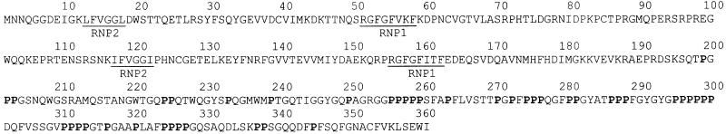
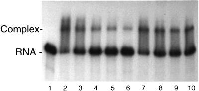
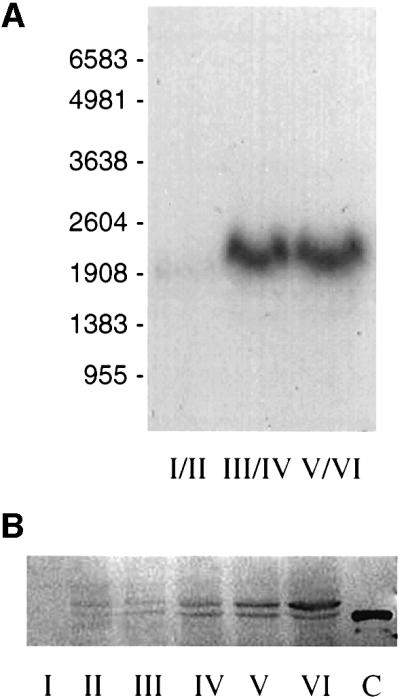
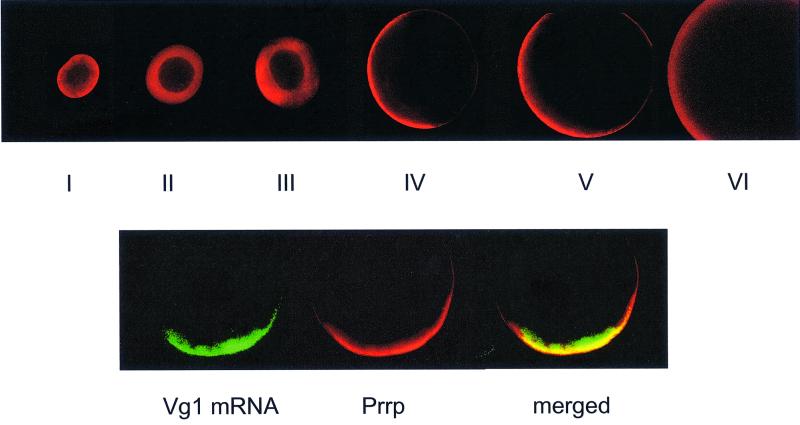
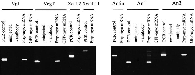
A 340 nucleotide element within the 3' untranslated region of Vg1 mRNA determines its localization to the vegetal cortex of Xenopus oocytes. To identify protein factors that bind to this region, we screened a cDNA expression library with an RNA probe containing this sequence. Five independent isolates encoded a protein (designated Prrp for proline-rich RNA binding protein) having two RNP domains followed by multiple polyproline segments. Prrp and Vg1 mRNAs are co-localized to the vegetal cortex of stage IV oocytes, substantiating an interaction between the two in vivo. Prrp also associates with VegT mRNA, which like Vg1 mRNA uses the late localization pathway, but not with Xcat-2 or Xwnt-11 mRNAs, which use the early pathway. The proline-rich domain of Prrp interacts with profilin, a protein that promotes actin polymerization. Prrp can also associate with the EVH1 domain of Mena, another microfilament-associated protein. Since the anchoring of Vg1 mRNA to the vegetal cortex is actin dependent, one function of Prrp may be to facilitate local actin polymerization, representing a novel function for an RNA binding protein.


Figures

None
Fig. 1. The predicted amino acid sequence of Prrp. The RNP1 and RNP2 core sequences that comprise the two RNP motifs are underlined and proline residues in the C-terminal half of the protein are indicated in bold. The entire nucleotide sequence, including flanking UTRs, can be found at DDBJ/EMBL/GenBank accession No. AY028920.
None
Fig. 2. Mobility shift assays for binding of Prrp to VLE RNA. Prrp (8 nM) and internally radiolabeled VLE RNA (1 nM) were incu bated with 0, 1, 5, 10 or 25 nM unlabeled VLE RNA (lanes 2–6, respectively) or 5, 25, 50 or 100 nM non-cognate RNA (lanes 7–10, respectively). Lane 1 contains VLE RNA only.
None
Fig. 3. Temporal expression of Prrp. (A) Oocytes were separated into three groups according to the designated Dumont stages and total RNA isolated for northern blot analysis. Each lane contains 10 oocyte equivalents of RNA. The positions of RNA size standards (nt) are indicated. (B) Staged oocytes were homogenized and two oocyte equivalents were run per lane in a western blot assay. The primary antibody is against a 22-amino-acid segment between the two RNP domains of Prrp. Lane C is a sample of Prrp expressed in E.coli and has six additional His residues at the C-terminal end of the protein.
None
Fig. 4. Localization of Prrp during oogenesis. Top panel: staged oocytes were processed for immunocytochemical analysis using a primary antibody prepared against a peptide derived from Prrp (amino acids 93–114) and visualization using a secondary antibody conjugated with Alex Fluor 568. In each case, the oocyte is oriented with the vegetal pole to the left. Bottom panel: stage IV oocytes were used for whole-mount in situ hybridization with an RNA probe labeled with Alexa Fluor 488-5-UTP. This was followed by immunocytochemical analysis with Prrp antibody. An optical confocal section was viewed in the green channel to detect Vg1 mRNA or in the red channel to detect Prrp. A merge of the two images demonstrates the co-localization of Prrp and Vg1 mRNA.
None
Fig. 5. In vivo RNA binding assays. Oocytes were injected with mRNA encoding myc-tagged Prrp. After an overnight incubation, cells were disrupted and Prrp was immunoprecipitated. Associated RNA was reverse transcribed into cDNA and then amplified by PCR using the gene-specific primers denoted by the bar above the relevant lanes in order to test for the presence of the indicated mRNA. For each mRNA tested, a standard was generated using total oocyte mRNA as the template for RT–PCR (PCR control). Control reactions included uninjected oocytes (uninjected), precipitation with protein A–Sepharose resin not coupled with myc antibody (– antibody) and oocytes injected with mRNA encoding myc-tagged GFP (GFP–myc mRNA).
None
Fig. 6. The proline-rich domain of Prrp interacts with profilin. Protein samples separated on an SDS–polyacrylamide gel (A) were transferred to a nitrocellulose filter that was incubated in a solution containing 1 µM Xenopus profilin. Profilin that remained associated with the filter after washing was detected by a colorimetric immunochemical assay (B). Lane 1, full-length Prrp with a C-terminal His tag; lane 2, RNA binding domain of Prrp fused to maltose binding domain; lane 3, ribosomal protein L5 with a C-terminal His tag; lane 4, L5 fused to maltose binding protein. (C) A colony-lift filter assay was used to measure interactions between the designated bait–prey combinations in a yeast two-hybrid system. Each column represents three independently selected colonies. 1, positive control (p53/T antigen); 2, Prrp/Xenopus profilin; 3, C-terminal (proline-rich) domain of Prrp/Xenopus profilin; 4, negative control (lamC/T antigen); 5, N-terminal (RNA binding) domain of Prrp/Xenopus profilin; 6, Prrp/prey vector; 7, bait vector/Xenopus profilin; 8, Prrp/yeast profilin.
None
Fig. 7. The interaction of Prrp with other polyproline binding domains. A colony-lift assay was used to measure the interaction between the C-terminal (proline-rich) domain of Prrp and the designated prey. Each column contains three independently selected colonies. 1, positive control (p53/T antigen); 2, WW domain of YAP; 3, SH3 domain of Abl; 4, EVH1 domain of Mena; 5, Xenopus profilin.

References

    1. Ahern-Djamali S.M., Bachmann,C., Hua,P., Reddy,S.K., Kastenmeier, A.S., Walter,U. and Hoffmann,F.M. (1999) Identification of profilin and src homology 3 domains as binding partners for Drosophila enabled. Proc. Natl Acad. Sci. USA, 96, 4977–4982. - PMC - PubMed
    1. Ashley C.T. Jr, Wilkinson,K.D., Reines,D. and Warren,S.T. (1993) FMR1 protein: conserved RNP family domains and selective RNA binding. Science, 262, 563–566. - PubMed
    1. Bashirullah A., Cooperstock,R.L. and Lipshitz,H.D. (1998) RNA localization in development. Annu. Rev. Biochem., 67, 335–394. - PubMed
    1. Bassell G. and Singer,R.H. (1997) mRNA and cytoskeletal filaments. Curr. Opin. Cell Biol., 9, 109–115. - PubMed
    1. Beach D.L., Salmon,E.D. and Bloom,K. (1999) Localization and anchoring of mRNA in budding yeast. Curr. Biol., 9, 569–578. - PubMed

Publication types

MeSH terms

Associated data