. 2001 May 1;20(9):2326-37.
doi: 10.1093/emboj/20.9.2326.

Multiple roles for the C-terminal domain of eIF5 in translation initiation complex assembly and GTPase activation

Affiliations

Multiple roles for the C-terminal domain of eIF5 in translation initiation complex assembly and GTPase activation

K Asano et al. EMBO J. .

Abstract

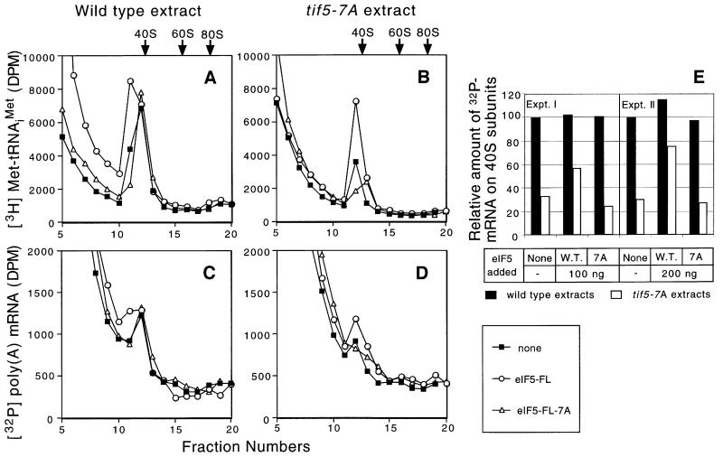
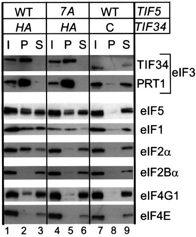
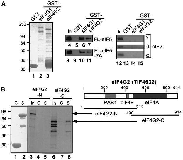
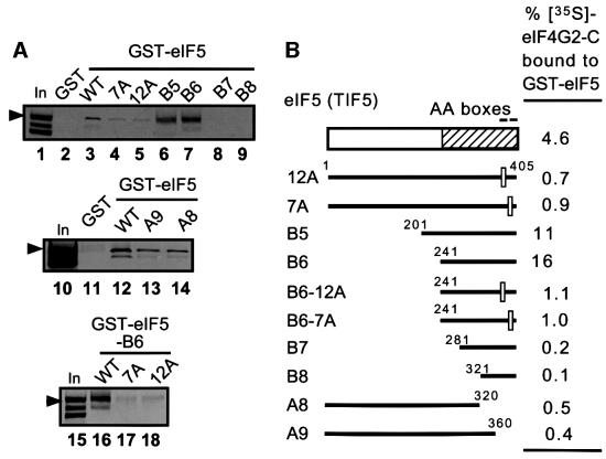
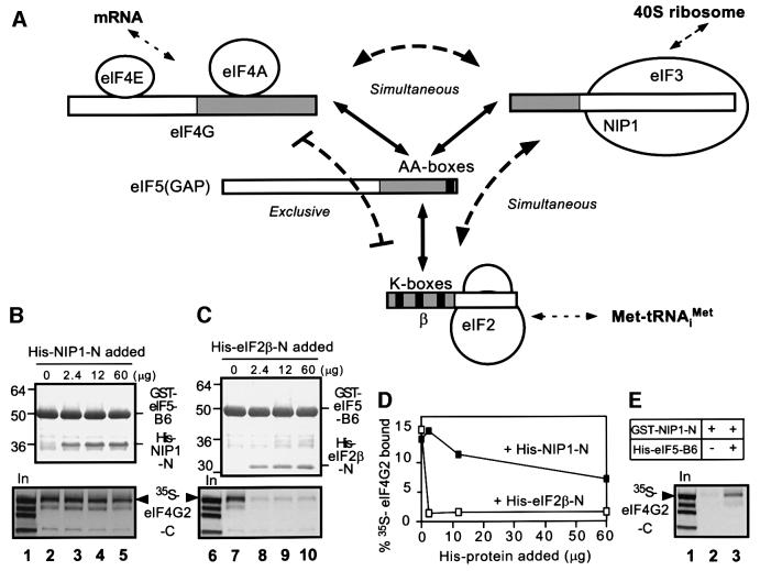
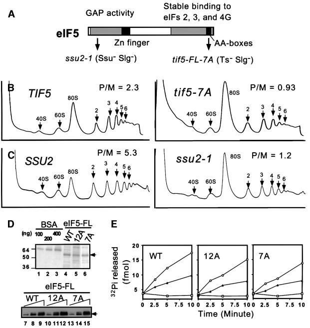
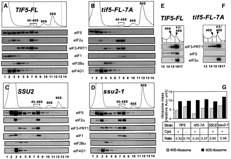
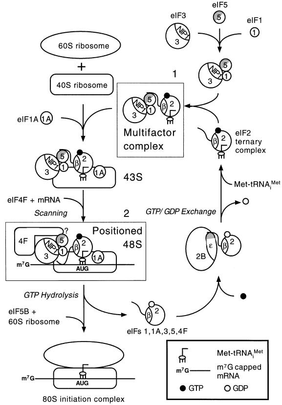
eIF5 stimulates the GTPase activity of eIF2 bound to Met-tRNA(i)(Met), and its C-terminal domain (eIF5-CTD) bridges interaction between eIF2 and eIF3/eIF1 in a multifactor complex containing Met-tRNA(i)(Met). The tif5-7A mutation in eIF5-CTD, which destabilizes the multifactor complex in vivo, reduced the binding of Met-tRNA(i)(Met) and mRNA to 40S subunits in vitro. Interestingly, eIF5-CTD bound simultaneously to the eIF4G subunit of the cap-binding complex and the NIP1 subunit of eIF3. These interactions may enhance association of eIF4G with eIF3 to promote mRNA binding to the ribosome. In vivo, tif5-7A eliminated eIF5 as a stable component of the pre-initiation complex and led to accumulation of 48S complexes containing eIF2; thus, conversion of 48S to 80S complexes is the rate-limiting defect in this mutant. We propose that eIF5-CTD stimulates binding of Met-tRNA(i)(Met) and mRNA to 40S subunits through interactions with eIF2, eIF3 and eIF4G; however, its most important function is to anchor eIF5 to other components of the 48S complex in a manner required to couple GTP hydrolysis to AUG recognition during the scanning phase of initiation.


Figures

None
Fig. 1. tif5-7A impairs the binding of Met-tRNAiMet and poly(A) mRNA to 40S ribosomes in vitro. Translation-competent extracts were prepared from KAY35 (TIF5) and KAY36 (tif5-7A) (Asano et al., 1999) and measured for the ability to transfer exogenous [3H]Met-tRNAiMet (A and B) and [32P]poly(A) MFA2 mRNA (C and D) to 40S ribosomes in the presence of 1.2 mM GMPPNP. Some of the reactions also contained 0.2 µg of purified native eIF5-FL or eIF5-FL-7A. The reactions were resolved by sucrose gradient–velocity sedimentation and the amounts of [3H]Met-tRNAiMet and [32P]poly(A) mRNA in each reaction were determined by scintillation counting. Arrows indicate the positions of 40S, 60S and 80S ribosomes, determined from the A254 profiles of the gradients. The data shown are typical results of several independent experiments. (E) The data from two independent experiments of the type described in (C) and (D) were quantified by summing the radioactivity in fractions 11–13 and subtracting the baseline values obtained with wild-type or mutant extracts to which [32P]poly(A) mRNA, but no eIF5, was added and the incubation at 26°C was omitted.
None
Fig. 2. The eIF5-CTD mediates interaction of eIF3 with eIF2 and eIF4G in vivo. Whole-cell extracts (WCEs) were prepared from strains KAY50 (TIF34-HA TIF5-FL) (lanes 1–3), KAY51 (TIF34-HA tif5-FL-7A) (lanes 4–6) and KAY37 (TIF34 TIF5-FL) (lanes 7–9) (Asano et al., 2000) grown in YPD medium at 30°C. Aliquots of WCEs were incubated with anti-HA affinity resin and, after extensive washing, the bound proteins were analyzed by SDS–PAGE and immunoblotting with antibodies against the proteins indicated on the right. Lanes 1, 4 and 7, 20% input (I) amounts of WCE; lanes 2, 5 and 8, the entire precipitated (P) fractions; lanes 3, 6 and 9, 10% of supernatant (S) fractions. The top panel describes the presence of wild-type (WT) or tif5-7A (7A) forms of eIF5 in the extracts, and the presence (HA) or absence (C) of the HA-epitope on eIF3-TIF34.
None
Fig. 3. eIF5 interacts with eIF4G in vitro. (A) Binding of GST–eIF4G to native eIF2 or recombinant eIF5-FL. Lanes 1–3, Coomassie Blue staining following SDS–PAGE of GST, GST–eIF4G1 and GST–eIF4G2 proteins used in the assays. Aliquots containing ∼5 µg of GST or ∼1 µg of the full-length GST–eIF4G fusion proteins were incubated with 100 ng of either recombinant eIF5-FL (lanes 4–7) or eIF5-FL-7A (lanes 8–11), or 1 µg of eIF2 (lanes 12–15). Proteins bound to the GST fusions were isolated with glutathione–Sepharose beads (GST pull-down) and analyzed by immunoblotting with the appropriate polyclonal antibodies, except that anti-FLAG antibodies were used for detecting eIF5-FL and eIF5-FL-7A. Lanes 4, 8, 12 and 16, 20% of input (In) amount of the indicated proteins. (B) Binding of GST–eIF5 to segments of eIF4G2 in GST pull-down assays. Aliquots containing ∼5 µg of GST (C) or GST–eIF5 (5), shown in a Coomassie Blue-stained gel following SDS–PAGE in lanes 1 and 2, were incubated with [35S]eIF4G2-N (lanes 3–5) or eIF4G2-C (lanes 6–8), and the bound proteins were separated by SDS–PAGE, followed by autoradiography and phosphoimaging analyses. In, 50% input amount. The amino acids present in the eIF4G2 segments are indicated on the bars shown beneath the box depicting the primary structure of yeast eIF4G2. Gray boxes in the latter denote the binding sites for the indicated proteins (Tarun and Sachs, 1996; Neff and Sachs, 1999).
None
Fig. 4. AA-boxes in eIF5-CTD mediate its binding to eIF4G2. (A) Binding of GST–eIF5 derivatives to eIF4G2-C. GST, GST–eIF5 and its derivatives described in (B) were tested for binding to [35S]eIF4G2-C, as in Figure 3B. In, 50% input amount. Arrowheads indicate full-length eIF4G2-C. (B) Summary of in vitro interactions between GST–eIF5 and [35S]eIF4G2-C. The box at the top indicates the primary structure of yeast eIF5, with the minimal binding domains for eIFs 2, 3 and 4G shown by a hatched box. Bars beneath it represent the eIF5 segments used as GST fusions for in vitro binding assays in (A), with their designations shown to the left. Empty squares on the bars represent 12 and 7 alanine substitutions for AA-boxes 1 and 2 in tif5-12A and tif5-7A, respectively (Asano et al., 1999).
None
Fig. 5. eIF5 bridges interaction between eIF3 and eIF4G in vitro. (A) Schematic illustration of interactions involving eIF2β, eIF3-NIP1, eIF5 and eIF4G. Boxes denote primary structures of the proteins involved and the gray regions denote the minimal binding domains. Filled boxes denote the conserved motifs responsible for protein–protein interactions. Solid arrows indicate direct interactions, whereas curved dotted lines indicate whether the interactions occur simultaneously or exclusively. Straight dotted arrows indicate interactions with the other major components of the translation initiation complex. (B and C) Competition experiments. GST pull-down assays were conducted using 5 µg of GST–eIF5-B6 and [35S]eIF4G2-C in the presence of the indicated amounts of His-NIP-N or His-eIF2β-N. Top panels: Coomassie Blue staining following SDS–PAGE of GST–eIF5-B6 and bound His-tagged proteins recovered in the pull-down assays. Bottom panels: autoradiograms showing bound [35S]eIF4G2-C. Lanes 1 and 6, 50% input amounts of [35S]eIF4G2-C. (D) Graph showing the binding of [35S]eIF4G2-C plotted against the amount of each His-tagged protein added to the binding reactions. (E) Bridging experiments. GST pull-down assays using 5 µg of GST–NIP-N and [35S]eIF4G2-C in the presence (60 µg) or absence of His-eIF5-B6. Bound [35S]eIF4G2-C is shown in the autoradiogram. Lane 1, 50% input (In) amount of [35S]eIF4G2-C.
None
Fig. 6. Effects of eIF5 mutations in separate domains on translation initiation in vivo and GAP activity in vitro. (A) Primary structure of yeast eIF5 (shown by box). Gray boxes denote the regions highly conserved among its eukaryotic homologs. Filled squares denote the zinc finger motif or AA-boxes, as indicated. Arrows indicate the positions of ssu2-1 (G62S) or tif5-7A mutations. The deduced roles of the N-terminal domain and CTD in GAP activity or initiation complex assembly, respectively, are indicated (see text). (B and C) Strains KAY50 (TIF5-FL), KAY51 (tif5-FL-7A) (B), or the transformants of ssu2-1 strain JRC179–4D (α ura3-53 his4 ssu2-1; T.F.Donahue, unpublished) carrying plasmid pKA235 (Table I) (SSU2) or YCplac33, the vector (ssu2-1) (C) were grown in YPD (B) or SC medium lacking uracil (C) at 30°C and cycloheximide was added just before harvesting the cells. WCEs were resolved on 5–45% sucrose gradients by centrifugation at 39 000 r.p.m. for 2.5 h, and the gradients were scanned continuously for A254. The A254 profiles of the gradients are shown from top (left) to bottom (right). The positions of 40S, 60S and 80S ribosomes and polysomes of different sizes are indicated, along with the mass ratios of polysomes to 80S ribosomes (P/M). Data for (B) were taken from Asano et al. (2000). (D) Wild-type (WT) or AA-box mutant (12A or 7A) versions of eIF5-FL were purified in one step from yeast WCEs with FLAG affinity resin (see Materials and methods). Lanes 4–6 show the Coomassie Blue staining following SDS–PAGE of the eIF5-FL preparations, along with bovine serum albumin loaded as a standard (lanes 1–3). Lanes 7–15 contain 1-, 2- and 4-fold amounts of each preparation analyzed by western blotting with anti-FLAG antibodies. (E) eIF5 GAP assay. About 100 fmol of 48S pre-initiation complexes containing the 40S ribosome, Met-tRNAiMet, rAUG, eIF2 and [γ-32P]GTP were incubated with wild-type (WT) or mutant (12A or 7A) eIF5-FL shown in (D). Aliquots were withdrawn at the indicated times and assayed for the amount of free phosphate released from the 48S complexes. Circles or triangles, 800 or 400 ng of WT or mutant eIF5-FL, respectively, were added to the reaction; squares, no eIF5 was added.
None
Fig. 7. The tif5-7A mutation impairs stable association of eIF5 with 43–48S initiation complexes and leads to accumulation of 48S complexes. (A–D) The TIF5-FL (A), tif5-FL-7A (B), SSU2 (C) and ssu2-1 (D) strains described in Figure 6 were grown in YPD (A and B) or SC medium lacking uracil (C and D) at 30°C, cycloheximide was added just prior to harvesting the cells and extracts were prepared in the presence of cycloheximide. Twenty A260 units of WCEs were fractionated on 15–40% sucrose gradients by centrifugation at 39 000 r.p.m. for 4.5 h. Top panels depict the A254 absorbance profiles of the gradients, and the panels below show the results of immunoblot analyses of the gradient fractions using antibodies against the factors listed next to the panels. Similar results were obtained when the strains shown in (A) and (B) were grown in SC versus YPD medium. (E and F) The same extracts analyzed in (A) and (B) were fractionated on 7.5–30% sucrose gradients by centrifugation at 41 000 r.p.m. for 5 h, and fractions 13–17 from the top of the gradient were analyzed by immunoblotting. Top panels show the A254 absorbance profiles for the 40–48S region of the gradient. (G) Histogram showing the free 40–48S and 60S subunit masses, quantitated by the area under the A254 profiles shown in (A)–(D) and from several independent experiments using 20 A260 units of extracts prepared in the presence or absence of cycloheximide. The bottom panel indicates the strains used (designated as in A–D), the presence (+) or absence (–) of cycloheximide in preparing the extracts, and the calculated 40–48S/60S ribosome mass ratio. In all these experiments, WCEs were prepared in the presence of heparin, an essential component to stabilize the 43–48S complex during sucrose gradient fractionation. The 43–48S complexes are unstable once isolated from yeast, and dissociate into the MFC and 40S subunits in the absence of heparin, as shown previously (Asano et al., 2000).
None
Fig. 8. Hypothetical model for the role of eIF5-CTD in assembling the translation initiation complex in S.cerevisiae. The CTD (gray half circle) of eIF5 (5), containing the conserved AA-boxes, bridges interaction between eIF3-NIP1 (3) and eIF2β (2) and mediates formation of the MFC, also containing Met-tRNAiMet and eIF1 (box 1) (Asano et al., 2000). The wavy line on eIF2β represents the K-box domain, the binding site for eIF5-CTD. As the MFC occurs free of the ribosomes, it could carry out the recruitment of TC, eIFs 1, 3 and 5 in a single step, to form the 43S complex. The eIF1A (1A) may bind directly to the 40S ribosome (Hershey and Merrick, 2000). Capped poly(A) mRNA bound to eIF4F is recruited to the 43S complex by interactions between eIF3 and the eIF4G subunit of eIF4F. The eIF2β–eIF5 interaction in the MFC may be replaced by eIF4G–eIF5 interaction in the 48S complex, possibly when eIF2β interacts with mRNA. The eIF4G–eIF5 and eIF2β–mRNA interactions help stabilize the 48S complex. The 48S complex scans to the AUG start codon. Base pairing between AUG and Met-tRNAiMet triggers hydrolysis of GTP bound to eIF2, dependent on the eIF5 N-terminal domain (white half circle), followed by ejection of eIF2-GDP and other eIFs. Joining of the 60S subunit is stimulated by eIF5B. The GDP bound to eIF2 is subsequently replaced with GTP by the guanine nucleotide exchange factor eIF2B (2B). This last interaction is mediated at least partly by a second AA-box-containing motif (shown as gray shading) in the catalytic (ε) subunit of eIF2B (Asano et al., 1999). Our results indicate that formation of the MFC is required for efficient binding of Met-tRNAiMet and mRNA to 40S ribosomes. It is also required for stable incorporation of eIF5 into 48S complexes and conversion of 48S to 80S complexes. The latter appears to be the rate-limiting defect in initiation produced by the tif5-7A mutation in eIF5-CTD, which destabilizes the MFC.

Similar articles

Cited by

References

    1. Asano K., Phan,L., Anderson,J. and Hinnebusch,A.G. (1998) Complex formation by all five homologues of mammalian translation initiation factor 3 subunits from yeast Saccharomyces cerevisiae. J. Biol. Chem., 273, 18573–18585. - PubMed
    1. Asano K., Krishnamoorthy,T., Phan,L., Pavitt,G.D. and Hinnebusch,A.G. (1999) Conserved bipartite motifs in yeast eIF5 and eIF2Bε, GTPase-activating and GDP–GTP exchange factors in translation initiation, mediate binding to their common substrate eIF2. EMBO J., 18, 1673–1688. - PMC - PubMed
    1. Asano K., Clayton,J., Shalev,A. and Hinnebusch,A.G. (2000) A multifactor complex of eukaryotic initiation factors eIF1, eIF2, eIF3, eIF5 and initiator tRNAMet is an important translation initiation intermediate in vivo. Genes Dev., 14, 2534–2546. - PMC - PubMed
    1. Benne R. and Hershey,J.W.B. (1978) The mechanism of action of protein synthesis initiation factors from rabbit reticulocytes. J. Biol. Chem., 253, 3078–3087. - PubMed
    1. Chakrabarti A. and Maitra,U. (1991) Function of eukaryotic initiation factor 5 in the formation of an 80S ribosomal polypeptide chain initiation complex. J. Biol. Chem., 266, 14039–14045. - PubMed

Publication types

MeSH terms