. 2001 May 1;20(9):2338-47.
doi: 10.1093/emboj/20.9.2338.

SRbeta coordinates signal sequence release from SRP with ribosome binding to the translocon

Affiliations

SRbeta coordinates signal sequence release from SRP with ribosome binding to the translocon

T A Fulga et al. EMBO J. .

Abstract

Protein targeting to the endoplasmic reticulum (ER) membrane is regulated by three GTPases, the 54 kDa subunit of the signal recognition particle (SRP) and the alpha- and beta-subunits of the SRP receptor (SR). Using a soluble form of SR and an XTP-binding mutant of SRbeta, we show that SRbeta is essential for protein translocation across the ER membrane. SRbeta can be cross-linked to a 21 kDa ribosomal protein in its empty and GDP-bound state, but not when GTP is bound. GTP binding to SRbeta is required to induce signal sequence release from SRP. This is achieved by the presence of the translocon, which changes the interaction between the 21 kDa ribosomal protein and SRbeta and thereby allows SRbeta to bind GTP. We conclude that SRbeta coordinates the release of the signal sequence from SRP with the presence of the translocon.


Figures
None
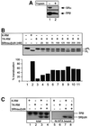
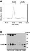
Fig. 1. Purification and characterization of recombinant SR. SRα and SRβ were co-expressed in E.coli and purified by metal chelate and ion exchange chromatography. The peak fractions were then analysed on a Superdex-200 gel filtration column (A). Samples from the two major peaks (fractions 18–21 and 29–30) were analysed by SDS–PAGE followed by staining with Coomassie Brilliant Blue (B).
None
Fig. 2. Functional analysis of recombinant SR (SRhisα/βΔN). (A) High-salt-washed membranes (K-RM; lane 1) were treated with 5 µg/ml trypsin (TK-RM; lane 2) to inactivate the endogenous SR. Equal aliquots of each membrane (10 eq.) were analysed by SDS–PAGE followed by immunoblotting with antibodies against the SRα and SRβ subunits. (B) Co-translational translocation of pre-prolactin (pPL) was assayed in a reticulocyte lysate supplemented with either K-RM (lane 2), TK-RM (lanes 3) or TK-RM with increasing concentrations of SRhisα/βΔN receptor (25–250 nM; lanes 4–8). The reactions were analysed by SDS–PAGE and phosphorimaging. The positions of the unprocessed pPL and the signal sequence-cleaved prolactin (PL) are indicated. The efficiency of translocation was deduced from quantification of the amounts of cleaved and non-cleaved pPL. (C) Translocation reactions were performed as in (B) with K-RM (lanes 1, 3, 5 and 7) or TK-RM (lanes 2, 4, 6 and 8) in the presence (lanes 3, 4, 7 and 8) or absence (lanes 1, 2, 5 and 6) of 50 nM SRhisα/βΔN. After translocation, the samples were analysed by immunoblotting with SRβ antibodies either directly (lanes 1–4) or after purification on Ni-NTA agarose (lanes 5–8). The positions of the endogenous SRβ and SRβΔN are indicated.
None
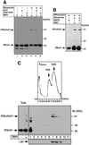
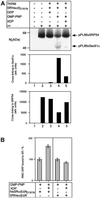
Fig. 3. Cross-linking of SRβΔN to a ribosomal protein. (A) SRhisα/βΔN was incubated in the presence or absence of high-salt-washed canine ribosomes either with no nucleotide, or with 200 µM GMP-PNP or GDP, and cross-linking was then induced with the reagent BMH (20 µM). The samples were analysed by SDS–PAGE followed by immunoblotting with anti-SRβ antibodies. The positions of the uncross-linked recombinant SRβ (SRβΔN) and the major ribosome-dependent cross-link of 47 kDa (SRβΔNx21) are indicated. (B) Cross-linking was performed as in (A) but with 20 µM of either BMH or BMOE. (C) Cross-linking reactions as in (A) with no nucleotide present (total fractions) were treated with puromycin and high salt and then separated by sucrose density gradient centrifugation. The gradients were then fractionated with continuous monitoring of the absorbance at 254 nm (upper panel) to determine the positions of the 40S and 60S subunits (arrows). The fractions were then analysed by SDS–PAGE followed by immunoblotting with antibodies against ribosomal protein L30 (lower panel) and against SRβ (middle panel).
None
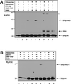
Fig. 4. Binding of ribosomes to Sec61p blocks cross-linking of SRβ to the ribosome. (A) SRhisα/βΔN was incubated with pancreatic ribosomes and/or EDTA high-salt-washed membranes (EK-RM) or liposomes. Where indicated, 200 µM GMP-PNP was also present in the reaction. Cross-linking was induced with BMH and the samples were analysed by SDS–PAGE and immunoblotting for SRβ. The positions of SRβΔN, the endogenous SRβ, the major ribosomal cross-link product (SRβΔNx21) and a cross-link to an unidentified protein of EK-RM (*) are indicated. (B) Cross-linking reactions were performed and analysed as in (A), except that purified Sec61p reconstituted into liposomes was used in place of EK-RM.
None
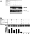
Fig. 5. Characterization of a mutant SR with altered nucleotide specificity in SRβΔN. (A) SRhisα/βΔN (upper panel) or SRhisα/βΔND181N (lower panel) was treated with BMH (20 µM) in the presence of different guanosine and xanthosine nucleotides (200 µM) as indicated. The reactions were then analysed by SDS–PAGE and immunoblotting with SRβ antibodies. Note the difference in internal cross-link (*) formation in the presence of the indicated nucleotides. (B) Co-translational translocation of pre-prolactin was assayed in a reticulocyte lysate supplemented with TK-RM and either 100 nM SRhisα/βΔN (lanes 1–3) or 100 nM SRhisα/βΔND181N (lanes 4–6). Where indicated, 500 µM XMP-PNP (lanes 2, 5 and 8) or 500 µM XDP (lanes 3, 6 and 9) was added in addition to the 100 µM GTP already present in the lysate.
None
Fig. 6. SRβ regulates a specific step in targeting. (A) Targeting reactions were performed with RNC–SRP complexes containing pPL86mer with a photoactivatable cross-linker ([Tmd]Phe) incorporated at position 18, TK-RM supplemented with SRhisα/βΔND181N (100 nM), 25 µM GMP-PNP or GDP, and, where indicated, 250 µM XDP. After targeting, the reactions were chilled on ice and irradiated with UV light (364 nm) prior to analysis by SDS–PAGE and phosphorimaging (upper panel). The positions of the pPL86xSRP54 and pPL86xSec61α cross-link products are indicated. The identity of the cross-links was confirmed by immunoprecipitation (data not shown). The amount of cross-linking to Sec61α and SRP54 was quantified directly from the phosphorimager and is displayed graphically (lower panel). (B) Binding of RNC–SRP complexes to SRhisα/βΔN. Targeting reactions were performed with purified pPL86mer RNC–SRP complexes, TK-RM supplemented with either SRhisα/βΔN or SRhisα/βΔND181N (100 nM), 100 µM GMP-PNP and, where indicated, 1 mM XDP. After targeting, the reactions were solubilized with 1% Nikkol and then the SRhisα/βΔN was re-isolated on an Ni-NTA matrix in the presence of high salt (300 mM KOAc). The amount of bound pPL86 was then quantified by SDS–PAGE and phosphorimaging; binding in the absence of XDP was set to 100%. Error bars indicate the standard deviation from experiments carriedout in triplicate.
None
Fig. 7. Proposed function of SRβ in coordinating release of the signal sequence with the presence of the Sec61p complex. The RNC–SRP complex binds to the SRP receptor via an interaction between SRP54 and SRα. Concomitantly, a 21 kDa ribosomal protein (21k) interacts with SRβ, and thereby promotes release of GDP, and stabilizes the empty form. Subsequent binding of the ribosome to the Sec61p complex (Sec61p) alters the interaction of SRβ with the 21 kDa protein, allowing SRβ to bind GTP. GTP binding to SR leads to the release of the signal peptide from the SRP–SR complex and its transfer to the Sec61p complex. Finally, GTP hydrolysis in SRβ should occur. However, the precise timing and regulation of this event remain to be characterized.

Similar articles

Cited by

References

    1. Andrews D.W., Lauffer,L., Walter,P. and Lingappa,V.R. (1989) Evidence for a two-step mechanism involved in assembly of functional signal recognition particle receptor. J. Cell Biol., 108, 797–810. - PMC - PubMed
    1. Bacher G., Lütcke,H., Jungnickel,B., Rapoport,T.A. and Dobberstein,B. (1996) Regulation by the ribosome of the GTPase of the signal recognition particle during protein targeting. Nature, 381, 248–251. - PubMed
    1. Bacher G., Pool,M. and Dobberstein,B. (1999) The ribosome regulates the GTPase of the β-subunit of the signal recognition particle receptor. J. Cell Biol., 146, 723–730. - PMC - PubMed
    1. Beckman R.D., Bubeck,R., Grassucci,P., Penczek,A., Verschoor,J., Blobel,G. and Frank,J. (1997) Alignment of conduits for the nascent polypeptide chain in the ribosome–Sec61 complex. Science, 278, 2123–2126. - PubMed
    1. Bernstein H.D., Poritz,M.A., Strub,K., Hoben,P.J., Brenner,S. and Walter,P. (1989) Model for signal sequence recognition from amino-acid sequence of 54K subunit of signal recognition particle. Nature, 340, 482–486. - PubMed

Publication types

MeSH terms