. 2001 May 1;15(9):1104-14.
doi: 10.1101/gad.879301.

Subcellular localization of the Snf1 kinase is regulated by specific beta subunits and a novel glucose signaling mechanism

Affiliations

Subcellular localization of the Snf1 kinase is regulated by specific beta subunits and a novel glucose signaling mechanism

O Vincent et al. Genes Dev. .

Abstract

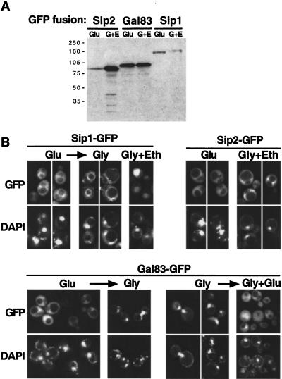
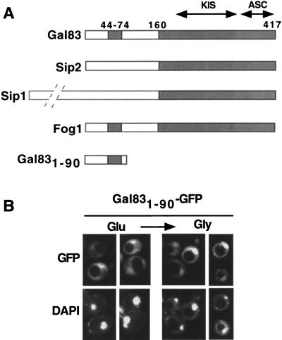
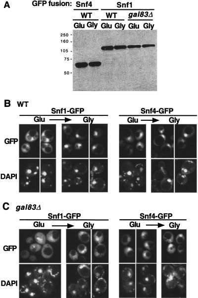
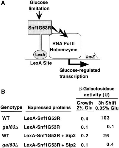
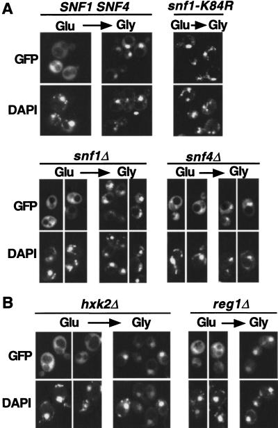
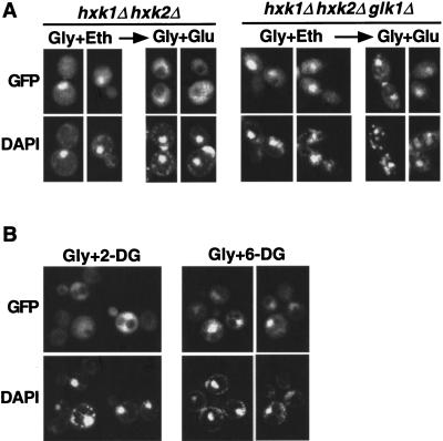
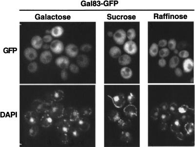
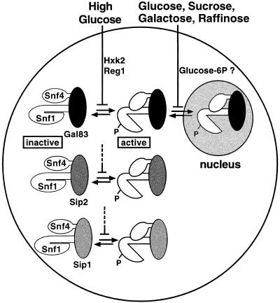
The Snf1/AMP-activated protein kinase family has broad roles in transcriptional, metabolic, and developmental regulation in response to stress. In Saccharomyces cerevisiae, Snf1 is required for the response to glucose limitation. Snf1 kinase complexes contain the alpha (catalytic) subunit Snf1, one of the three related beta subunits Gal83, Sip1, or Sip2, and the gamma subunit Snf4. We present evidence that the beta subunits regulate the subcellular localization of the Snf1 kinase. Green fluorescent protein fusions to Gal83, Sip1, and Sip2 show different patterns of localization to the nucleus, vacuole, and/or cytoplasm. We show that Gal83 directs Snf1 to the nucleus in a glucose-regulated manner. We further identify a novel signaling pathway that controls this nuclear localization in response to glucose phosphorylation. This pathway is distinct from the glucose signaling pathway that inhibits Snf1 kinase activity and responds not only to glucose but also to galactose and sucrose. Such independent regulation of the localization and the activity of the Snf1 kinase, combined with the distinct localization of kinases containing different beta subunits, affords versatility in regulating physiological responses.


Figures

Figure 1
Figure 1
Subcellular localization of Sip1, Sip2, and Gal83. Strains MCY2649, MCY2700 (sip2Δ), and MCY4458 (gal83Δ) were transformed with centromeric plasmids expressing Sip1–GFP, Sip2–GFP, or Gal83–GFP, respectively; plasmids were pOV90, pRT9, and pRT12. (A) Cells were grown in synthetic medium with 2% glucose (Glu) or 2% glycerol plus 3% ethanol (G + E). Protein extracts were prepared, separated by SDS-PAGE, and immunoblotted with anti-GFP antibody. (B) The subcellular localization of each GFP fusion protein was examined by fluorescence microscopy in cells grown on synthetic medium with 2% glucose (Glu), 2% glycerol plus 3% ethanol (Gly + Eth), or 5% glycerol (Gly). The arrows indicate that cells were shifted from the growth medium to a different carbon source and incubated for 20 min before observation. Cells grown in glucose were shifted to 2% or 5% glycerol, and cells grown in glycerol were shifted to 5% glycerol plus 2% glucose. Nuclear export of Gal83 also was observed when cells were shifted from 5% glycerol to 2% glucose (data not shown). A low level of autofluorescence was observed with untransformed strains (not shown). DNA was stained with DAPI. (GFP) green fluorescent protein.
Figure 2
Figure 2
N terminus of Gal83 confers regulated localization. (A) Sip1, Sip2, Gal83, and Fog1 are represented schematically. Shading indicates conserved sequences, which include two domains that interact with Snf1 and Snf4, designated KIS and ASC, respectively (Yang et al. 1994; Jiang and Carlson 1997). Numbers indicate amino acid residues in Gal83. Sip1 is substantially larger than the other subunits, with 863 residues, as indicated by the broken lines. (B) Fluorescence microscopy was performed with strain MCY4458 (gal83Δ) expressing Gal831–90–GFP from the GAL83 promoter on the centromeric plasmid pOV92. Cells were grown to mid-log phase on synthetic medium with 2% glucose (Glu) and shifted for 20 min to 5% glycerol (Gly). Immunoblot analysis confirmed that Gal831–90–GFP is intact. (GFP) green fluorescent protein.
Figure 3
Figure 3
Glucose-regulated localization of Snf1–GFP and Snf4–GFP. Strains MCY4455 (wild type, WT) and MCY4458 (gal83Δ) were transformed with centromeric plasmids pOV84 and pOV76, expressing Snf1–GFP and Snf4–GFP, respectively. Cells were grown to mid-log phase on synthetic medium with 2% glucose (Glu) and shifted to 2% glycerol (Gly) for 20 min. (A) Protein extracts were prepared from the indicated cultures, separated by SDS-PAGE, and immunoblotted with anti-GFP antibody. (B,C) Wild-type (B) and gal83Δ mutant (C) cells expressing the indicated proteins were examined by fluorescence microscopy. (GFP) green fluorescent protein.
Figure 4
Figure 4
Gal83 is required for transcriptional activation of a reporter by LexA–Snf1G53R. (A) The hyperactive kinase LexA–Snf1G53R is bound to LexA sites 5′ to the promoter of a lacZ reporter. Previous studies showed that when cells are limited for glucose, transcription of the reporter is stimulated, dependent on Snf1 catalytic activity (Kuchin et al. 2000). (B) CTY10–5d (WT) and its isogenic derivative MCY4024 (gal83Δ) were transformed with pRJ216 expressing LexA–Snf1G53R (Kuchin et al. 2000) and either pOV65, which overexpresses Sip2 from the ADH1 promoter or its parent vector pSK134, which does not express any protein (Vincent and Carlson 1999). Transformants were grown selectively to mid-log phase in 2% glucose and shifted to 0.05% glucose for 3 hr. β-galactosidase activity was assayed in permeabilized cells and expressed in Miller units (Miller 1972). Values are the average β-galactosidase activity of at least three transformants. Standard errors were <8% for values >0.5. Immunoblot analysis confirmed the stable expression of LexA–Snf1G53R in a gal83Δ mutant.
Figure 5
Figure 5
Glucose-regulated localization of Gal83–GFP in mutant strains. (A) The subcellular localization of Gal83–GFP expressed from the 2-μm plasmid pRT14 was monitored in strains with the indicated genotypes (MCY4458, MCY2634, MCY2916, and MCY2693). Cells were grown to mid-log phase on synthetic medium with 2% glucose (Glu) and shifted for 20 min to 5% glycerol (Gly). (B) Gal83–GFP was expressed from pRT12 in strain MCY3541 (hxk2Δ) and from pRT14 in MCY4408 (reg1Δ). Cells were grown in 2% glucose and shifted for 20 min to 2% glycerol. (GFP) green fluorescent protein.
Figure 6
Figure 6
Glucose phosphorylation is required for nuclear export of Gal83–GFP. (A) Strains MCY4043 (hxk1Δ hxk2Δ) and WAY.78-1 (hxk1Δ hxk2Δ glk1Δ) expressed Gal83–GFP from pRT12. Cells were grown to mid-log phase on synthetic medium with 2% glycerol plus 3% ethanol (Gly + Eth) and shifted to 2% glycerol + 2% glucose (Gly + Glu) for 20 min. (B) Strain MCY4458 (gal83Δ) expressing Gal83–GFP from pRT12 was grown on 5% glycerol and shifted to 5% glycerol + 0.02% 2-deoxyglucose (Gly + 2-DG) or 5% glycerol + 0.02% 6-deoxyglucose (Gly + 6-DG) for 20 min. Nuclear export of Gal83–GFP occurred normally in wild-type cells isogenic to WAY.78-1 (ENY-WA-1A; not shown). (GFP) green fluorescent protein.
Figure 7
Figure 7
Localization of Gal83–GFP in cells grown in different fermentable carbon sources. Fluorescence microscopy was performed with strain MCY4416 (gal83Δ) expressing Gal83–GFP from plasmid pRT12. Cells were grown to mid-log phase on synthetic medium with either 2% sucrose, 2% raffinose, or 2% galactose. (GFP) green fluorescent protein.
Figure 8
Figure 8
Model for regulation of Snf1 kinase activity and localization by two distinct glucose signaling pathways. Snf1 complexes contain one of the three β subunits: Gal83 (black), Sip2 (dark gray), or Sip1 (light gray). Inactive Snf1 kinase complexes are depicted in a closed conformation in which the Snf1 catalytic domain is autoinhibited by the Snf1 regulatory domain. Active Snf1 complexes are shown in an open conformation and are phosphorylated (P). High glucose inhibits (indicated by a bar) the activation of the Snf1 kinase, and Hxk2 and Reg1 are required for this inhibition. Glucose and other fermentable carbon sources inhibit the nuclear localization of Snf1 complexes containing Gal83, and evidence cited in the text suggests that glucose-6-phosphate (Glucose-6P) is necessary and sufficient for this inhibition. This pathway also may regulate the vacuolar localization of Sip1 (not shown; see text).

Similar articles

Cited by

References

    1. Alepuz PM, Cunningham KW, Estruch F. Glucose repression affects ion homeostasis in yeast through the regulation of the stress-activated ENA1 gene. Mol Microbiol. 1997;26:91–98. - PubMed
    1. Ashrafi K, Farazi TA, Gordon JI. A role for Saccharomyces cerevisiae fatty acid activation protein 4 in regulating protein N-myristoylation during entry into stationary phase. J Biol Chem. 1998;273:25864–25874. - PubMed
    1. Ashrafi K, Lin SS, Manchester JK, Gordon JI. Sip2p and its partner Snf1p kinase affect aging in S. cerevisiae. Genes & Dev. 2000;14:1872–1885. - PMC - PubMed
    1. Beck T, Hall MN. The TOR signalling pathway controls nuclear localization of nutrient-regulated transcription factors. Nature. 1999;402:689–692. - PubMed
    1. Carlson M. Glucose repression in yeast. Curr Opin Microbiol. 1999;2:202–207. - PubMed

Publication types

MeSH terms

Substances