. 2001 May 1;15(9):1128-39.
doi: 10.1101/gad.200201.

The chimeric leucine-rich repeat/extensin cell wall protein LRX1 is required for root hair morphogenesis in Arabidopsis thaliana

Affiliations

The chimeric leucine-rich repeat/extensin cell wall protein LRX1 is required for root hair morphogenesis in Arabidopsis thaliana

N Baumberger et al. Genes Dev. .

Abstract

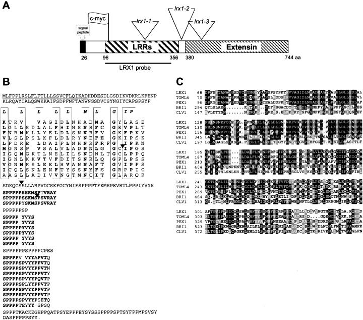
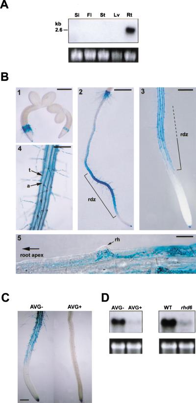
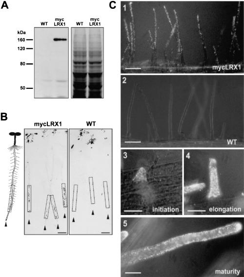
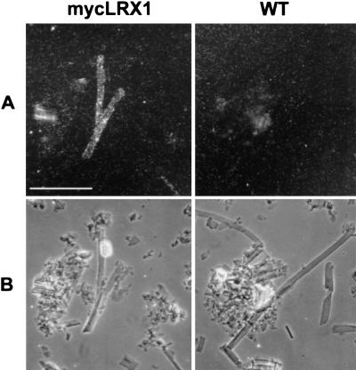
In plants, the cell wall is a major determinant of cell morphogenesis. Cell enlargement depends on the tightly regulated expansion of the wall, which surrounds each cell. However, the qualitative and quantitative mechanisms controlling cell wall enlargement are still poorly understood. Here, we report the molecular and functional characterization of LRX1, a new Arabidopsis gene that encodes a chimeric leucine-rich repeat/extensin protein. LRX1 is expressed in root hair cells and the protein is specifically localized in the wall of the hair proper, where it becomes insolubilized during development. lrx1-null mutants, isolated by a reverse-genetic approach, develop root hairs that frequently abort, swell, or branch. Complementation and overexpression experiments using modified LRX1 proteins indicate that the interaction with the cell wall is important for LRX1 function. These results suggest that LRX1 is an extracellular component of a mechanism regulating root hair morphogenesis and elongation by controlling either polarized growth or cell wall formation and assembly.


Figures

Figure 1
Figure 1
Structure of the LRX1 gene. (A) Schematic representation of the gene indicating the different domains of the encoded protein, the En-1 element insertion sites in the three mutant lines (triangles), the position of the c-myc tag used for immunolocalization of LRX1 (flag), and the fragment used as probe in hybridization experiments (LRX1 probe). (B) Deduced amino acid sequence of the LRX1 gene. The predicted signal peptide is underlined and the different domains have been arranged to highlight their organization. The LRRs are aligned on the plant extracellular LRR consensus sequence (first line in the frames). The conserved amino acids are indicated in bold. The diverse higher-order repeats of the extensin domain are aligned and the amino acids conserved within those repeats are indicated in bold. Black arrowheads indicate the position of the three En-1 insertions shown in A. (C) Alignment of the LRR domains of LRX1, PEX1 (maize, Z34465), TOML-4 (tomato, M76671), CLV1 (Arabidopsis, U96879), and BRI1 (Arabidopsis, AF017056). Identity and similarity are indicated by black and grey shading, respectively.
Figure 2
Figure 2
LRX1 expression pattern (A) Organ-specific expression of LRX1. Total RNA was extracted from green siliques (Si), flowers and flower buds (Fl), inflorescence stems (St), and rosette leaves (Lv), of 35–40-day-old wild-type Columbia plants. Roots (Rt) were harvested from 14-day-old seedlings grown vertically on MS medium. For Northern analysis, 10 μg of total RNA was hybridized with a 32P-labeled LRX1 probe and 25S rRNA was used as loading control (bottom). (B) Transgenic seedlings containing the LRX1 promoter fused to the uidA gene were histochemically stained at different developmental stages to reveal the tissue-specific expression of LRX1. GUS activity (blue staining) was first detected in the epidermal root hair forming cells of the collet region, 2 d after germination (1). In 4-day-old seedlings, GUS activity was mainly detected in rhizodermal cells of the root differentiation zone (rdz) and in the root cap (2,3). In the differentiation zone, only trichoblast cell files (t), which alternate with atrichoblast cell files (a), show GUS activity (4). GUS expression is first visible within cells undergoing root hair (rh) initiation (5). Bars, 400 μm (1,3), 800 μm (2), 200 μm (4), and 20 μm (5). (C) GUS expression in AVG-treated seedlings. AVG treatment (AVG+) prevented root hair formation and blocked GUS expression compared with control seedlings (AVG−). Bar, 250 μm. (D) LRX1 expression in AVG-treated seedlings and rhd6 mutants. Total RNA was extracted from seedlings grown on MS medium containing the ethylene biosynthesis inhibitor L-α-(2-amino-ethoxyvinyl)glycine (left, AVG+), from rhd6 mutants, which do not develop root hairs (right, rhd6), and from wild-type control plants grown on normal MS medium (left, AVG−; right, WT). Total RNA (10 μg) was used for Northern analysis. 25S rRNA was used as loading control (bottom).
Figure 3
Figure 3
Identification and immunolocalization of the LRX1 protein by epitope tagging. (A) Protein gel blot of root extracts from wild-type (WT) and transgenic (mycLRX1) plants expressing the c-myc-tagged LRX1 protein under control of the LRX1 promoter. Blots were incubated with the mouse myc-mAbs (left) and a duplicate gel was stained with Coomassie blue to test for equal loading (right). (B) Tissue print immunoblot. Four-day-old wild-type (WT) and transgenic mycLRX1 (mycLRX1) seedlings were pressed on nitrocellulose membranes to transfer soluble proteins. A scheme of seedlings at the same scale as the blots is represented at left of the panels for orientation. Arrowheads indicate the position of each root tip and frames delimit the root differentiation zone. The dark spots at the top of the membrane were caused by anthocyanins of the cotyledons also transferred onto the membrane. Signals were visible in the differentiation zone of mycLRX1 plants (mycLRX1) but not in controls (WT). Bars, 1 mm. (C) Whole-mount immunolocalization of mycLRX1. Three-day-old wild-type (WT) and mycLRX1 (mycLRX1) seedlings were fixed and immunolabeled with myc-mAbs. The c-myc epitope was found to be associated with root hairs in mycLRX1 (1), but not in wild-type (2) seedlings. Labeling was visible from root hair initiation (3), throughout root hair elongation (4), and after root hair maturation (5). Bars, 100 μm (1,2), 25 μm (35).
Figure 4
Figure 4
Immunodetection of mycLRX1 in purified cell walls. Wild-type (WT) and mycLRX1 (mycLRX1) roots were ground in liquid nitrogen and cell walls were purified before immunolabeling with myc-mAbs and gold-conjugated secondary antibodies. After silver enhancement, the preparation was observed under epipolarized light (A) to reveal the labeling, or transmitted light (B) to identify cell types. Tubular structures, identified as root hair debris were regularly labeled in mycLRX1 material, whereas such structures were not decorated in wild-type preparations. Bar, 100 μm.
Figure 5
Figure 5
LRX1 expression in lrx1 mutants. Total RNA was extracted from 14-d-old seedlings grown vertically on the surface of MS plates. Seedlings from wild-type Columbia (WT) and from each of the three lrx1 mutant lines (lrx1-1, lrx1-2, and lrx1-3) were used. Total RNA (20 μg) was loaded per lane and the membrane was hybridized with a 32P-labeled LRX1 probe (see Fig. 1A). 25S rRNA was used as loading control (bottom).
Figure 6
Figure 6
lrx1-1 mutant phenotype. Wild-type and lrx1-1 mutant seedlings were grown vertically for 3 d on the surface of MS plates and were either observed under a stereomicroscope with dark field illumination (A,B), transmitted light (C,D), or frozen in liquid nitrogen and investigated at low temperature in a scanning electron microscope (E,F). Compared with wild type (A,C,E), the lrx1-1 mutant (B,D,F) has fewer fully elongated root hairs (B), and mutant root hairs frequently have a swollen basis (D,F), show an irregular diameter and often branch (F). The three lrx1 mutant lines had identical phenotypes. Bar, 1 mm (A,B), 350 μm (C,D), 250 μm (E,F).
Figure 7
Figure 7
Overexpression of the complete (35S-LRX1) and truncated (35S-N/LRX1) LRX1 protein. The sequences coding for the full-length or truncated LRX1 protein were transformed into Arabidopsis under the control of the 35S CaMV promoter. (A) Total RNA was extracted from roots of 14-d-old wild-type seedlings (WT), transgenic seedlings overexpressing the truncated LRX1 (35S-N/LRX1) and transgenic seedlings overexpressing the complete LRX1 (35S-LRX1). Total RNA (5 μg) was loaded per lane and the membrane was hybridized with a 32P- labeled LRX1 probe (Fig. 1A). The blot was exposed first for 2 h (1) and the framed area in 1 was further exposed for 3 d (2). Transgene transcript levels were more than 100-fold higher than the endogenous LRX1 transcript. The bottom panel shows 25S ribosomal RNA as a loading control. (B) Immunoblot of root protein extracts from wild-type and 35S-LRX1 plants (1) and wild-type and 35S-N/LRX1 plants (2). The membrane was immunoreacted with anti-LRX1 antiserum. The endogenous LRX1 protein was not detectable with these antibodies. A duplicate gel was stained with Coomassie blue as loading control (right). (C) Phenotype of the lines used in A and B. 35S-LRX1 plants (2) are indistinguishable from wild-type plants (1), whereas 35S-N/LRX1 plants (3) show the same phenotype as lrx1 mutants (4). Bar, 350 μm.

Similar articles

Cited by

References

    1. Baumann E, Lewald J, Saedler H, Schulz B, Wisman E. Successful PCR-based reverse genetic screens using an En-1–mutagenised Arabidopsis thaliana population generated via single-seed descent. Theor Appl Genet. 1998;97:729–734.
    1. Becker D, Kemper E, Schell J, Masterson R. New plant binary vectors with selectable markers located proximal to the left T-DNA border. Plant Mol Biol. 1992;20:1195–1197. - PubMed
    1. Bibikova TN, Zhigilei A, Gilroy S. Root hair growth in Arabidopsis thaliana is directed by calcium and an endogenous polarity. Planta. 1997;203:495–505. - PubMed
    1. Bibikova TN, Blancaflor EB, Gilroy S. Microtubules regulate tip growth and orientation in root hairs of Arabidopsis thaliana. Plant J. 1999;17:657–665. - PubMed
    1. Bradley DJ, Kjellbom P, Lamb CJ. Elicitor- and wound-induced oxidative cross-linking of a proline-rich plant cell wall protein: A novel, rapid defense response. Cell. 1992;70:21–30. - PubMed

Publication types

Associated data