. 2001 May 1;15(9):1140-51.
doi: 10.1101/gad.871201.

Sharp, an inducible cofactor that integrates nuclear receptor repression and activation

Affiliations

Sharp, an inducible cofactor that integrates nuclear receptor repression and activation

Y Shi et al. Genes Dev. .

Abstract

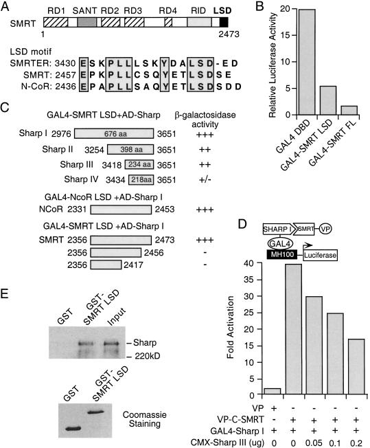
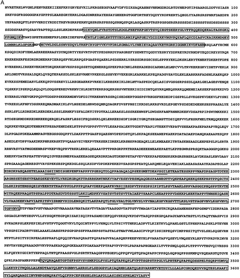
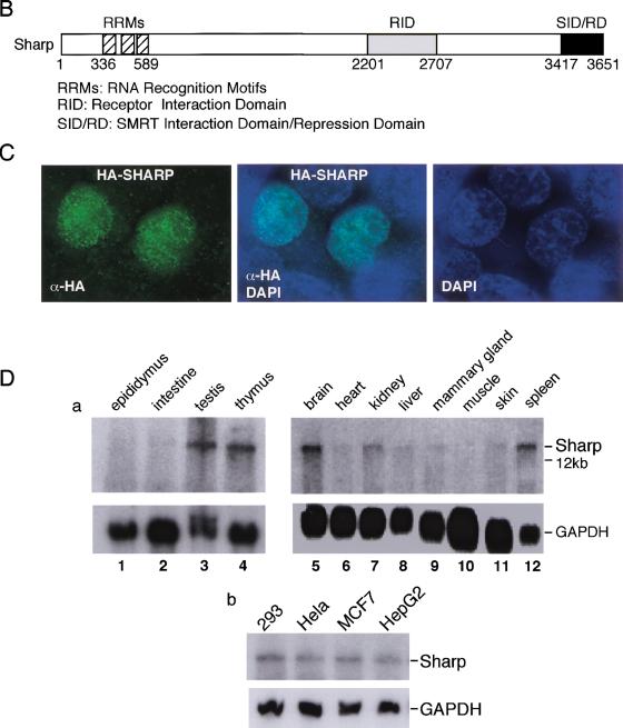
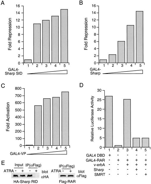
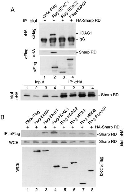
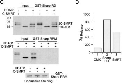
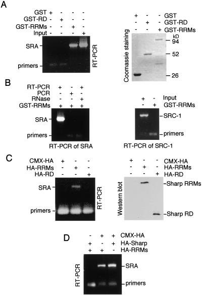
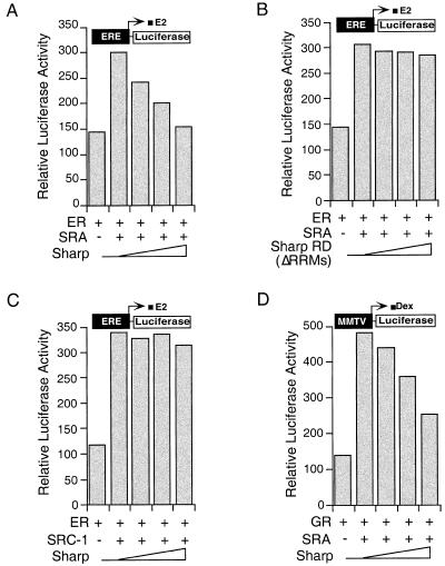
A yeast two-hybrid screen using the conserved carboxyl terminus of the nuclear receptor corepressor SMRT as a bait led to the isolation of a novel human gene termed SHARP (SMRT/HDAC1 Associated Repressor Protein). SHARP is a potent transcriptional repressor whose repression domain (RD) interacts directly with SMRT and at least five members of the NuRD complex including HDAC1 and HDAC2. In addition, SHARP binds to the steroid receptor RNA coactivator SRA via an intrinsic RNA binding domain and suppresses SRA-potentiated steroid receptor transcription activity. Accordingly, SHARP has the capacity to modulate both liganded and nonliganded nuclear receptors. Surprisingly, the expression of SHARP is itself steroid inducible, suggesting a simple feedback mechanism for attenuation of the hormonal response.


Figures

Figure 1
Figure 1
Isolation of SHARP as a SMRT-interacting protein. (A) Schematic representation of SMRT domain structure and the LSD motif sequences. (B) The SMRT LSD domain represses basal transcription. CV-1 cells were transfected with GAL4 DBD or GAL4-fusion constructs, together with MH100-tk-luc reporter and CMX-β-gal. The reporter luciferase activity was normalized with the internal control β-galactosidase activity. The results represent the average of triplicate assays. (C) SHARP interacts with the LSD domain of SMRT and N-CoR. GAL4 fusion of SMRT LSD (residues 2356–2473), N-CoR LSD (residues 2331–2453), or the SMRT LSD deletion mutants were transformed into yeast cells along with an activation domain (AD) fusion of various SHARP fragments (I to IV), followed by a liquid β-galactosidase assay. Two viable transformants were picked for each assay. (D) SMRT–SHARP interaction in mammalian two-hybrid assay. CV-1 cells were cotransfected with GAL4–SHARP I and VP alone or VP-C-SMRT with the indicated amounts of CMX-SHARP III, together with MH100-tk-luc and CMX-β-gal. Fold activation is determined by the relative reporter luciferase activity normalized with the β-galactosidase activity and represents an average of three independent experiments. (E) Full-length SHARP interacts with SMRT. 35S-labeled in vitro translated full-length SHARP was incubated with purified GST or GST–SMRT LSD fusion on glutathione beads. The bound proteins were analyzed by SDS-PAGE and visualized by fluorography. Lower panel is the Coomassie staining of GST and GST–SMRT LSD fusion protein.
Figure 2
Figure 2
The amino acid sequence and expression pattern of SHARP. (A) The amino acid sequence of SHARP predicted from the cDNA sequence. The amino-terminal three RRMs, the receptor interaction domain (RID, aa 2201–2707), and the carboxy-terminal SID/RD are boxed and shaded individually. The IXXI/V motifs (or its variants) in the RID are underlined. (B) Schematic representation of the SHARP functional domains. (C) Nuclear-localization of SHARP. CMX-HA-SHARP was transfected into 293 cells. The transfected cells were immuno-stained with HA-specific antibody and FITC-conjugated secondary antibody. Also shown are nuclear staining by DAPI and merged FITC and DAPI staining. No FITC staining was seen in HA antibody-incubated nontransfected cells. (D) Expression pattern of SHARP. (a) Mouse tissue Northern blots were probed with a SHARP cDNA probe. (b) Expression of SHARP in human tumor cells.
Figure 2
Figure 2
The amino acid sequence and expression pattern of SHARP. (A) The amino acid sequence of SHARP predicted from the cDNA sequence. The amino-terminal three RRMs, the receptor interaction domain (RID, aa 2201–2707), and the carboxy-terminal SID/RD are boxed and shaded individually. The IXXI/V motifs (or its variants) in the RID are underlined. (B) Schematic representation of the SHARP functional domains. (C) Nuclear-localization of SHARP. CMX-HA-SHARP was transfected into 293 cells. The transfected cells were immuno-stained with HA-specific antibody and FITC-conjugated secondary antibody. Also shown are nuclear staining by DAPI and merged FITC and DAPI staining. No FITC staining was seen in HA antibody-incubated nontransfected cells. (D) Expression pattern of SHARP. (a) Mouse tissue Northern blots were probed with a SHARP cDNA probe. (b) Expression of SHARP in human tumor cells.
Figure 3
Figure 3
SHARP represses transcription. (A) SHARP SID represses basal transcription. Increasing amounts of GAL4–SHARP SID were transfected into CV-1 cells along with MH100-tk-luc and CMX-β-gal. Fold repression was determined relative to the activity of GAL4 DBD and represents an average of triplicate assays. (B) The full-length SHARP represses basal transcription. The experimental procedure was the same as in A except that the GAL4 fusion of full-length SHARP was used. (C) GAL4-VP does not repress basal transcription. The experimental procedure was the same as in A except that the GAL4 fusion of VP16 activation domain was used. (D) SHARP restores the repression activity of RAR. GAL4–RAR was transfected into CV-1 cells with v-erbA, a combination of v-erbA and SHARP, or v-erbA and SMRT, together with MH100-luc and CMX-β-gal. The transcriptional activity of GAL4–DBD and GAL4–RAR was determined as relative luciferase reporter activity and represents an average of triplicate assays. (E) SHARP interacts with RAR. 293 cells transfected with CMX-HA-SHARP RID and CMX-Flag-RAR were treated with or without ATRA. The cell lysates were immunoprecipitated with an anti-Flag antibody. The immunoprecipitates (IP) were immunoblotted with an anti-HA or an anti-Flag antibody. Cell lysate inputs were included in the anti-HA blot.
Figure 4
Figure 4
SHARP is associated with SMRT and HDAC1. (A) SHARP interacts with HDAC1. Human 293 cells were transfected with CMX-Flag, or Flag tagged-HDAC1, HDAC3, and HDAC7, each along with CMX-HA-SHARP RD. Half of the cell lysates were immunoprecipitated with anti-HA agarose and the immunoprecipitates were subjected to Western blot analysis with Flag-specific antibody. Another half of the cell lysates were immunoprecipitated with anti-Flag agarose and immunoblotted with HA-specific antibody. One aliquot of the cell lysates was assayed directly by anti-Flag Western blot. An aliquot of the cell lysates and anti-HA immunoprecipitates were assayed by anti-HA Western blot. (B) SHARP is associated with components of the NuRD complex. Flag-tagged Sin3A, SMRT (short version of human SMRT, corresponding to mSMRT amino acids 1060–2473; Chen and Evans 1995), HDAC1, HDAC2, MTA2, MBD3, RbAp48, or CMX-Flag were transfected into 293 cells along with CMX-HA-SHARP RD. The cell lysates were immunoprecipitated with anti-Flag agarose and the immunoprecipitates were analyzed by Western blot with HA-specific antibody. Aliquots of the cell lysates were also assayed directly by Western blot with HA-specific antibody or Flag-specific antibody. (C) SHARP interacts with SMRT and HDAC1 in vitro. 35S-labeled in vitro translated HDAC1 and SMRT (residues 1851–2473) were incubated with purified recombinant GST–SHARP RD or GST–SHARP RRMs on glutathione beads individually, or together. The bound proteins were analyzed by SDS-PAGE and fluorography. Lower panel is Coomassie staining of the GST–SHARP RRMs protein. (D) SHARP is associated with histone deacetylase activity. Lysates prepared from cells expressing vector alone, HA-SHARP RD, or Flag-SMRT were immunoprecipitated with anti-HA or anti-Flag agarose. The immunoprecipitates were resuspended in deacetylase assay buffer for histone deacetylase assays.
Figure 4
Figure 4
SHARP is associated with SMRT and HDAC1. (A) SHARP interacts with HDAC1. Human 293 cells were transfected with CMX-Flag, or Flag tagged-HDAC1, HDAC3, and HDAC7, each along with CMX-HA-SHARP RD. Half of the cell lysates were immunoprecipitated with anti-HA agarose and the immunoprecipitates were subjected to Western blot analysis with Flag-specific antibody. Another half of the cell lysates were immunoprecipitated with anti-Flag agarose and immunoblotted with HA-specific antibody. One aliquot of the cell lysates was assayed directly by anti-Flag Western blot. An aliquot of the cell lysates and anti-HA immunoprecipitates were assayed by anti-HA Western blot. (B) SHARP is associated with components of the NuRD complex. Flag-tagged Sin3A, SMRT (short version of human SMRT, corresponding to mSMRT amino acids 1060–2473; Chen and Evans 1995), HDAC1, HDAC2, MTA2, MBD3, RbAp48, or CMX-Flag were transfected into 293 cells along with CMX-HA-SHARP RD. The cell lysates were immunoprecipitated with anti-Flag agarose and the immunoprecipitates were analyzed by Western blot with HA-specific antibody. Aliquots of the cell lysates were also assayed directly by Western blot with HA-specific antibody or Flag-specific antibody. (C) SHARP interacts with SMRT and HDAC1 in vitro. 35S-labeled in vitro translated HDAC1 and SMRT (residues 1851–2473) were incubated with purified recombinant GST–SHARP RD or GST–SHARP RRMs on glutathione beads individually, or together. The bound proteins were analyzed by SDS-PAGE and fluorography. Lower panel is Coomassie staining of the GST–SHARP RRMs protein. (D) SHARP is associated with histone deacetylase activity. Lysates prepared from cells expressing vector alone, HA-SHARP RD, or Flag-SMRT were immunoprecipitated with anti-HA or anti-Flag agarose. The immunoprecipitates were resuspended in deacetylase assay buffer for histone deacetylase assays.
Figure 5
Figure 5
SHARP binds to SRA. (A) SHARP interacts with SRA in vitro. In vitro-transcribed SRA mRNA was incubated with GST, GST–SHARP RD, or GST–SHARP RRMs on glutathione beads. After extensive washes, the beads were added to a RT–PCR reaction with SRA specific primers. The RT–PCR products (700 bp) were analyzed with 1% TAE agarose gel. Right panel is Coomassie staining of an aliquot of the GST proteins. (B) Specificity of the SHARP–SRA interaction. In vitro-transcribed SRA mRNA was incubated with GST–SHARP RRMs on glutathione beads. After extensive washes, the beads were either added to a RT–PCR reaction, or to a PCR reaction without reverse transcriptase, or incubated with RNase prior to RT–PCR. In a parallel experiment, in vitro-transcribed SRC-1 mRNA was incubated with GST–RRMs and subjected to RT–PCR using SRC-1 specific primers. An aliquot of the SRC-1 RNA input was included in the RT–PCR reaction. (C) The SHARP RRMs interact with SRA in vivo. 293 cells were transfected with CMX, CMX-HA SHARP RRMs, or CMX-HA SHARP RD, along with SCT1–SRA2 (Lanz et al. 1999). The cell lysates were incubated with anti-HA agarose and the immunoprecipitates were analyzed by RT–PCR with SRA specific primers. Right panel is anti-HA Western blot of the cell lysates. (D) The full-length SHARP interacts with SRA. The experimental procedure is the same as in C except that CMX-HA full-length SHARP was transfected.
Figure 6
Figure 6
SHARP represses SRA-potentiated ER and GR activity. (A–C) SHARP represses SRA- but not SRC-1-potentiated ER activity. CMX-ER, ERE-luc, and CMX-β-gal were transfected into CV-1 cells along with SCT1–SRA2 or CMX-SRC-1, and increasing amount of CMX–SHARP or CMX–SHARP RD (ΔRRMs). The transfected cells were treated with 10 nM 17β-estradiol (E2) for 24 h before luciferase assay. (D) SHARP represses SRA-stimulated GR activity. CMX-GR, MMTV-luc, and CMX-β-gal were transfected into CV-1 cells along with SCT1–SRA2 and increasing amount of CMX–SHARP. The transfected cells were treated with 50 nM dexamethasone (Dex) for 24 h before luciferase assay.
Figure 7
Figure 7
SHARP expression is estrogen-inducible. (A) MCF-7 cells were treated with E2 for indicated periods of time. Northern blot on total RNA was probed for SHARP and GAPDH. (B) Fold induction of SHARP was determined by PhosphorImager quantitation of the SHARP message level at each time point and normalized by the mRNA level of GAPDH.

References

    1. Alland L, Muhle R, Hou H, Jr, Potes J, Chin L, Schreiber-Agus N, DePinho RA. Role for N-CoR and histone deacetylase in Sin3-mediated transcriptional repression. Nature. 1997;387:49–55. - PubMed
    1. Ayer DE, Lawrence QA, Eisenman RN. Mad-Max transcriptional repression is mediated by ternary complex formation with mammalian homologs of yeast repressor Sin3. Cell. 1995;80:767–776. - PubMed
    1. Bannister AJ, Kouzarides T. The CBP co-activator is a histone acetyltransferase. Nature. 1996;384:641–643. - PubMed
    1. Blanco JC, Minucci S, Lu J, Yang XJ, Walker KK, Chen H, Evans RM, Nakatani Y, Ozato K. The histone acetylase PCAF is a nuclear receptor coactivator. Genes & Dev. 1998;12:1638–1651. - PMC - PubMed
    1. Brown AM, Jeltsch JM, Roberts M, Chambon P. Activation of pS2 gene transcription is a primary response to estrogen in the human breast cancer cell line MCF-7. Proc Natl Acad Sci. 1984;81:6344–6348. - PMC - PubMed

Publication types

MeSH terms