. 2001 May 1;15(9):1152-66.
doi: 10.1101/gad.191301.

Xenopus Sprouty2 inhibits FGF-mediated gastrulation movements but does not affect mesoderm induction and patterning

Affiliations

Xenopus Sprouty2 inhibits FGF-mediated gastrulation movements but does not affect mesoderm induction and patterning

S L Nutt et al. Genes Dev. .

Abstract

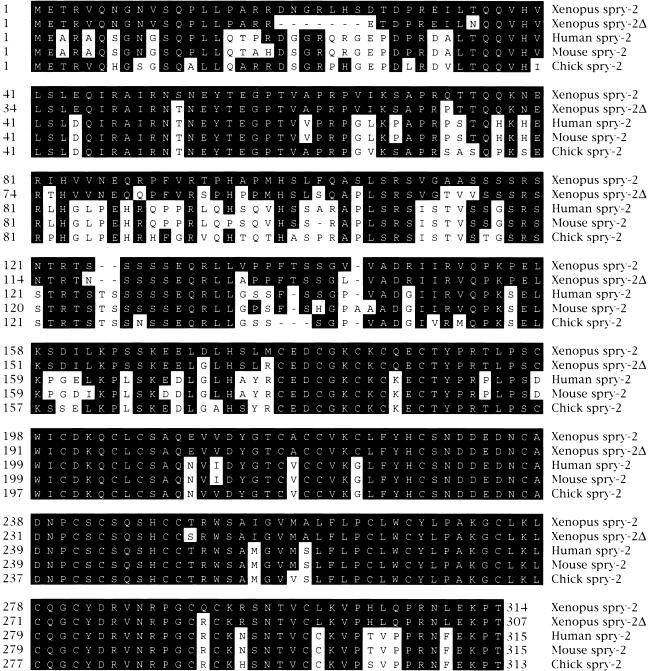
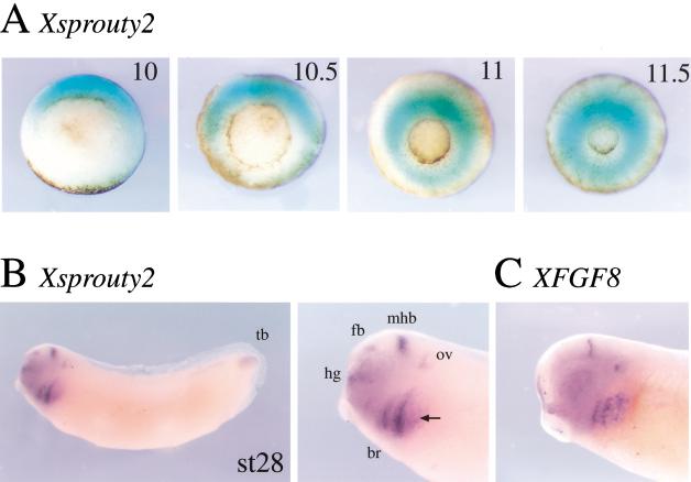
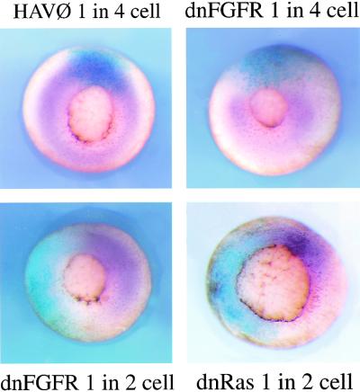
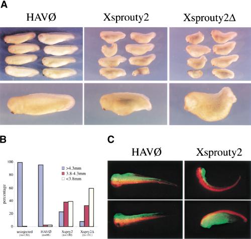
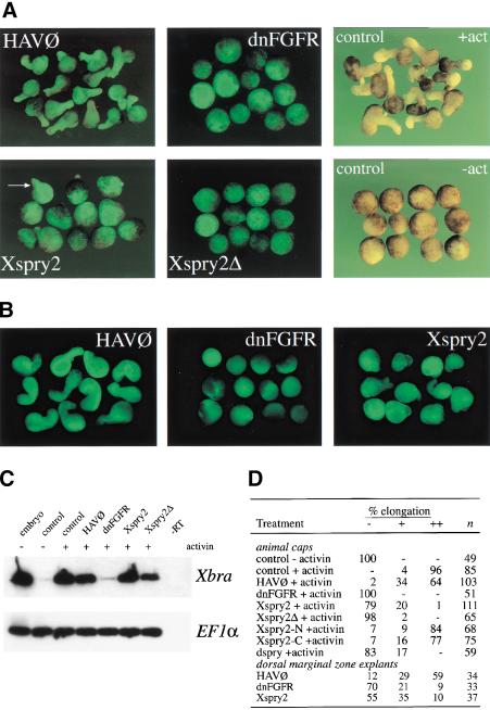
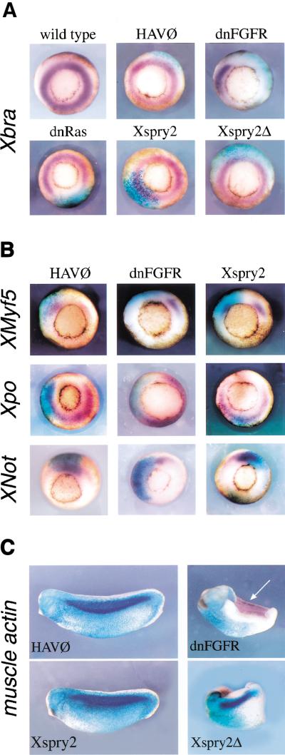
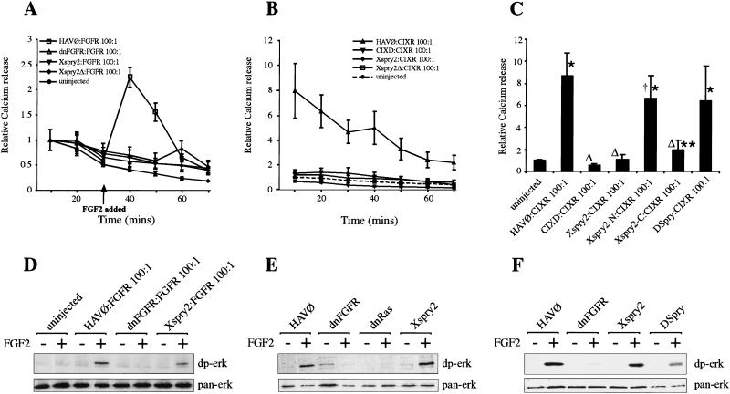
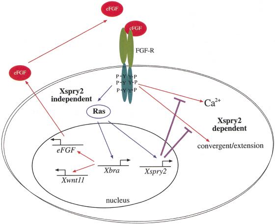
Signal transduction through the FGF receptor is essential for the specification of the vertebrate body plan. Blocking the FGF pathway in early Xenopus embryos inhibits mesoderm induction and results in truncation of the anterior-posterior axis. The Drosophila gene sprouty encodes an antagonist of FGF signaling, which is transcriptionally induced by the pathway, but whose molecular functions are poorly characterized. We have cloned Xenopus sprouty2 and show that it is expressed in a similar pattern to known FGFs and is dependent on the FGF/Ras/MAPK pathway for its expression. Overexpression of Xsprouty2 in both embryos and explant assays results in the inhibition of the cell movements of convergent extension. Although blocking FGF/Ras/MAPK signaling leads to an inhibition of mesodermal gene expression, these markers are unaffected by Xsprouty2, indicating that mesoderm induction and patterning occurs normally in these embryos. Finally, using Xenopus oocytes we show that Xsprouty2 is an intracellular antagonist of FGF-dependent calcium signaling. These results provide evidence for at least two distinct FGF-dependent signal transduction pathways: a Sprouty-insensitive Ras/MAPK pathway required for the transcription of most mesodermal genes, and a Sprouty-sensitive pathway required for coordination of cellular morphogenesis.


Figures

Figure 1
Figure 1
Amino acid sequence comparison of vertebrate Sprouty proteins. The deduced amino acid sequences of Xenopus Xspry2 and Xspry2Δ were aligned with the human, mouse, and chick Spry2 proteins (Hacohen et al. 1998; Minowada et al. 1999). Dashes represent gaps inserted to maximize alignment. Amino acids conserved with Xspry2 are indicated by bold overlay.
Figure 2
Figure 2
Xsprouty2 is expressed dynamically during early development. Whole mount in situ hybridization of Xspry2 and XFGF8. (A) Vegetal view of gastrula stage embryos (stages 10–11.5). Xspry2 expression is induced in the dorsal marginal zone. Expression expands ventrally around the marginal zone during mid to late gastrula stages. (B) Xspry2 expression in a stage 28 albino embryo and close-up of the head region. Xspry2 can be detected primarily in anterior neural structures, such as the mid-brain/hind-brain isthmus (mhb), otic vesicle (ov), forebrain (fb), and hatching gland (hg). Lateral expression is seen in the branchial arches (br) and the tail bud (tb). (C) Close-up of head region of a stage 28 embryo stained for XFGF8. XFGF8 has a similar expression profile to Xspry2, although with more sharply defined boundaries. Black arrow indicates expression in the third branchial arch, which is absent from the Xspry2 expression profile.
Figure 3
Figure 3
Xsprouty2 expression requires FGF signaling. Vegetal view of in situ hybridization to Xspry2 (purple) at the mid-to-late gastrula stages (stages 11–11.5). Embryos were injected into one blastomere at the two- or four-cell stages with 1 ng of HAV∅, dnRas, or dnFGFR together with 500 pg of Lacz mRNA, and stained for β-galactosidase activity (blue) as a lineage tracer.
Figure 4
Figure 4
Overexpression of Xsprouty2 leads to a shortened A-P axis. (A) Embryos were injected once into the dorsal marginal zone at the two-cell stage, with 1 ng of the indicated mRNA plus 100 pg of GAP43-GFP mRNA and analyzed at stage 25. (B) Quantitation of the truncated phenotype of injected embryos. Embryos were measured and grouped into three 5-mm categories. Whereas virtually all noninjected and HAV∅-injected embryos were more than 4.3 mm, the majority of Xspry2 and Xspry2Δ overexpressing embryos were less than 3.8 mm. (n) the number of embryos examined for each treatment. (C) Representative embryos injected with RLDX and Xspry2/GAP43-GFP or HAV∅/GAP43-GFP into the dorsal blastomeres at the four-cell stage and allowed to development until stage 40. The majority of HAV∅/GFP embryos showed a full extension of GFP-labeled cells dorsally along the A-P axis, whereas Xspry2/GFP-injected embryos showed a kinking of the body toward the injected side (top) or a truncation and generalized disorganization of the GFP-positive cells (bottom).
Figure 5
Figure 5
Xsprouty2 inhibits convergent extension but not mesoderm induction. (A) Embryos were injected as indicated with 500 pg of mRNA along with 100 pg of GAP43-GFP mRNA into the animal pole of both blastomeres at the two-cell stage. Animal caps were excised at stage 8 and cultured until stage 18 in the presence of 10 ng/ml activin, before being examined for GFP fluorescence and elongation. Control noninjected caps were incubated with (+act) or without (−act) activin. All images, except for the control animal caps were taken under fluorescence. (B) Injections were carried out as in A, with the exception that they were targeted to the DMZ. DMZ explants were excised at stage 10 and cultured until stage 19 before being measured for elongation. All images were taken under fluorescence. (C) RT-PCR analysis of Xbra transcripts from animal caps injected as in A, and cultured in the presence or absence of activin until stage 11. Whereas dnFGFR completely blocked activin-induced mesoderm formation as measured by Xbra expression, Xbra was expressed in Xspry2 and Xspry2Δ overexpressing caps. (-RT) whole embryo controls without reverse transcriptase. EF1α was amplified as a loading control. (D) Quantitation of the explant elongation assays from A and B. (n) number of animal caps examined; (−) rounded explants; (+) partial elongation; (++) full extension.
Figure 6
Figure 6
Xsprouty2 overexpression does not block mesoderm induction, patterning, or maintenance. (A) Vegetal views of in situ hybridization (purple) to (A) Xbra and (B) Xmyf5, Xpo, and Xnot at mid-gastrulation (stage 11). Embryos were injected at the four-cell stage into a dorsal blastomere with 1 ng of mRNA along with 250 pg LacZ mRNA. Embryos were stained for β-galactosidase activity (blue) as a lineage marker. (C) In situ hybridization to muscle actin (purple) were performed on stage 25 embryos injected at the two-cell stage into a single blastomere with 1 ng of either HAV∅, dnFGFR, Xspry2, or Xspry2Δ together with 250 pg of β-galactosidase mRNA (blue). Approximately 10% of Xspry2 and Xspry2Δ-injected embryos failed to close the blastopore yet retained muscle differentiation as assessed by coexpression of injected cells with muscle actin mRNA. Arrow indicates muscle actin expression on the noninjected side of dnFGFR embryos, which is absent of the β-galactosidase lineage tracer.
Figure 7
Figure 7
Xsprouty2 proteins function as intracellular inhibitors of the FGF signaling pathway. (A) Xspry2 proteins block FGF signaling. FGF-stimulated calcium efflux from oocytes was measured after injection of 250 pg of Xenopus FGFR1 (FGFR) and 25 ng of the indicated mRNA (100-fold excess). Oocytes were loaded with 45Ca2+ and the media collected at 10-min intervals, before and after the addition of FGF2 (100 ng/ml) and subjected to scintillation counting. All points represent the mean of at least nine wells (10 oocytes per well), derived from three independent experiments. For standardization, the counts obtained in the first 10 min for each condition was given a value of 1 and all other points are shown relative to the counts measured for each condition in the first 10 min. Error bars indicate ±S.E.M. (B) Xspry2 functions downstream of the FGFR. Oocytes were injected as in A with 250 pg of the constitutively active RTK, CIXR, and the indicated mRNA at 25 ng. CIXD is a dominant negative form of CIXR; CIXR constitutively dimerizes and signals in the absence of ligand, as seen by the FGF-independent calcium efflux. For standardization, all values are shown relative to the counts obtained from uninjected oocytes in the first 10 min. (C) DSpry does not inhibit Ca2+ signaling in oocytes, but the carboxyl terminus of Xspry2 does inhibit Ca2+ signaling. Oocytes were injected and assayed as in B, except that only the relative 45Ca2+ release for the first 10 min is shown. Error bars indicate ±S.D. *P < 0.001 and **P < 0.01 compared to uninjected. ΔP < 0.001 and P < 0.05 compared to HAV∅:CIXR. (D) Xspry2 does not inhibit FGF-dependent MAPK activation in ovo. Oocytes were injected and cultured as in A, before being incubated with or without FGF2 (100 ng/ml) for 15 min. Protein extracts were then analyzed for activated (dp-erk) and total (pan-erk) MAPK levels. (E) MAPK in animal cap explants is not sensitive to Xspry2 overexpression. Animal caps injected with the indicated mRNA were incubated for 15 min with or without FGF2 before being subjected to immunoblotting as in D. (F) DSpry does not inhibit MAPK activation in FGF-treated animal caps. Animal caps were injected with the indicated mRNAs and treated as in E.
Figure 8
Figure 8
Model for the function of Xsprouty2 in Xenopus gastrulation. At least two signaling pathways emanate from the FGFR during gastrulation: a Xsprouty2-insensitive signal through the Ras/MAPK pathway results in mesoderm induction/maintenance as well as Xsprouty2, Xbra, and indirectly eFGF and Xwnt11 expression. The second Xsprouty2-sensitive pathway results in Ca2+ mobilization and convergent extension, two processes known to require Xwnt11 signaling. Thus, Xsprouty2 functions to coordinate the distinct roles of the FGFR in mesoderm induction and morphogenesis. See discussion for further details.

Similar articles

Cited by

References

    1. Amaya E, Musci TJ, Kirschner MW. Expression of a dominant negative mutant of the FGF receptor disrupts mesoderm formation in Xenopus embryos. Cell. 1991;66:257–270. - PubMed
    1. Amaya E. FGF signalling in early Xenopus development. Phd Thesis. San Francisco: University of California; 1992.
    1. Amaya E, Stein PA, Musci TJ, Kirschner MW. FGF signalling in the early specification of mesoderm in Xenopus. Development. 1993;118:477–487. - PubMed
    1. Beiman M, Shilo BZ, Volk T. Heartless, a Drosophila FGF receptor homolog, is essential for cell migration and establishment of several mesodermal lineages. Genes & Dev. 1996;10:2993–3002. - PubMed
    1. Carballada R, Yasuo H, Lemaire P. Phosphatidylinositol-3 kinase acts in parallel to the ERK MAP kinase in the FGF pathway during Xenopus mesoderm induction. Development. 2001;128:35–44. - PubMed

Publication types

MeSH terms

Associated data

LinkOut - more resources