. 2001 May 8;98(10):5578-83.
doi: 10.1073/pnas.091100898. Epub 2001 May 1.

Tryptophan zippers: stable, monomeric beta -hairpins

Affiliations

Tryptophan zippers: stable, monomeric beta -hairpins

A G Cochran et al. Proc Natl Acad Sci U S A. .

Erratum in

  • Proc Natl Acad Sci U S A 2002 Jun 25;99(13):9081

Abstract

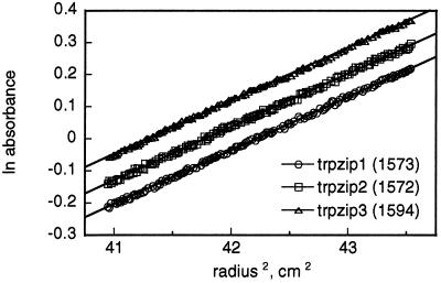
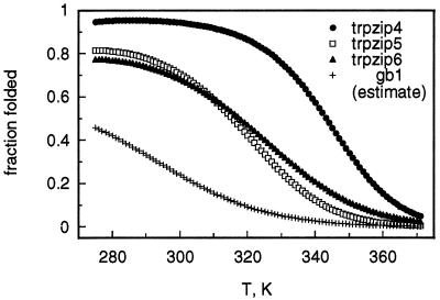
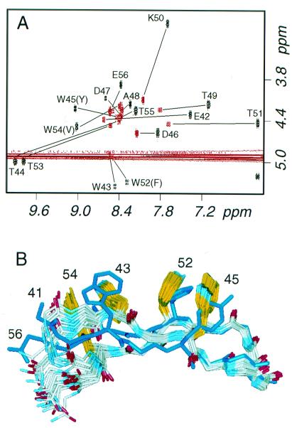
A structural motif, the tryptophan zipper (trpzip), greatly stabilizes the beta-hairpin conformation in short peptides. Peptides (12 or 16 aa in length) with four different turn sequences are monomeric and fold cooperatively in water, as has been observed previously for some hairpin peptides. However, the folding free energies of the trpzips exceed substantially those of all previously reported beta-hairpins and even those of some larger designed proteins. NMR structures of three of the trpzip peptides reveal exceptionally well-defined beta-hairpin conformations stabilized by cross-strand pairs of indole rings. The trpzips are the smallest peptides to adopt an unique tertiary fold without requiring metal binding, unusual amino acids, or disulfide crosslinks.


Figures

Figure 1
Figure 1
Folding of trpzips 1–3. (A) CD spectrum of trpzip1. (Inset) The near UV region is shown with a 10-fold expanded y axis. (B) Thermal denaturation of trpzip1 (20 μM) monitored by CD. The forward melting curve is shown as open circles, while the reverse melting curve is shown as the error bars associated with signal averaging during data acquisition. (Inset) The first derivatives of melting curves (20, 50, 100, and 150 μM peptide) are overlaid. (C) Temperature dependence of folding for trpzips 1–3 (calculated from the thermodynamic parameters listed in Table 2).
Figure 2
Figure 2
Equilibrium ultracentrifugation of trpzips 1–3. The data shown are for 60 μM peptide samples and a rotor speed of 40 krpm. Apparent molecular weights obtained from the slopes (assuming ideal behavior) are shown; calculated formula weights are 1,608 for trpzips 1 and 2 and 1,648 for trpzip3. Trpzip1 data are offset vertically (ln absorbance −0.085) for clarity.
Figure 3
Figure 3
NMR structures of trpzips 1 and 2. (A) Ensemble of 20 structures of trpzip1 optimally aligned by using backbone atoms of residues 2–11. (B) Representative structures of trpzips 1 and 2 aligned on the backbone atoms of residues 2–5 and 8–11 (rmsd of the mean coordinates of the aligned backbone atoms in the two ensembles is 0.37 Å); the bottom view is rotated 90° relative to the top view. The backbone carbonyl of residue 6 is indicated to emphasize the difference in turn geometry between the two structures (type II′ for trpzip1 vs. type I′ for trpzip2).
Figure 4
Figure 4
Temperature dependence of folding for trpzips 4–6 (calculated from the thermodynamic parameters listed in Table 2). Our estimated curve for gb1 was calculated by assuming that mutations in trpzip4 (i.e., those present in trpzips 5 and 6) have independent and additive effects on hairpin stability [ΔGunf,gb1 = ΔGunf,trpzip5 − (ΔGunf,trpzip4 − ΔGunf, trpzip6)] (12) and is nearly identical in shape to previously reported gb1 denaturation curves based on fluorescence (7) or NMR (8) measurements. However, our estimated population for the folded conformation of gb1 is much closer to that in the original report (28) than to later estimates (7, 8).
Figure 5
Figure 5
NMR analysis of trpzip4. (A) Overlay of the COSY fingerprint regions for the wild-type gb1 peptide (red) and trpzip4 (black); the location of crosspeaks are indicated for both peptides and are labeled for trpzip4. (B) Ensemble of 20 structures of trpzip 4 compared with the minimized mean structure of the B1 domain of protein G (Protein Data Bank code 2GB1) (30); backbone atoms of protein residues 46–52 were superposed on the mean coordinates of the ensemble, yielding a rmsd of 0.67 Å.

References

    1. Struthers M D, Cheng R P, Imperiali B. Science. 1996;271:342–345. - PubMed
    1. Dahiyat B I, Sarisky C A, Mayo S L. J Mol Biol. 1997;273:789–796. - PubMed
    1. Dahiyat B I, Mayo S L. Science. 1997;278:82–87. - PubMed
    1. Kortemme T, Ramirez-Alvarado M, Serrano L. Science. 1998;281:253–256. - PubMed
    1. Gellman S H. Curr Opin Chem Biol. 1998;2:717–725. - PubMed

LinkOut - more resources