. 2001 May 22;98(11):6372-7.
doi: 10.1073/pnas.091113598. Epub 2001 May 1.

Glutamate antagonists limit tumor growth

Affiliations

Glutamate antagonists limit tumor growth

W Rzeski et al. Proc Natl Acad Sci U S A. .

Erratum in

  • Proc Natl Acad Sci U S A 2001 Jul 17;98(15):8921

Abstract

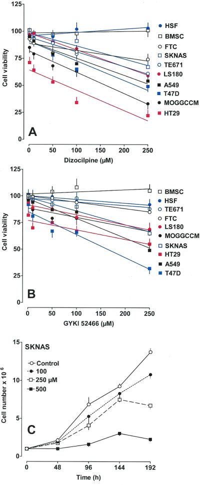
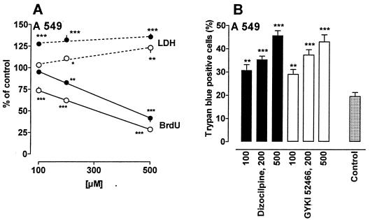
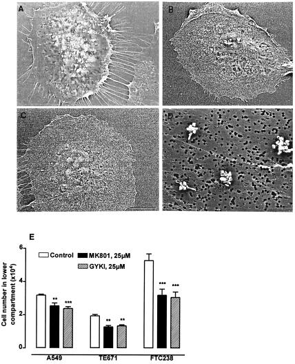
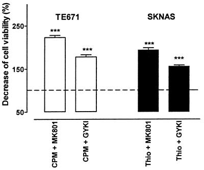
Neuronal progenitors and tumor cells possess propensity to proliferate and to migrate. Glutamate regulates proliferation and migration of neurons during development, but it is not known whether it influences proliferation and migration of tumor cells. We demonstrate that glutamate antagonists inhibit proliferation of human tumor cells. Colon adenocarcinoma, astrocytoma, and breast and lung carcinoma cells were most sensitive to the antiproliferative effect of the N-methyl-d-aspartate antagonist dizocilpine, whereas breast and lung carcinoma, colon adenocarcinoma, and neuroblastoma cells responded most favorably to the alpha-amino-3-hydroxy-5-methyl-4-isoxazole-propionate antagonist GYKI52466. The antiproliferative effect of glutamate antagonists was Ca(2+) dependent and resulted from decreased cell division and increased cell death. Morphological alterations induced by glutamate antagonists in tumor cells consisted of reduced membrane ruffling and pseudopodial protrusions. Furthermore, glutamate antagonists decreased motility and invasive growth of tumor cells. These findings suggest anticancer potential of glutamate antagonists.


Figures

Figure 1
Figure 1
The NMDA antagonist dizocilpine (A) and the AMPA antagonist GYKI52466 (B) exert concentration-dependent antiproliferative effect in human tumor cell lines but not in human skin fibroblasts and bone marrow stromal cells. Cells were exposed to either culture medium alone (control), dizocilpine (1–250 μM), or GYKI52466 (1–250 μM) for 96 h, and viability was measured photometrically by means of the MTT assay. Data represent mean normalized optical densities ± SEM of 6–8 trials and were analyzed by means of linear regression. (C) Growth inhibition of neuroblastoma cells in three-dimensional cultures by dizocilpine (100–500 μM) is shown. Numbers of cells were assessed by counting, after trypsinization. ANOVA showed that the effect of treatment was significant [F(3,36) = 49.33, P < 0.001], with multiple comparisons revealing that dizocilpine induced antiproliferative action in a dose-dependent manner. The effect of time was also significant [F(4,36) = 296.69, P < 0.001], indicating that the antiproliferative effect of dizocilpine was more pronounced with time. SKNAS, human neuroblastoma; TE671, human rhabdomyosarcoma/medulloblastoma; MOGGCCM, human brain astrocytoma; FTC238, human thyroid carcinoma; A549, human Caucasian lung carcinoma; LS180, human Caucasian colon adenocarcinoma; T47D, human breast carcinoma; HT29, human colon adenocarcinoma; HSF, human skin fibroblasts; BMSC, human bone marrow stromal cells.
Figure 2
Figure 2
The antiproliferative effect of dizocilpine (●) and GYKI52466 (○) is attributed to decreased cell division and to increased cell death. (A) Human lung carcinoma (A549) cells were grown in culture medium only (control), in the presence of dizocilpine (100–500 μM), or in the presence of GYKI52466 (100–500 μM) for 96 h. BrdUrd incorporation was used as a marker for cell division. Cell death was quantified by measuring LDH release from damaged cells after 96 h in culture. The data represent mean normalized optical densities ± SEM of 6–8 trials. *, P < 0.05; **, P < 0.01; ***, P < 0.001 vs. control, Student's t test. (B) The effect of exposure of human lung carcinoma (A549) cells to different concentrations of dizocilpine or GYKI52466 on the numbers of trypan blue-accumulating cells is presented. Bars represent mean ± SEM number of trypan blue-positive cells/100 counted cells after 96 h in culture in the absence (control) or in the presence of different concentrations (μM) of glutamate antagonists (n = 6). **, P < 0.01; ***, P < 0.001, Student's t test.
Figure 3
Figure 3
The antiproliferative effect of dizocilpine and GYKI52466 can be reproduced by other NMDA and AMPA antagonists in human lung carcinoma (A549) and human rhabdomyosarcoma/medulloblastoma (TE671) cells and is reversed by Ca2+ deprivation. A549 (A) and TE671 (C) cells were exposed to either culture medium alone (control), or the NMDA antagonists (+)dizocilpine, (−)dizocilpine, ketamine, or memantine in concentrations ranging from 1 to 500 μM for 96 h, and viability was measured photometrically by means of the MTT assay. Data represent mean normalized optical densities ± SEM of 4–6 trials and were analyzed by means of analysis of variance. The antiproliferative effect of dizocilpine was abolished in Ca2+-free medium in lung carcinoma (A) [FA549(1,37) = 77.10, P < 0.001] and rhabdomyosarcoma/medulloblastoma cells (C) [FTE671(1,50) = 42.11, P < 0.001]. The antiproliferative effect of (−)dizocilpine was less pronounced in both tumor cell lines compared to the effect of (+)dizocilpine [FA549(1,80) = 128.52, P < 0.001; FTE671(1,80) = 268.60, P < 0.001]. (B and D) A549 and TE671 cells were exposed to either culture medium alone (control) or the AMPA antagonists CFM-2 and NBQX in concentrations ranging from 1 to 500 μM for 96 h, and viability was measured photometrically by means of the MTT assay. Data represent mean normalized optical densities ± SEM of 4–6 trials and were analyzed by means of ANOVA. The antiproliferative effect of GYKI52466 was significantly reduced in Ca2+-free medium in lung carcinoma (B) [FA549(1,39) = 51.27, P < 0.001] and abolished in rhabdomyosarcoma/medulloblastoma cells (D) [FTE671(1,45) = 17.34, P < 0.001].
Figure 4
Figure 4
Glutamate antagonists alter tumor cell morphology and decrease tumor cell motility. Scanning electron micrographs of thyroid carcinoma cells (FTC238) under control conditions (A) and after exposure to dizocilpine (B, 100 μM) or GYKI52466 (C, 100 μM). Tumor cells display numerous pseudopodia (A), which are far less prominent after exposure to glutamate antagonists (B and C). (D) Protrusion of tumor cell pseudopodia of FTC238 cells through the 0.4-μm pores of a polycarbonate filter are shown under control conditions. After exposure to dizocilpine (100 μM) or GYKI52466 (100 μM), protrusion of cell pseudopodia was nearly absent. (E) Glutamate antagonists decrease migration of A549, TE671, and FTC238 cells. The 1 × 10⁵ cells were placed on the upper chambers of 12-well transwells (polycarbonate filter with 3-μm pore size) and allowed to grow for 96 h alone or in the presence of dizocilpine (25 μM) or GYKI52466 (25 μM). The cells, which migrated into the lower chamber through the 3-μm pores of the polycarbonate filter, were counted. Results represent mean ± SEM of six measurements. **, P < 0.01; ***, P < 0.001 vs. control, Student's t test. (Scale bars = 10 μm in A–C and 5 μm in D.)
Figure 5
Figure 5
Glutamate antagonists enhance antiproliferative effect of cytostatic drugs. Cyclophosphamide (CPM; 1.5 mM) and thiotepa (100 μM) decreased viability of TE671 and SKNAS cells as measured by means of the MTT assay. The effect of cyclophosphamide and thiotepa on viability of TE671 and SKNAS cells was enhanced by dizocilpine (100 μM) and GYKI52466 (100 μM). Tumor cells were incubated for 96 h in culture medium (control) and with either cytostatic drug in the absence or presence of glutamate antagonists. Decrease of cell viability by the cytostatic drug in control cultures was set as 100% (dotted line). Enhancement of cytostatic effect by glutamate antagonists is expressed as % of the cell viability decrease achieved with the cytostatic drug alone. Bars represent mean increase of cytostatic effect expressed in percentage ± SEM (n = 6). ***, P < 0.001 vs. cytostatic drug alone, Student's t test.

Comment in

  • Glutamate antagonists: deadly liaisons with cancer.
    Cavalheiro EA, Olney JW. Cavalheiro EA, et al. Proc Natl Acad Sci U S A. 2001 May 22;98(11):5947-8. doi: 10.1073/pnas.121179198. Proc Natl Acad Sci U S A. 2001. PMID: 11371628 Free PMC article. No abstract available.

Similar articles

Cited by

References

    1. Sporn M B. Lancet. 1996;347:1377–1381. - PubMed
    1. Vijayakumar S, Hellman S. Lancet. 1997;349:S1–S3. - PubMed
    1. Vokes E V. Lancet. 1997;349:S4–S6.
    1. Scott A M, Cebon J. Lancet. 1997;349:S19–S22.
    1. Cavalheiro E A, Lehmann J, Turski L. Frontiers in Excitatory Amino Acid Research. New York: Liss; 1988.

MeSH terms

LinkOut - more resources