. 2001 May 8;98(10):5619-24.
doi: 10.1073/pnas.091502398. Epub 2001 May 1.

SNARE proteins are highly enriched in lipid rafts in PC12 cells: implications for the spatial control of exocytosis

Affiliations

SNARE proteins are highly enriched in lipid rafts in PC12 cells: implications for the spatial control of exocytosis

L H Chamberlain et al. Proc Natl Acad Sci U S A. .

Abstract

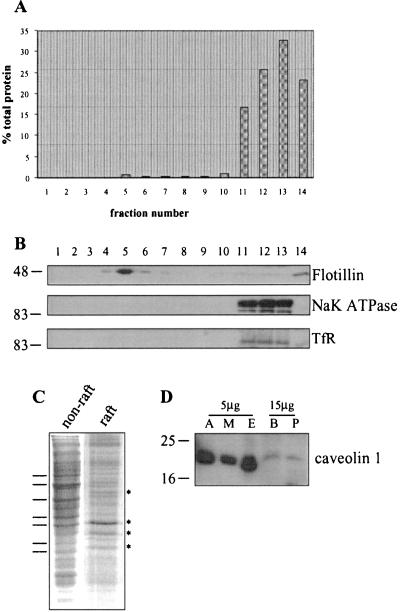
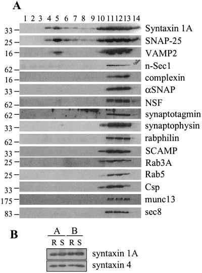
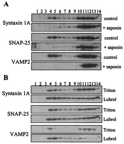
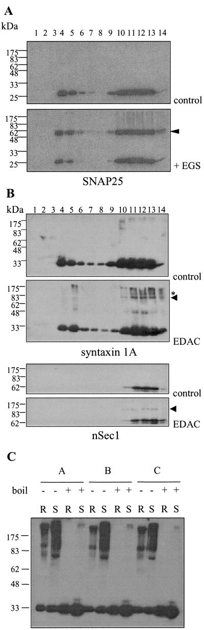
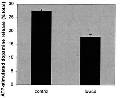
Lipid rafts are microdomains present within membranes of most cell types. These membrane microdomains, which are enriched in cholesterol and glycosphingolipids, have been implicated in the regulation of certain signal transduction and membrane traffic pathways. To investigate the possibility that lipid rafts organize exocytotic pathways in neuroendocrine cells, we examined the association of proteins of the exocytotic machinery with rafts purified from PC12 cells. The target soluble N-ethylmaleimide-sensitive factor attachment protein receptor (tSNARE) proteins syntaxin 1A and synaptosomal-associated protein of 25 kDa (SNAP-25) were both found to be highly enriched in lipid rafts ( approximately 25-fold). The vesicle SNARE vesicle-associated membrane protein (VAMP)2 was also present in raft fractions, but the extent of this recovery was variable. However, further analysis revealed that the majority of VAMP2 was associated with a distinct class of raft with different detergent solubility characteristics to the rafts containing syntaxin 1A and SNAP-25. Interestingly, no other studied secretory proteins were significantly associated with lipid rafts, including SNARE effector proteins such as nSec1. Chemical crosslinking experiments showed that syntaxin1A/SNAP-25 heterodimers were equally present in raft and nonraft fractions, whereas syntaxin1A/nSec1 complexes were detected only in nonraft fractions. SDS-resistance assays revealed that raft-associated syntaxin1A/SNAP-25 heterodimers were able to interact with VAMP2. Finally, reduction of cellular cholesterol levels decreased the extent of regulated exocytosis of dopamine from PC12 cells. The results described suggest that the interaction of SNARE proteins with lipid rafts is important for exocytosis and may allow structural and spatial organization of the secretory machinery.


Figures

Figure 1
Figure 1
Characterization of raft and nonraft fractions isolated from PC12 cells and caveolin expression in PC12 cells. PC12 cells were solubilized in 1% Triton X-100 and fractionated on a discontinuous sucrose gradient, as detailed in Materials and Methods. (A) The protein content of gradient fractions shown as a percentage of the total protein on the gradient. (B) Equal volumes of the gradient fractions were separated by SDS/PAGE and transferred to nitrocellulose for immunoblotting analysis by using antibodies specific for flotillin, α1 subunit of the NaK ATPase and the transferrin receptor (TfR). (C) Similar amounts of protein from fraction 12 (nonraft) and fraction 5 (raft) were separated by SDS/PAGE and stained with Coomassie blue. Differences between the protein profiles are highlighted by lines and asterisks. (D) Homogenates prepared from 3T3-L1 adipocytes (A), L6 skeletal muscle cells (M), endothelium (E), brain (B) and PC12 cells (P) were separated by SDS/PAGE and transferred to nitrocellulose for immunoblotting analysis by using a caveolin-1 specific antibody. Molecular weight markers are shown on Left in B and D.
Figure 2
Figure 2
Analysis of raft-associated proteins in PC12 cells. Cells were solubilized in 1% Triton X-100 and fractionated on a discontinuous sucrose gradient, as detailed in Materials and Methods. (A) Equal volumes of the recovered fractions were separated by SDS/PAGE and transferred to nitrocellulose for immunoblotting analysis by using antibodies against the indicated proteins. Molecular weight markers are indicated on the Left of all blots. This analysis was repeated on four separate gradients with similar results. Shown is a representative experiment. (B) Comparison of syntaxin 1A and syntaxin 4 association with isolated raft (R) and solubilized (S) fractions from two separate experiments (A and B).
Figure 3
Figure 3
Cholesterol dependence of raft association of SNARE proteins, and analysis of SNARE association with Lubrol-insoluble rafts. PC12 cells were solubilized in (A) either 1% Triton X-100 (control) or 0.5% Triton + 0.5% saponin (+ saponin); or (B) 1% Triton X-100 (Triton) or 1% Lubrol WX (Lubrol), and fractionated on a discontinuous sucrose gradient, as detailed in Materials and Methods. Equal volumes of the recovered fractions were separated by SDS/PAGE and transferred to nitrocellulose for immunoblotting analysis by using antibodies specific for syntaxin 1A, SNAP-25 and VAMP2.
Figure 4
Figure 4
Analysis of protein complexes in raft and nonraft fractions. Membranes were prepared from PC12 cells and incubated in the presence or absence of 10 mM EGS (A) or 10 mM EDAC (B) for 30 min at room temperature. The membranes were washed, solubilized in 1% Triton X-100, and fractionated on sucrose gradients, as described in Materials and Methods. Equal volumes of the recovered fractions were separated by SDS/PAGE and transferred to nitrocellulose for immunoblotting analysis by using the antibodies indicated. Arrowheads indicate putative SNAP-25/syntaxin 1A heterodimers (A) and nSec1/syntaxin 1A complex (B). Asterisk in B highlights an unknown syntaxin 1A-containing complex. (C) Raft fractions (R) and solubilized fractions (S) were identified and separated by SDS/PAGE with (+) or without (−) previous boiling of the samples. Immunoblotting analysis with syntaxin 1A antibody detected monomeric syntaxin 1A and syntaxin 1A-containing SDS-resistant complexes in nonboiled samples. This analysis was performed on fractions prepared from three individual experiments (A, B, and C). Molecular weight markers are indicated on the Left of all blots.
Figure 5
Figure 5
Effect of cellular cholesterol depletion on ATP-stimulated dopamine release from PC12 cells. Cells were treated without (control) or with lovastatin and methyl-β-cyclodextrin (lov/cd) as detailed in Materials and Methods. Cells were loaded with [3H] dopamine, and release was assayed in response to 300 μM ATP for 10 min. The total cellular [3H] dopamine content of each sample was also measured and dopamine release in response to ATP expressed as a percentage of total dopamine content. [3H] dopamine release in the absence of ATP was assayed and subtracted from the values shown. The data shown are averaged from three separate experiments (n = 18).

References

    1. Weber T, Zemelman B V, McNew J A, Westermann B, Gmachl M, Parfiati F, Sollner T H, Rothman J E. Cell. 1998;92:759–772. - PubMed
    1. Sudhof T C. Nature (London) 1995;375:645–653. - PubMed
    1. Chamberlain L H, Roth D, Morgan A, Burgoyne R D. J Cell Biol. 1995;130:1063–1071. - PMC - PubMed
    1. Ungermann C, Nichols B J, Pelham H R B, Wickner W. J Cell Biol. 1998;140:61–69. - PMC - PubMed
    1. Xu T, Ashery U, Burgoyne R D, Neher E. EMBO J. 1999;18:3293–3304. - PMC - PubMed

Publication types

LinkOut - more resources