. 2001 May 8;98(10):5509-14.
doi: 10.1073/pnas.091613398. Epub 2001 May 1.

Structure of Hjc, a Holliday junction resolvase, from Sulfolobus solfataricus

Affiliations

Structure of Hjc, a Holliday junction resolvase, from Sulfolobus solfataricus

C S Bond et al. Proc Natl Acad Sci U S A. .

Abstract

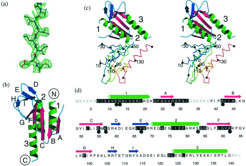
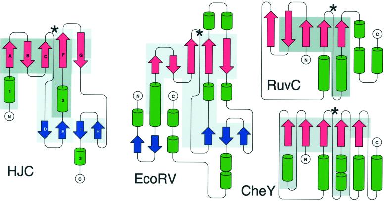
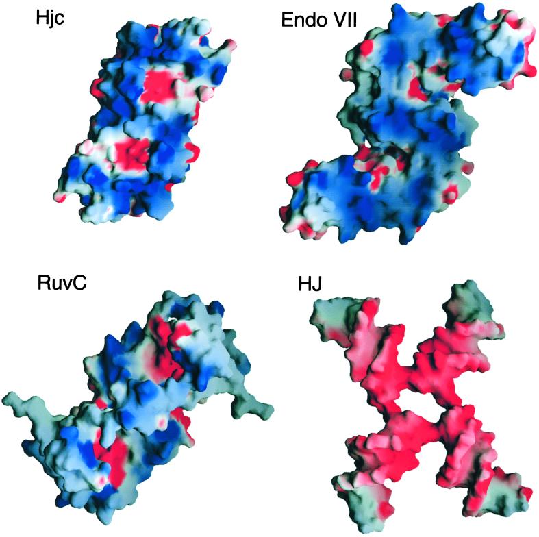
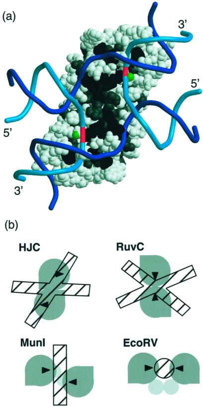
The 2.15-A structure of Hjc, a Holliday junction-resolving enzyme from the archaeon Sulfolobus solfataricus, reveals extensive structural homology with a superfamily of nucleases that includes type II restriction enzymes. Hjc is a dimer with a large DNA-binding surface consisting of numerous basic residues surrounding the metal-binding residues of the active sites. Residues critical for catalysis, identified on the basis of sequence comparisons and site-directed mutagenesis studies, are clustered to produce two active sites in the dimer, about 29 A apart, consistent with the requirement for the introduction of paired nicks in opposing strands of the four-way DNA junction substrate. Hjc displays similarity to the restriction endonucleases in the way its specific DNA-cutting pattern is determined but uses a different arrangement of nuclease subunits. Further structural similarity to a broad group of metal/phosphate-binding proteins, including conservation of active-site location, is observed. A high degree of conservation of surface electrostatic character is observed between Hjc and T4-phage endonuclease VII despite a complete lack of structural homology. A model of the Hjc-Holliday junction complex is proposed, based on the available functional and structural data.


Figures

Figure 1
Figure 1
The structure of S. solfataricus Hjc. (a) Solvent-flattened experimental electron density (1.3σ) for residues in strand C superimposed on the final model. (b) Ribbon representation of the Hjc monomer. The core β-sheet is shown as magenta arrows, α-helices are shown as green ribbon, and the peripheral β-sheet is shown as blue arrows. (c) Stereo representation of the Hjc dimer. One subunit is colored as above, with helices 1–3 labeled. The second subunit is a trace colored from blue at the N terminus to red at the C terminus. Every tenth Cα atom is marked by a black sphere and every twentieth is labeled. (d) The amino acid sequence of Hjc. Secondary structure elements are colored as above. Black shading marks residues that are highly conserved among Hjc family members. Large black circles mark residues for which mutation is deleterious to function. ad were prepared by using MOLSCRIPT (36), RASTER3D (37), and ALSCRIPT (38). Secondary structure was assigned by using DSSP (39).
Figure 2
Figure 2
The topologies of Hjc, EcoRV, Thermotoga maritima CheY, and E. coli RuvC. Asterisks mark the position of the divalent metal-binding site of each enzyme. Shading marks regions where Hjc shows structural similarity to the other enzymes. Prepared by using PROMOTIF (40) and TOPDRAW (available from authors).
Figure 3
Figure 3
The DNA-binding surfaces of Hjc, T4 endonuclease VII, and RuvC colored by electrostatic potential (blue represents positive and red represents negative charge). For comparison, the surface of a Holliday junction (HJ) computer-modeled in the X-shaped global structure predicted for junction bound by Hjc also is shown. Prepared by using GRASP (41).
Figure 4
Figure 4
(a) A model of Holliday junction DNA bound to Hjc. Hjc is shown as Corey–Pauling–Koltun spheres with residues conserved among Hjc sequences (gray) and residues for which mutants are inactive (black). A green ball marks the proposed metal-binding site. DNA is shown as a phosphate-backbone trace (exchange strand, cyan; continuous strand, blue), with the cleavage point highlighted in red. Prepared by using MOLSCRIPT (36). (b) Varying arrangements of similar nuclease domains produce different DNA-nicking patterns for the resolving enzymes Hjc and RuvC and the endonucleases EcoRV and MunI. Gray shapes indicate nuclease domains, with black triangles at the nicking site. DNA is represented by hatched rectangles or a circle.

References

    1. West S C. Annu Rev Genet. 1997;31:213–244. - PubMed
    1. Shinagawa H, Iwasaki H. Trends Biochem Sci. 1996;21:107–111. - PubMed
    1. Lilley D M J, White M F. Proc Natl Acad Sci USA. 2000;97:9351–9353. - PMC - PubMed
    1. White M F, Giraud-Panis M J, Pohler J R, Lilley D M J. J Mol Biol. 1997;269:647–664. - PubMed
    1. Ariyoshi M, Vassylyev D G, Iwasaki H, Nakamura H, Shinagawa H, Morikawa K. Cell. 1994;78:1063–1072. - PubMed

Publication types

Substances

Associated data

LinkOut - more resources