. 2001 May 8;98(10):5693-8.
doi: 10.1073/pnas.101049998. Epub 2001 May 1.

Trans-spliced leader addition to mRNAs in a cnidarian

Affiliations

Trans-spliced leader addition to mRNAs in a cnidarian

N A Stover et al. Proc Natl Acad Sci U S A. .

Abstract

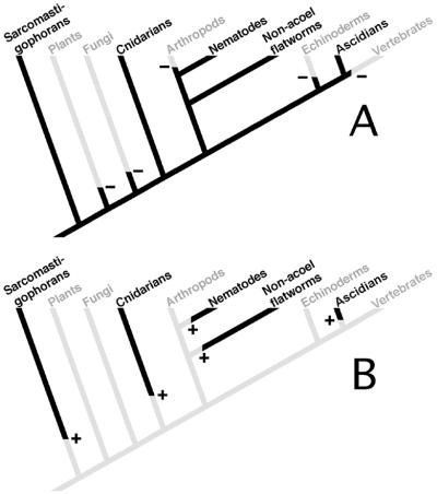
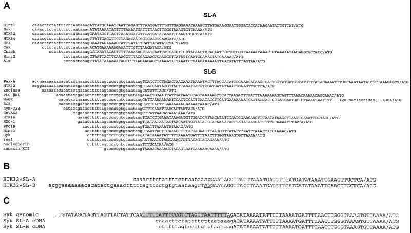
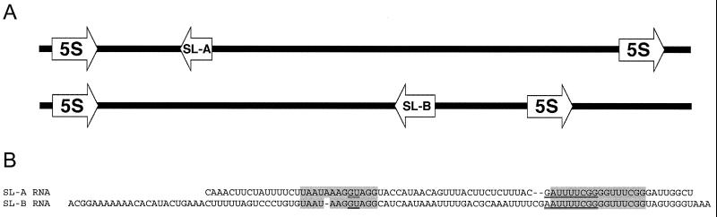
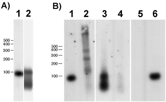
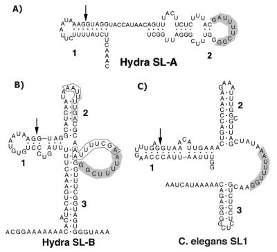
A search of databases with the sequence from the 5' untranslated region of a Hydra cDNA clone encoding a receptor protein-tyrosine kinase revealed that a number of Hydra cDNAs contain one of two different sequences at their 5' ends. This finding suggested the possibility that mRNAs in Hydra receive leader sequences by trans-splicing. This hypothesis was confirmed by the finding that the leader sequences are transcribed as parts of small RNAs encoded by genes located in the 5S rRNA clusters of Hydra. The two spliced leader (SL) RNAs (SL-A and -B) contain splice donor dinucleotides at the predicted positions, and genes that receive SLs contain splice acceptor dinucleotides at the predicted positions. Both of the SL RNAs are bound by antibody against trimethylguanosine, suggesting that they contain a trimethylguanosine cap. The predicted secondary structures of the Hydra SL RNAs show significant differences from the structures predicted for the SLs of other organisms. Messenger RNAs have been identified that can receive either SL-A or -B, although the impact of the two different SLs on the function of the mRNA is unknown. The presence and features of SL addition in the phylum Cnidaria raise interesting questions regarding the evolution of this process.


Figures

Figure 1
Figure 1
Phylogenetic distribution of spliced leader addition to mRNAs. Phylogenetic relationships of only those taxa in which spliced leader addition is known to be present or likely to be absent are shown. The phylogenetic relationships between the taxa are based on multiple molecular studies (–44). (A) A version of the tree in which spliced leader addition arose in a unicellular eukaryote and was subsequently lost from the various taxa indicated in gray. Minuses indicate points at which loss occurred. (B) A version of the tree in which spliced leader addition arose independently in the various phyla indicated in black. +, points at which origin of spliced leader addition occurred.
Figure 2
Figure 2
(A) Sequence identities at the 5′ ends of cDNA clones from Hydra genes. Identical 5′ sequences are in lowercase; divergent downstream sequences are in uppercase. The translation start ATG codon is separated from the 5′ untranslated region sequence by a slash. The upper group of sequences contains the spliced leader A (SL-A) sequence; the lower group contains the spliced leader B (SL-B) sequence. GenBank accession nos. for the sequences are as follows: Hint, M64611; Syk, AF060949; HTK32, AF123442; HTK54, U24116; HFZ, AF209200; Csk, AF067775; Cnash, U36275; Alx, AF295531; Pax-A, U96193; enolase, U85827; PLC-βHI, AB017511; hyGK, AF031931; ECE, AF162671; hym-323, AB40074; HTK16, U00936; HZO-1, AF230482; PKC1B, Y12857; ras1, X78597; nucleoporin, U85827; annexin XII, M83736. All genes are from H. vulgaris except hyGK (Hydra oligactis), enolase (H. oligactis), Pax-A (Hydra littoralis), and PLC-βI (Hydra magnipapillata). The three different Hint sequences (labeled Hint 1–3) arise because of alternative splicing. Hint produces a long transcript with SL-A at the 5′ end and a shorter transcript that can contain either SL-A or -B (45). The single nucleotide difference (T>G) in the SL-A sequence of HTK54 may be because of an error during cDNA synthesis or the presence of multiple alleles of SL-A. A complete copy of the SL-B sequence is located internally in a H. vulgaris cDNA for cAMP-response element-binding protein (CREB) (46), where it results in the truncation of a highly conserved portion of the CREB coding sequence. We have attempted, without success, to confirm this arrangement by amplification of the corresponding region from first-strand cDNA made from H. vulgaris polyA+ RNA. We therefore believe that this clone is a hybrid produced during cDNA library construction by ligation of the 3′ end of a partial CREB cDNA to the 5′ end of a cDNA derived from an SL-B-containing mRNA. (B) Alignment of sequences from the 5′ ends of HTK32 cDNA clones containing SL-A or -B sequences. The clone containing SL-B includes four nucleotides that are not present in the SL-A-containing message. The splice acceptor dinucleotide is underlined. The SL-A-containing sequence is from a clone isolated from a cDNA library. The SL-B-containing sequence was obtained by 5′ RACE (see Materials and Methods). (C) Genomic sequence from the Hydra Syk gene (24). The splice acceptor dinucleotide is indicated by double underlining. The genomic sequence is aligned with the sequences from Syk cDNAs containing either SL-A or -B (see A). The pyrimidine-rich sequence upstream of the splice acceptor dinucleotide is shaded.
Figure 3
Figure 3
SL-A and -B RNA sequences and gene arrangement. (A) Arrangement of the SL-A and -B genes in the 5S rRNA clusters. The 5′ and 3′ ends of the 5S gene were identified by comparison to available cnidarian 5S rRNA sequences (–23). (B) The SL-A and -B RNA sequences were aligned manually. Conserved sequences surrounding the splice donor site and the predicted Sm-binding site are shaded. The predicted Sm-binding sequence and the splice donor dinucleotide are doubly underlined.
Figure 4
Figure 4
(A) Northern blot of Hydra total RNA hybridized with probes to the intron region of SL-A (lane 1) and the exon region of SL-B (lane 2). Both probes hybridize to RNA of the predicted sizes as described in the text. (B) TMG-containing RNA was isolated from an aliquot of total RNA by using an anti-TMG antibody–agarose conjugate. Bound (lanes 1, 3, and 5) and unbound (lanes 2, 4, and 6) RNAs were hybridized with the SL-A (lanes 1 and 2) and -B (lanes 3 and 4) probes used in A and with a probe for 5S rRNA (lanes 5 and 6).
Figure 5
Figure 5
Predicted secondary structures of SL-A RNA (A), SL-B RNA (B), and the C. elegans SL1 RNA (C). The structures were generated by using Version 2.3 of the mfold RNA secondary structure modeling program (27, 28). The parameters and constraints used for folding are detailed in the text. The stem–loop structures are numbered for reference. Arrows in Stem 1 indicate the 3′ end of the exon sequence in each RNA. Putative Sm protein-binding sequences are shaded. Two other possible Sm-binding sequences discussed in the text are boxed in B. The diagrams were produced by inputting the structural data generated by mfold into RnaViz (47).

References

    1. Vandenberghe A E, Meedel T H, Hastings K E M. Genes Dev. 2001;15:294–303. - PMC - PubMed
    1. Davis R E. Parasitol Today. 1996;12:33–40. - PubMed
    1. Nilsen T W. Infect Agents Dis. 1992;1:212–218. - PubMed
    1. Nilsen T W. Annu Rev Microbiol. 1993;47:413–440. - PubMed
    1. Agabian N. Cell. 1990;61:1157–1160. - PubMed

Publication types

Associated data

LinkOut - more resources