

Hydrogen peroxide mediates the cell growth and transformation caused by the mitogenic oxidase Nox1
- PMID: 11331784
- PMCID: PMC33250
- DOI: 10.1073/pnas.101505898
Hydrogen peroxide mediates the cell growth and transformation caused by the mitogenic oxidase Nox1
Abstract
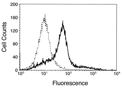
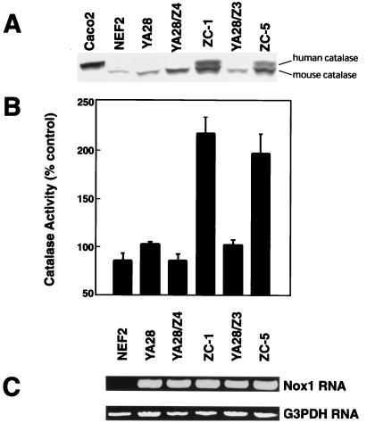
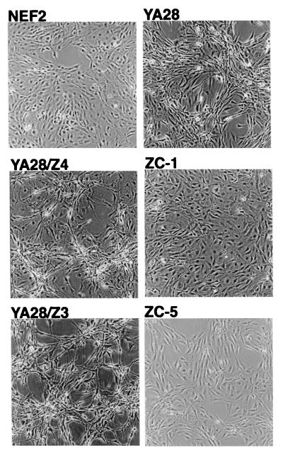
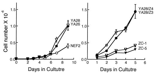
Nox1, a homologue of gp91phox, the catalytic moiety of the superoxide (O(2)(-))-generating NADPH oxidase of phagocytes, causes increased O(2)(-) generation, increased mitotic rate, cell transformation, and tumorigenicity when expressed in NIH 3T3 fibroblasts. This study explores the role of reactive oxygen species (ROS) in regulating cell growth and transformation by Nox1. H(2)O(2) concentration increased approximately 10-fold in Nox1-expressing cells, compared with <2-fold increase in O(2)(-). When human catalase was expressed in Nox1-expressing cells, H(2)O(2) concentration decreased, and the cells reverted to a normal appearance, the growth rate normalized, and cells no longer produced tumors in athymic mice. A large number of genes, including many related to cell cycle, growth, and cancer (but unrelated to oxidative stress), were expressed in Nox1-expressing cells, and more than 60% of these returned to normal levels on coexpression of catalase. Thus, H(2)O(2) in low concentrations functions as an intracellular signal that triggers a genetic program related to cell growth.
Figures
References
-
- Burdon R. Free Radical Biol Med. 1995;18:775–794. - PubMed
-
- Sundaresan M, Yu Z-X, Ferrans V J, Irani K, Finkel T. Science. 1995;270:296–299. - PubMed
-
- Chen Q M. Ann N Y Acad Sci. 2000;908:111–125. - PubMed
-
- Bladier C, Wolvetang E J, Hutchinson P, de Haan J B, Kola I. Cell Growth Differ. 1997;8:589–598. - PubMed
Publication types
MeSH terms
Substances
Grants and funding
LinkOut - more resources
Full Text Sources
Other Literature Sources
Molecular Biology Databases
