. 2001 May 2;93(9):691-9.
doi: 10.1093/jnci/93.9.691.

Epigenetic inactivation of RASSF1A in lung and breast cancers and malignant phenotype suppression

Affiliations

Epigenetic inactivation of RASSF1A in lung and breast cancers and malignant phenotype suppression

D G Burbee et al. J Natl Cancer Inst. .

Abstract

Background: The recently identified RASSF1 locus is located within a 120-kilobase region of chromosome 3p21.3 that frequently undergoes allele loss in lung and breast cancers. We explored the hypothesis that RASSF1 encodes a tumor suppressor gene for lung and breast cancers.

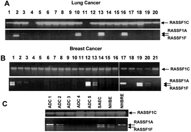
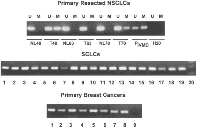
Methods: We assessed expression of two RASSF1 gene products, RASSF1A and RASSF1C, and the methylation status of their respective promoters in 27 non-small-cell lung cancer (NSCLC) cell lines, in 107 resected NSCLCs, in 47 small-cell lung cancer (SCLC) cell lines, in 22 breast cancer cell lines, in 39 resected breast cancers, in 104 nonmalignant lung samples, and in three breast and lung epithelial cultures. We also transfected a lung cancer cell line that lacks RASSF1A expression with vectors containing RASSF1A complementary DNA to determine whether exogenous expression of RASSF1A would affect in vitro growth and in vivo tumorigenicity of this cell line. All statistical tests were two-sided.

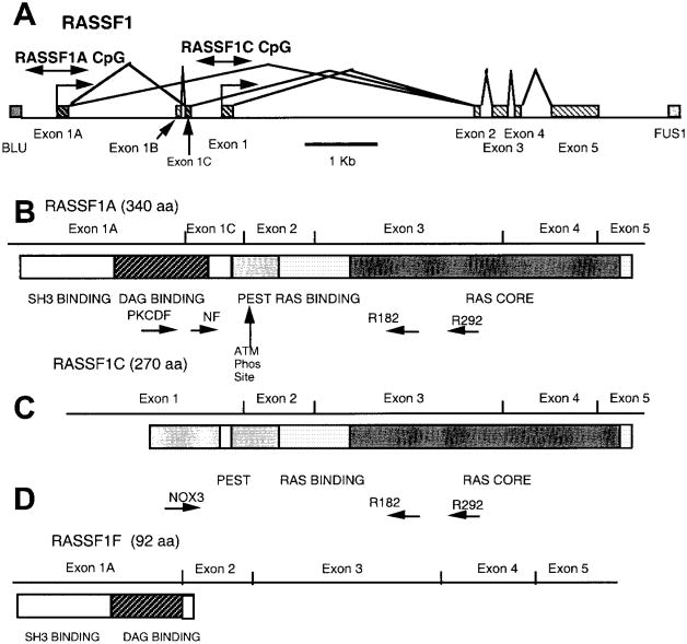
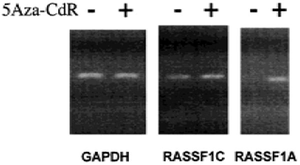
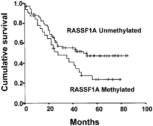
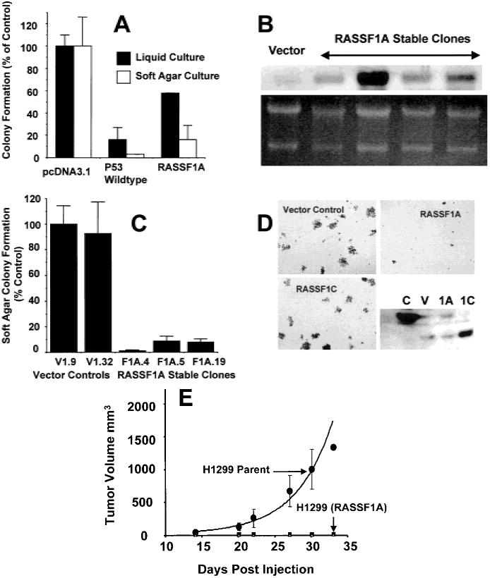
Results: RASSF1A messenger RNA was expressed in nonmalignant epithelial cultures but not in 100% of the SCLC, in 65% of the NSCLC, or in 60% of the breast cancer lines. By contrast, RASSF1C was expressed in all nonmalignant cell cultures and in nearly all cancer cell lines. RASSF1A promoter hypermethylation was detected in 100% of SCLC, in 63% of NSCLC, in 64% of breast cancer lines, in 30% of primary NSCLCs, and in 49% of primary breast tumors but in none of the nonmalignant lung tissues. RASSF1A promoter hypermethylation in resected NSCLCs was associated with impaired patient survival (P =.046). Exogenous expression of RASSF1A in a cell line lacking expression decreased in vitro colony formation and in vivo tumorigenicity.

Conclusion: RASSF1A is a potential tumor suppressor gene that undergoes epigenetic inactivation in lung and breast cancers through hypermethylation of its promoter region.


Figures

Fig. 1
Fig. 1
Map of the RASSF1 locus, transcripts, and protein domains. A) The exon–intron structure of the RASSF1 locus with the location of the CpG islands in the predicted promoter regions (the locations of which are shown by double-headed arrows) of RASSF1A and RASSF1C. RASSF1A transcription is predicted to come from the most centromeric promoter region located within a CpG island and begins with exon 1A. RASSF1F also commences at this promoter but is missing exon 1C. Transcription of RASSF1C is predicted to begin in the most telomeric promoter region, which is approximately 2 kilobases from that of RASSF1A and begins with exon 1. Blocks represent exons; lines represent introns. B) Schematic of the RASSF1A transcript and predicted protein-sequence domains. The location of the various primers (PKCDF, NF, R182, and R292) used for isoform-specific reverse transcription (RT)–polymerase chain reaction (PCR) analyses are indicated. Tick marks identify the exon boundaries. The potential src homology 3 (SH3)-binding region, putative diacylglycerol (DAG)-binding domain, PEST sequence, Ras-association domain, and ataxia-telangiectasia-mutated (ATM) phosphorylation site are labeled. C) Schematic of the RASSF1C transcript and predicted protein-sequence domains. The locations of the various primers (NOX3, R182, and R292) used for isoform-specific RT–PCR analyses are indicated. D) Schematic of the RASSF1F transcript and predicted protein-sequence domains.
Fig. 2
Fig. 2
RASSF1A and RASSF1C messenger RNA levels detected by isoform-specific reverse transcription–polymerase chain reaction (RT–PCR) in a sampling of lung cancer cell lines (A), breast cancer lines (B), and resected lung tumors and normal human lung and breast epithelial cultures (C). All RT– PCR products were separated on 2% agarose gels and were identified by staining with ethidium bromide. Arrows indicate location of transcripts. A) Lung cancer lines tested in lanes: 1 = H157; 2 = H358; 3 = H727; 4 = H740; 5 = H748; 6 = H838; 7 = H1184; 8 = H1299; 9 = H1304; 10 = H1437; 11 = H1450; 12 = H1770; 13 = H1792; 14 = H1963; 15 = H1993; 16 = H2009; 17 = H2077; 18 = H2108; 19 = HHCC44; and 20 = HCC78. B) Breast cancer lines tested in lanes: 1 = HCC38; 2 = HCC1187; 3 = HTB19; 4 = HTB20; 5 = HTB22; 6 = HTB23; 7 = HTB24; 8 = HTB25; 9 = HTB26; 10 = HTB27; 11 = HTB121; 12 = HTB129; 13 = HTB130; 14 = HTB131; 15 = HTB132; 16 = HTB133; 17 = HCC1395; 18 = HCC1428; 19 = HCC1569; 20 = HCC1806; and 21 = HCC2157. C) Resected lung adenocarcinoma samples (ADC 1–5) and cultures of normal small-airway epithelial cells (SAECs), normal human bronchial epithelial (NHBE) cultures, and normal human breast epithelial (NHBRE) cultures.
Fig. 3
Fig. 3
Expression of RASSF1A after treatment of lung cancer cells with 5-aza-2′-deoxycytidine (5Aza-CdR). NCI-H157, a non-small-cell lung carcinoma (NSCLC) cell line that expresses RASSF1C but not RASSF1A, was grown in the presence (+ lanes) and absence (− lanes) of 0.5 μM 5Aza-CdR for 48 hours. Total RNA was isolated, complementary DNA was prepared, and isoform-specific reverse transcription–polymerase chain reaction was performed for RASSF1A, RASSF1C, and glyceraldehyde-3-phosphate dehydrogenase (GAPDH) as a control.
Fig. 4
Fig. 4
Methylation-specific polymerase chain reaction (PCR) for the detection of methylated RASSF1A 5′ CpG sequences in primary resected non-small-cell lung carcinomas (NSCLCs) and their accompanying normal lung tissue (upper panel), small-cell lung carcinoma (SCLC) cell lines (middle panel), and primary breast cancers (lower panel). Representative samples are shown. For resected NSCLCs, U = results with primers specific for unmethylated sequences; M = results with primers specific for methylated sequences. NL = normal lung tissue; T = tumor; P = results with peripheral blood lymphocyte DNA, which is unmethylated or in vitro methylated (IVMD); and H20 = negative controls with water blanks. For SCLCs, each lane shows the PCR results for the methylated sequences from a different cell line. Lane 20 is negative control. For the breast cancers, each lane shows the PCR results for methylated sequences from a different sample. PCR products were separated on 2% agarose gels, and bands were detected after staining with ethidium bromide.
Fig. 5
Fig. 5
Kaplan-Meier survival curve for 107 patients with resected non-small-cell lung carcinomas based on RASSF1A methylation status (32 methylated and 75 not methylated). For the patients with unmethylated RASSF1A alleles, the number of cases = 75, censored = 39, and events = 36, with a mean overall survival of 52 months (95% confidence interval [CI] = 44 to 59) and a median overall survival of 49 months (95% CI = 44 to 59); for the patients with methylated RASSF1A alleles, the number of cases = 32, censored = nine, and events = 23, with a mean overall survival of 37 months (95% CI = 27 to 46) and a median overall survival of 28 months (95% CI = 9 to 47). The log-rank test statistic for equality of survival distributions for RASSF1A methylation was 3.97, with df 1, P = .0463. The patients at risk for each group were: RASSF1A unmethylated—12 months (n = 63), 36 months (n = 34), and 60 months (n = 16); RASSF1A methylated—12 months (n = 24), 36 months (n = 13), and 60 months (n = 5).
Fig. 6
Fig. 6
Effect of RASSF1A on the in vitro and in vivo growth of the non-small-cell lung carcinoma (NSCLC) cell line NCI-H1299. A) Anchorage-dependent and anchorage-independent colony formation after transfection of NCI-H1299 cells with the empty vector (pcDNA3.1+) or pcDNA3.1+ expression vectors containing wild-type p53 or RASSF1A. For analysis of anchorage-dependent growth, after 2 days in nonselective growth medium, transfected NCI-H1299 cells were diluted into 100-mm2 dishes with selective medium. Transfected cells were plated in liquid medium (for anchorage-dependent assays) or soft agar (for anchorage-independent assays) containing 800 μg/mL of G418. Colonies were stained with methylene blue in anchorage-dependent experiments after 14 days. Results represent the average of eight to 12 experiments in liquid medium and three soft-agar experiments. Standard deviations are shown or are less than 2%. Solid bars = anchorage-dependent growth (95% confidence interval [CI] = 0 to 36 for wt-p53 (wild-type) and 52 to 60 for RASSF1A); open bars = anchorage-independent growth (95% CI = 0 to 6 for wild-type (wt)-p53 and 0 to 39 for RASSF1A). B) Northern blot analysis of the RASSF1A expression in stable clones of NCI-H1299 cells transfected with the pcDNA3.1+ vector or pcDNA3.1+ containing RASSF1A complementary DNA (cDNA). The vector control (vector) and four separate clones with various RASSF1A messenger RNA levels are shown. Several of these clones were used in the anchorage-independent growth assay shown in D. Ethidium bromide staining of the ribosomal RNA is shown as a loading control. The clones were also verified to express the RASSF1A isoform by reverse transcription–polymerase chain reaction with the use of isoform-specific primers (data not shown). C) Soft-agar (anchorage-independent) colony formation in stable clones of NCI-H1299 cells transfected with the pcDNA3.1+ vector or pcDNA3.1+ containing RASSF1A cDNA. The means and standard deviations are shown. For each of the RASSF1A-expressing clones, the 95% CI = 0 to 4 for F1A.4, 2 to 16 for F1A.5, and 3 to 14 for F1A.19. D) NCI-H1299 cells were infected with the pBABEpuro retrovirus expression vectors containing either the vector control or the RASSF1A or RASSF1C cDNAs. Infected cells (10 000 per plate) were suspended in 0.33% agar, and the suspension was layered over a 0.5% agar base. Colonies greater than 0.2 mm in diameter were counted after 21 days. The lower right panel shows a representative western blot, developed with a rabbit antibody to the RASSF1-glutathione S-transferase fusion protein, to verify the expression of the RASSF1 proteins. C = positive control generated by transient transfection of NCI-H1299 cells with pcDNA3.1+ containing RASSF1A cDNA; V = infection of NCI-H1299 cells with the retroviral vector control (note runover from positive control); 1A = infection of NCI-H1299 cells with the retroviral vector containing RASSF1A; and 1C = infection of NCI-H1299 cells with the retroviral vector containing RASSF1C. E) Effect of RASSF1A on the in vivo growth of NCI-H1299 cells. Approximately 107 viable NCI-H1299 cells expressing RASSF1A were injected into the flanks of each of five previously irradiated BALB/c (nu/nu) nude mice. Tumor size was monitored over time, and size is shown in cubic millimeters. The average volume of tumors grown in more than 20 mice that were given an injection of vector-transfected NCI-H1299 cells is shown (H1299 parent). Mice that were given an injection of RASSF1A-infected NCI-H1299 cells grew no measurable tumors.

Comment in

References

    1. Wistuba I, Behrens C, Virmani A, Mele G, Milchgrub S, Girard L, et al. High resolution chromosome 3p allelotyping of human lung cancer and preneoplastic/preinvasive bronchial epithelium reveals multiple, discontinuous sites of 3p allele loss and three regions of frequent breakpoints. Cancer Res. 2000;60:1949–60. - PubMed
    1. Sekido Y, Fong KM, Minna JD. Progress in understanding the molecular pathogenesis of human lung cancer. Biochim Biophys Acta. 1998;1378:F21–59. - PubMed
    1. Wistuba II, Lam S, Behrens C, Virmani AK, Fong KM, LeRiche J, et al. Molecular damage in the bronchial epithelium of current and former smokers. J Natl Cancer Inst. 1997;89:1366–73. - PMC - PubMed
    1. Wistuba II, Behrens C, Milchgrub S, Bryant D, Hung J, Minna JD, et al. Sequential molecular abnormalities are involved in the multistage development of squamous cell lung carcinoma. Oncogene. 1999;18:643–50. - PubMed
    1. Kok K, Naylor SL, Buys CH. Deletions of the short arm of chromosome 3 in solid tumors and the search for suppressor genes. Adv Cancer Res. 1997;71:27–92. - PubMed

Publication types